. 2012 Mar 16;287(12):9525-33.
doi: 10.1074/jbc.M111.327064. Epub 2012 Jan 31.

Lipid polarity is maintained in absence of tight junctions

Affiliations

Lipid polarity is maintained in absence of tight junctions

Junichi Ikenouchi et al. J Biol Chem. .

Abstract

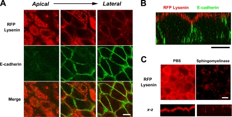
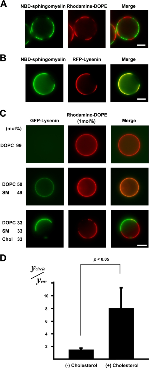
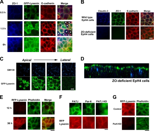
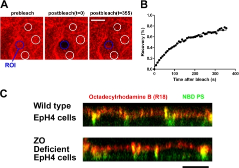
The role of tight junctions (TJs) in the establishment and maintenance of lipid polarity in epithelial cells has long been a subject of controversy. We have addressed this issue using lysenin, a toxin derived from earthworms, and an influenza virus labeled with a fluorescent lipid, octadecylrhodamine B (R18). When epithelial cells are stained with lysenin, lysenin selectively binds to their apical membranes. Using an artificial liposome, we demonstrated that lysenin recognizes the membrane domains where sphingomyelins are clustered. Interestingly, lysenin selectively stained the apical membranes of epithelial cells depleted of zonula occludens proteins (ZO-deficient cells), which completely lack TJs. Furthermore, the fluorescent lipid inserted into the apical membrane by fusion with the influenza virus did not diffuse to the lateral membrane in ZO-deficient epithelial cells. This study revealed that sphingomyelin-cluster formation occurs only in the apical membrane and that lipid polarity is maintained even in the absence of TJs.


Figures

FIGURE 1.
FIGURE 1.
Selective recognition of the apical membrane of epithelial cells by lysenin. A, EpH4 cells were fixed and stained with RFP-lysenin (red) and anti-E-cadherin mAb (green) (scale bar, 10 μm). B, xz section of a confocal image demonstrates that lysenin selectively recognized the apical membrane (scale bar, 10 μm). C, cells were treated with bacterial sphingomyelinase before fixation for 15 min at 37 °C. In bacterial sphingomyelinase-treated cells, staining of lysenin in the apical membrane disappeared (scale bar, 5 μm).
FIGURE 2.
FIGURE 2.
MS profiling of PC and SM molecular species in the apical and basolateral membranes. A, shown is the scheme of apical and basolateral membrane isolation for epithelial cell monolayers cultured in dishes. For details, see “Experimental Procedures.” B, shown is an immunoblot analysis of the apical membrane fraction, basolateral membrane fraction, and floating fraction. 5 μg of each membrane fraction was separated by SDS-PAGE (Coomassie Brilliant Blue staining in the left panel), transferred to nitrocellulose membranes, and probed with antibodies against the indicated marker proteins (right panel). The results are representative of three independent experiments. C, intact EpH4 cells and basolateral membrane were fixed and stained with the indicated antibodies. Note that signals of GM130/DAPI/claudin-3 were little observed in the basolateral membrane (scale bar, 10 μm). D, shown is scanning electron micrographs of isolated apical and basolateral membranes (white bar, 1 μm; black bar, 3 μm). E, shown are negative ion mass spectra of the m/z range 500–1000 of lipid extracts prepared from the apical and basolateral membranes. The peak assignment to sphingomyelin molecular species is indicated (red). Compared with the amount of phosphatidylcholine (34:1), sphingomyelin (d18:1–16:0) and sphingomyelin (d18:1–24:1) were enriched in both the apical membrane and basolateral membrane. For additional peak assignments, see “Results.” The total carbon chain length (x) and number of carbon-carbon double bonds (y) of individual lipid molecular species are specified as (x:y). The results are representative of three independent experiments.
FIGURE 3.
FIGURE 3.
Binding of lysenin to sphingomyelin-containing GUVs. A, shown is the observation of a GUV containing 5 mol% NBD-sphingomyelin (green) (NBD-C12-SM/DOPC/SM(d18:1–16:0)/cholesterol 6:33:27:33) and RFP-lysenin (red) (scale bar, 10 μm). B, shown is the observation of binding of GFP-lysenin (green) to a GUV of DOPC/rhodamine-DOPE (molar ratio, 99:1) (upper panel), SM (d18:1–16:0)/DOPC/rhodamine-DOPE (molar ratio, 49:50:1) (middle panel), or SM(d18:1–16:0)/DOPC/cholesterol/rhodamine-DOPE (molar ratio, 33:33:33:1) (lower panel). Each GUV contained 1 mol% rhodamine-DOPE (scale bar, 10 μm). C, the fluorescence of GFP-lysenin associated with liposome (Ycircle) and the fluorescence of GFP-lysenin outside of the liposome (Yenv) were quantitatively measured in each GUV. The value of Ycircle/Yenv of the SM(d18:1- 16:0)/DOPC/cholesterol (Chol)/rhodamine-DOPE (molar ratio, 33:33:33:1) and SM (d18:1–16:0)/DOPC/rhodamine-DOPE (molar ratio, 49:50:1) GUVs was calculated. Data are the means ± S.D. of three independent experiments. p < 0.05 by Student's t test.
FIGURE 4.
FIGURE 4.
Asymmetric sphingomyelin clusters are retained in the absence of TJs. A, EpH4 cells were cultured overnight in low Ca2+ medium, and their polarization was initiated by transferring to normal Ca2+ medium. After a 0.5-, 1.5-, or 6-h incubation, cells were fixed and stained with anti-ZO-1 mAb (blue), GFP-lysenin (green), and anti-E-cadherin mAb (red). In some cells TJs were formed based on the staining of ZO-1 before apical sphingomyelin cluster formation (asterisks, middle panel) (scale bar, 10 μm). B, EpH4 cells and ZO-deficient EpH4 cells were fixed and stained with anti-claudin-3 polyclonal antibody (blue), anti-ZO-1 mAb (green), and anti-E-cadherin mAb (red) (scale bar, 10 μm). C, ZO-deficient EpH4 cells were fixed and stained with GFP-lysenin (green) and anti-GM130 mAb (blue). The asymmetric lysenin staining was maintained in ZO-deficient EpH4 cells (scale bar, 10 μm). D, xz section of a confocal image of B is shown. E, ZO-deficient cells were cultured in low Ca2+ medium overnight, and their polarization was initiated by transferring to normal Ca2+ medium. After a 12- or 36-h incubation, cells were fixed and stained with RFP-lysenin (red) and phalloidin (green) (scale bar, 20 μm). F, EpH4 cells were transfected with a Myc-tagged PATJ expression vector (top), a Myc-tagged Par-6A expression vector (middle), or a vector that simultaneously produces shRNA against PATJ and expresses GFP (bottom). Cells were stained with anti-Myc mAb (green) (top, middle) and RFP-lysenin (red) (scale bar, 10 μm). G, EpH4 cells were transfected with a control H1 promoter vector (top) or a vector that produces shRNA against Par-6 (bottom). KD, knock down. Cells were fixed and stained with RFP-lysenin (red) and phalloidin (green) (scale bar, 10 μm).
FIGURE 5.
FIGURE 5.
TJs are not essential for the diffusion barrier of lipids in epithelial cells. A, the apical membranes of wild-type EpH4 cells were fused with the R18-labeled influenza virus. The selected region of interest (blue circle) was then allowed to recover. The fluorescence intensities of three independent regions that had not been photobleached (white circles) were utilized to correct the photo damage (scale bar, 10 μm). ROI, region of interest. B, R18 fluorescence recovery at the apical membrane after photobleaching was calculated as detailed under “Experimental Procedures.” The R18 fluorescent lipid inserted into the apical membrane exhibited mobile fractions (fraction value, 73.9 ± 4.2%). C, wild-type EpH4 cells and ZO-deficient EpH4 cells were incubated with the R18-labeled influenza virus (red) and NBD-PS (100 μm) (green) on ice for 30 min. After the induction of viral fusion, the cells were incubated at 15 °C for 5 min, after which the distributions of R18 and NBD-PS were observed under a confocal microscope. R18 fluorescent lipid (red) was retained at the apical membrane even in ZO-deficient EpH4 cells (scale bar, 10 μm).

Similar articles

Cited by

References

    1. Nelson W. J. (2003) Adaptation of core mechanisms to generate cell polarity. Nature 422, 766–774 - PMC - PubMed
    1. Gassama-Diagne A., Yu W., ter Beest M., Martin-Belmonte F., Kierbel A., Engel J., Mostov K. (2006) Phosphatidylinositol 3,4,5-trisphosphate regulates the formation of the basolateral plasma membrane in epithelial cells. Nat. Cell Biol. 8, 963–970 - PubMed
    1. Martin-Belmonte F., Gassama A., Datta A., Yu W., Rescher U., Gerke V., Mostov K. (2007) PTEN-mediated apical segregation of phosphoinositides controls epithelial morphogenesis through Cdc42. Cell 128, 383–397 - PMC - PubMed
    1. van Meer G., Simons K. (1982) Viruses budding from either the apical or the basolateral plasma membrane domain of MDCK cells have unique phospholipid compositions. EMBO J. 1, 847–852 - PMC - PubMed
    1. van Meer G., Simons K. (1986) The function of tight junctions in maintaining differences in lipid composition between the apical and the basolateral cell surface domains of MDCK cells. EMBO J. 5, 1455–1464 - PMC - PubMed

Publication types

LinkOut - more resources