. 2012 Mar 16;287(12):9052-60.
doi: 10.1074/jbc.M111.306407. Epub 2012 Jan 30.

Internalization of proprotein convertase PC7 from plasma membrane is mediated by a novel motif

Affiliations

Internalization of proprotein convertase PC7 from plasma membrane is mediated by a novel motif

Jeroen Declercq et al. J Biol Chem. .

Abstract

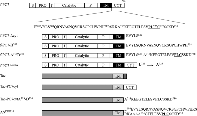
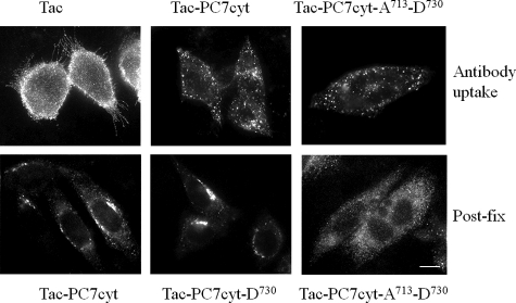
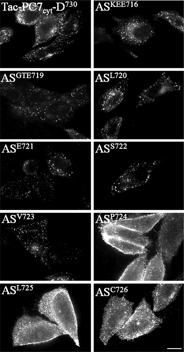
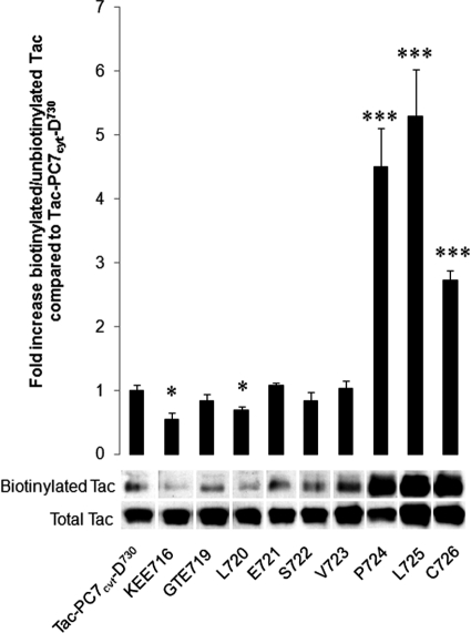
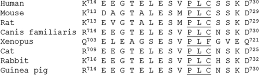
Proprotein convertase 7 (PC7) is a member of the subtilisin-like proprotein convertase family, which is involved in the endoproteolysis of a variety of precursor proteins. Under steady state conditions, PC7 is mainly localized in the trans-Golgi network, but a small fraction is found at the cell surface. So far, no sorting signals for membrane trafficking have been identified in PC7. In this study, we have examined the internalization of PC7 from the plasma membrane. Our results show that internalization of PC7 is mediated by clathrin-coated vesicles. After inhibition of clathrin-mediated endocytosis using hypertonic conditions or the small molecule inhibitor, Pitstop 2, PC7 accumulated at the plasma membrane. Furthermore, PC7 was present in isolated clathrin-coated vesicles. To determine the internalization motif, constructs were generated in which parts of the N and C terminus of the cytoplasmic tail of PC7 were deleted, and chimeric proteins were constructed consisting of the luminal and transmembrane domains of Tac (CD25) and parts of the cytoplasmic domain of PC7. Antibody uptake experiments as well as surface biotinylation experiments demonstrated that the region between Ala(713) and Cys(726) in the cytoplasmic domain of PC7 is essential and sufficient for the internalization of PC7 but not for trans-Golgi network localization. Individual amino acids in this region were substituted with alanine, which identified Pro, Leu, and Cys as the essential amino acids. In conclusion, internalization of PC7 depends on a short transferable sequence in the cytoplasmic tail, which contains the three crucial amino acids PLC.


Figures

FIGURE 1.
FIGURE 1.
Schematic overview of different types of constructs. The different domain structures are indicated (S = signal peptide; PRO = propeptide; P = processing domain, f = FLAG tag; TM = transmembrane; CYT = cytoplasmic). In deletion constructs, a stop codon was introduced after the indicated amino acid (f-PC7-H708 is included as example). In the chimeric proteins, the cytoplasmic tail of Tac was swapped with (part of) the cytoplasmic tail of PC7. In the AS constructs, individual amino acids or groups of three amino acids were substituted with alanine. ASKEE716 is shown as example.
FIGURE 2.
FIGURE 2.
PC7 recycles from PM to TGN via CCVs. A, CHO-K1 cells transfected with f-PC7 were incubated with the anti-FLAG antibody M2 for 30 min at 4 °C and 1 h at 37 °C, fixed, and stained with a TRITC-conjugated secondary antibody to visualize PC7. B, immunofluorescent post-fix staining of the same cells for TGN38 to visualize the TGN. C, merged picture of those cells demonstrates that f-PC7 was transported from the PM to the TGN. D, CCVs were purified using standard procedures as described under ”Experimental Procedures.“ Each fraction from the second density gradient was separated by SDS-PAGE and analyzed by Western blot analysis for α- and γ-adaptin and PC7. E, antibody uptake experiment (30 min 4 °C, 15 min 37 °C) in hypertonic medium on CHO-K1 cells transfected with f-PC7 using the anti-FLAG antibody M2. A double immunofluorescent staining was performed to visualize intracellular PC7 in red and to visualize cell surface-localized PC7 in green. PC7 mainly localizes at the PM under these conditions. F, antibody uptake experiment (30 min 4 °C, 15 min 37 °C) on CHO-K1 cells transfected with f-PC7 and incubated with 30 μm Pitstop 2, using the anti-FLAG antibody M2. A double immunofluorescent staining was performed to visualize intracellular PC7 in red and to visualize cell surface-localized PC7 in green. PC7 mainly localizes at the PM under these conditions. G, CHO-K1 cells transfected with f-PC7 were incubated for 30 min at 4 °C with serum-free medium containing anti-FLAG antibody M2. A double immunofluorescent staining was performed to visualize intracellular PC7 in red and cell surface-localized PC7 in green. PC7 mainly localizes at the PM under these conditions. Scale bar = 10 μm.
FIGURE 3.
FIGURE 3.
Region between Ala713 and Cys726 in cytoplasmic domain of PC7 is essential for endocytosis of PC7. Antibody uptake experiments (30 min 4 °C, 15 min 37 °C) using the anti-FLAG antibody M2 were performed on CHO-K1 cells transfected with f-PC7 or different truncation constructs in which parts of the cytoplasmic tail of PC7 were deleted. Construct f-PC7L725A contains the entire cytoplasmic tail in which Leu725 was substituted with alanine. Double immunofluorescent staining was performed to stain cell surface-localized PC7 in green and to stain internalized PC7 in red. Scale bar = 10 μm.
FIGURE 4.
FIGURE 4.
Truncation of cytoplasmic tail of PC7 beyond Ser727 results in increased surface localization. CHO-K1 cells transfected with the same constructs as in Fig. 3. Surface molecules were biotinylated with sulfo-NHS-LC-biotin, and the ratio of biotinylated/unbiotinylated PC7 was determined by Western blot analysis. A representative figure of this Western blot analysis is shown. Quantification of biotinylated and unbiotinylated PC7 was performed using the Image Station 440 (Kodak Digital Science). Statistical analysis was performed using the Student's t test. Values were considered to be significantly different from f-PC7 when p value < 0.05. ***, p value < 0.001. Error bars indicate S.E.
FIGURE 5.
FIGURE 5.
Region encoded by amino acids Ala713–Asp730 is sufficient for endocytosis but not for TGN localization. Upper panels, Chimeric proteins were constructed composed of the luminal and transmembrane domains of Tac (CD25) and (parts of) the cytoplasmic domain of PC7, and antibody uptake experiments were performed (30 min 4 °C, 15 min 37 °C). When the cytoplasmic tail of Tac was swapped with the cytoplasmic tail of PC7 or only with part of the cytoplasmic tail of PC7 (between Ala713 and Asp730), PC7 was still endocytosed. Lower panels, transfected cells were fixed, and Tac was subsequently visualized by immunofluorescent staining. Both Tac-PC7cyt and TaPC7cyt-D730 were found to be concentrated in the TGN, but Tac-PC7cyt-A713-D730 was not. Scale bar = 10 μm.
FIGURE 6.
FIGURE 6.
A newly defined PLC motif is essential and sufficient for endocytosis. Chimeric proteins were constructed consisting of the luminal and transmembrane domains of Tac (CD25) and part of the cytoplasmic domain of PC7 and in which individual amino acids or groups of three amino acids were substituted with alanine to determine the exact TGN sorting motif. Antibody uptake experiments (30 min 4 °C, 15 min 37 °C) using an anti-Tac antibody were performed on CHO-K1 cells transfected with those constructs. Three amino acids (PLC) were essential for the internalization of the chimeric construct. Scale bar = 10 μm.
FIGURE 7.
FIGURE 7.
Surface biotinylation assay. CHO-K1 cells transfected with the same constructs as in Fig. 6. Surface molecules were biotinylated with sulfo-NHS-LC-biotin, and the ratio of biotinylated/unbiotinylated Tac chimeras was determined by Western blot analysis. A representative figure of this Western blot analysis is shown. Quantification of biotinylated and unbiotinylated Tac chimeras was performed using the Image Station 440 (Kodak Digital Science). Statistical analysis was performed using the Student's t test. Values were considered to be significantly different from Tac-PC7cyt-D730 when p value < 0.05. *, p value < 0.05, ***, p value < 0.001. Error bars indicate S.D.
FIGURE 8.
FIGURE 8.
Sequence alignment of part of cytoplasmic tail of PC7, containing internalization motif, from different organisms. Alignment of the amino acid sequence corresponding to the region Lys714–Asp730 of the cytoplasmic tail of human PC7 in mouse, rat, dog, Xenopus, cat, rabbit, and guinea pig is shown.

References

    1. Seidah N. G. (2011) What lies ahead for the proprotein convertases? Ann. N.Y. Acad. Sci. 1220, 149–161 - PubMed
    1. Taylor N. A., Van De Ven W. J., Creemers J. W. (2003) Curbing activation: proprotein convertases in homeostasis and pathology. FASEB J. 17, 1215–1227 - PubMed
    1. Creemers J. W., Siezen R. J., Roebroek A. J., Ayoubi T. A., Huylebroeck D., Van de Ven W. J. (1993) Modulation of furin-mediated proprotein processing activity by site-directed mutagenesis. J. Biol. Chem. 268, 21826–21834 - PubMed
    1. Thomas G. (2002) Furin at the cutting edge: from protein traffic to embryogenesis and disease. Nat. Rev. Mol. Cell Biol. 3, 753–766 - PMC - PubMed
    1. Meerabux J., Yaspo M. L., Roebroek A. J., Van de Ven W. J., Lister T. A., Young B. D. (1996) A new member of the proprotein convertase gene family (LPC) is located at a chromosome translocation breakpoint in lymphomas. Cancer Res. 56, 448–451 - PubMed

Publication types

LinkOut - more resources