. 2012;7(1):e30344.
doi: 10.1371/journal.pone.0030344. Epub 2012 Jan 25.

Nlrp2, a maternal effect gene required for early embryonic development in the mouse

Affiliations

Nlrp2, a maternal effect gene required for early embryonic development in the mouse

Hui Peng et al. PLoS One. 2012.

Abstract

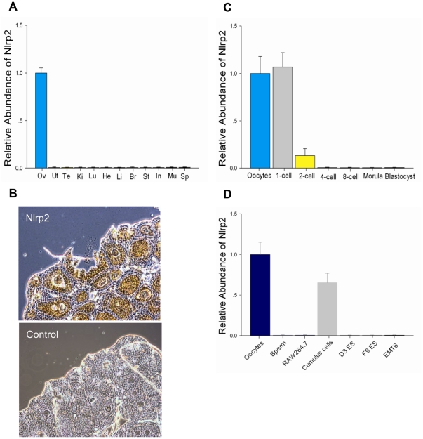
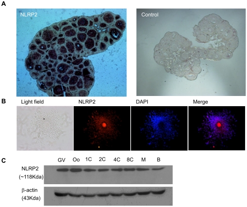
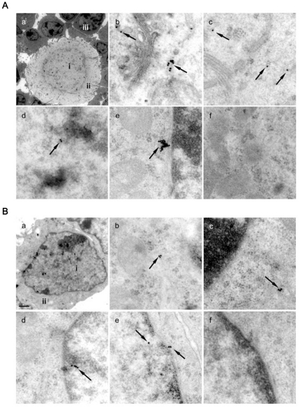
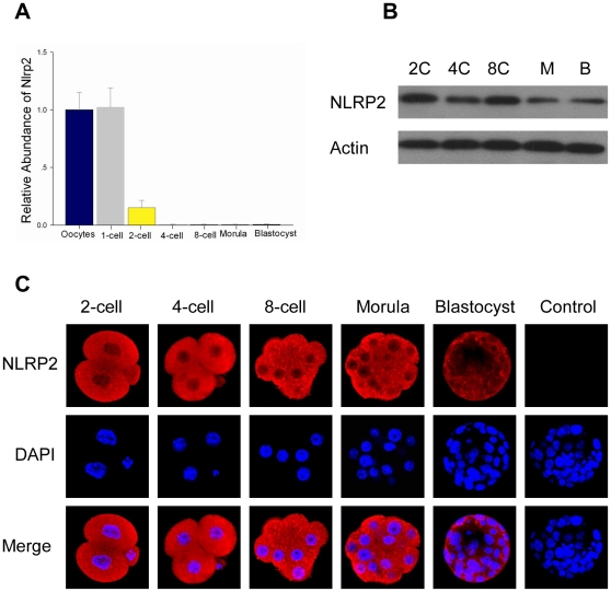
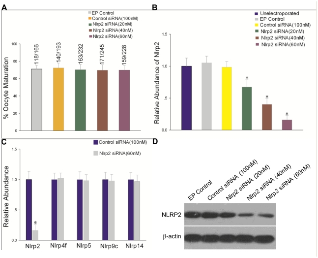
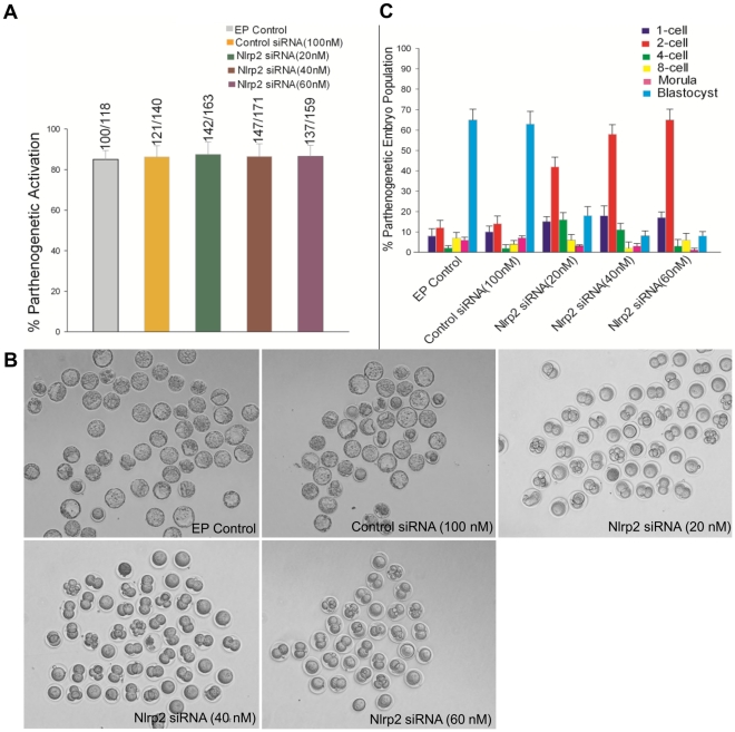
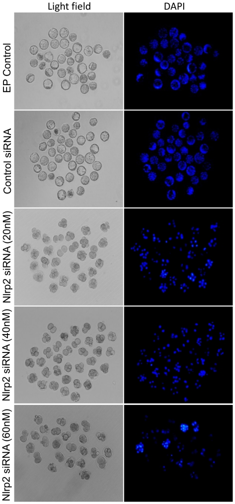
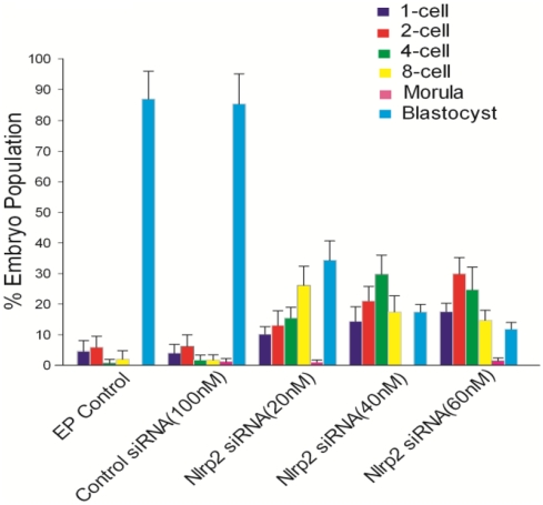
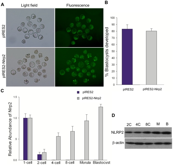
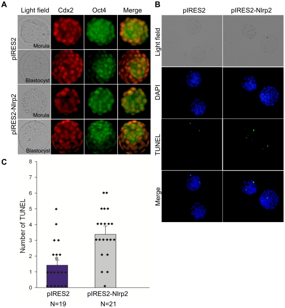
Maternal effect genes encode proteins that are produced during oogenesis and play an essential role during early embryogenesis. Genetic ablation of such genes in oocytes can result in female subfertility or infertility. Here we report a newly identified maternal effect gene, Nlrp2, which plays a role in early embryogenesis in the mouse. Nlrp2 mRNAs and their proteins (∼118 KDa) are expressed in oocytes and granulosa cells during folliculogenesis. The transcripts show a striking decline in early preimplantation embryos before zygotic genome activation, but the proteins remain present through to the blastocyst stage. Immunogold electron microscopy revealed that the NLRP2 protein is located in the cytoplasm, nucleus and close to nuclear pores in the oocytes, as well as in the surrounding granulosa cells. Using RNA interference, we knocked down Nlrp2 transcription specifically in mouse germinal vesicle oocytes. The knockdown oocytes could progress through the metaphase of meiosis I and emit the first polar body. However, the development of parthenogenetic embryos derived from Nlrp2 knockdown oocytes mainly blocked at the 2-cell stage. The maternal depletion of Nlrp2 in zygotes led to early embryonic arrest. In addition, overexpression of Nlrp2 in zygotes appears to lead to normal development, but increases blastomere apoptosis in blastocysts. These results provide the first evidence that Nlrp2 is a member of the mammalian maternal effect genes and required for early embryonic development in the mouse.


Conflict of interest statement

Competing Interests: The authors have declared that no competing interests exist.

Figures

Figure 1
Figure 1. Developmental expression of Nllrp2 in the mouse.
(A) Quantitative reverse-transcription polymerase chain reaction (qRT–PCR) with total RNA extracted from 4-week-old mouse ovary (Ov), uterus (Ut), testis (Te), kidney (Ki), lung (Lu), heart (He), liver (Li), brain (Br), stomach (St), intestines (In), muscle (Mu), spleen (Sp) were performed. Results were normalized to the abundance in the ovary and expressed as the mean ± SEM. (B) In situ hybridization of fixed, paraffin wax-embedded 6 µm ovary sections probed with DIG-labeled Nlrp2 oligonucleotide probes. The original magnification was ×100. (C) The relative abundance of Nlrp2 transcripts in mouse oocytes and preimplantation embryos. (D) The relative abundance of Nlrp2 transcripts in different mouse cells.
Figure 2
Figure 2. Developmental expression of NLRP2 protein in mouse.
(A) Immunohistochemical analysis of sequential sections from a 3-week-old mouse ovary using an anti-NLRP2 antibody. The original magnification was ×40. (B) Immunofluorescent detection of NLRP2 in cumulus–oocyte complexes after permeabilization and incubation with an anti-NLRP2 antibody. The original magnification was ×100. (C) Immunoblots of lysates isolated from oocytes and preimplantation embryos. Molecular masses (KDa) are indicated on the left; β-actin was used as a control.
Figure 3
Figure 3. Cellular localization of NLRP2 protein.
Confocal microscopic images of oocytes and preimplantation embryos. Each sample was counterstained with DAPI to visualize DNA (blue). The original magnification was ×200.
Figure 4
Figure 4. Subcellular localization of NLRP2 protein.
(A) Subcellular localization of NLRP2 protein in immature mouse oocytes. Using an anti-NLRP2 antibody and ultrathin ovarian sections of 10-day-old mice, immunogold reactions were examined by transmission electron microscopy. Black spots with arrows are immunogold particles indicating the presence of NLRP2 protein. a, Oocytes and surrounding granulosa cells (×4,000). The positions of nucleus (i), cytoplasm (ii) and granulosa cells (iii) are indicated. b and c, Oocyte cytoplasm with immunogold particles (×50,000). d, Nucleus with immunogold particles (×50,000). e, Nuclear pore with immunogold particles nearby (×50,000). f, Control oocyte without immunogold particles in the absence of the primary antibody (×50,000). (B) Subcellular localization of NLRP2 protein in mouse granulosa cells. a, Granulosa cell (×15,000). The positions of the nucleus (i) and cytoplasm (ii). b and c, Cytoplasm with immunogold particles (×50,000). d, Nucleus with immunogold particles (×50,000). e, Nucleus and nuclear pore with immunogold particles (×50,000). f, Granulosa cell without immunogold particles in the absence of primary antibody (×50,000).
Figure 5
Figure 5. Developmental expression and localization of Nlrp2 in parthenogenetic embryos.
(A) The relative abundance of Nlrp2 transcripts in mouse oocytes and parthenogenetic embryos. (B) Immunoblots of lysates isolated from parthenogenetic embryos. (C) Confocal microscopic images of parthenogenetic embryos. Each sample was counterstained with DAPI to visualize DNA (blue). The original magnification was ×200.
Figure 6
Figure 6. GV-stage oocyte maturation after electroporation with Nlrp2 siRNA.
(A) Oocyte maturation rate following electroporation (EP) of GV-stage oocytes in the presence or absence of control and Nlrp2 siRNA. The numbers on top of each bar indicate the numbers of oocyte matured/numbers of oocytes electroporated. (B) The relative abundance of Nlrp2 transcripts after electroporation with Nlrp2 siRNA. The data have been normalized to untreated oocytes. Statistical comparisons were made using ANOVA and LSD tests (* p<0.05). (C) Nlrp2, Nlrp4f, Nlrp5, Nlrp9c and Nlrp14 gene expression by qRT–PCR in mouse oocytes at 24 h after electroporation with Nlrp2 siRNA (60 nM). Results were normalized to control siRNA (100 nM) group. * p<0.05. (D) Immunoblots of mouse oocytes at 24 h after electroporation in the presence or absence of control and Nlrp2 siRNA.
Figure 7
Figure 7. Development of parthenogenetic embryos derived from EP control, control siRNA and Nlrp2 siRNA-treated groups (20, 40 and 60 nM).
(A) Parthenogenetic activation rate of electroporated oocytes. (B) Morphological appearance of parthenogenetic embryos after being cultured for 3.5 days. The original magnification was ×100. (C) Percentage of parthenogenetic embryos at different stages after being cultured for 3.5 days.
Figure 8
Figure 8. Nlrp2 mRNA and protein levels in Nlrp2 knockdown embryos.
(A) The relative abundance of Nlrp2 transcripts in mouse embryos collected at 2 h, 11 h, 20 h, 29 h and 38 h after electroporation. Results have been normalized to the abundance in untreated zygotes and are expressed as the mean ± SEM. (B) Immunoblots of mouse embryos at 4 h (1-cell), 28 h (2-cell) and 52 h (8-cell) after electroporation.
Figure 9
Figure 9. Morphology of Nlrp2 knockdown embryos after being cultured for 3.5 days.
Morphology (left) and DAPI staining (right). The original magnification was ×100.
Figure 10
Figure 10. Developmental stages of Nlrp2 knockdown embryos.
Percentage of embryos at different stages after being cultured for 3.5 days.
Figure 11
Figure 11. Overexpression of Nlrp2 in zygotes.
(A) Representative micrographs of blastocysts developing from zygotes that had been microinjected with pIRES2 or pIRES2-Nlrp2. Fluorescence images show the expression of GFP. The original magnification was ×100. (B) The blastocyst formation rate of zygotes microinjected with pIRES2 or pIRES2-Nlrp2. (C) The relative abundance of Nlrp2 transcripts after microinjection. (D) Immunoblots of mouse embryos at different stages after microinjection.
Figure 12
Figure 12. Effects of Nlrp2 overexpression on the expression of CDX2 and OCT3/4 and incidence of apoptosis in blastocysts.
(A) Immunostaining of CDX2 (red) and OCT3/4 (green) in morulae and blastocysts developing from zygotes that had been microinjected with pIRES2 or pIRES2-Nlrp2. The original magnification was ×200. (B) TUNEL apoptosis assay of blastocysts (green). Each sample was counterstained with DAPI to visualize DNA (blue). The original magnification was ×200. (C) Number of apoptotic cells in each blastocyst. *p<0.05.

References

    1. Latham KE. Mechanisms and control of embryonic genome activation in mammalian embryos. Int Rev Cytol. 1999;193:71–124. - PubMed
    1. Christians E, Davis AA, Thomas SD, Benjamin IJ. Maternal effect of Hsf1 on reproductive success. Nature. 2000;407:693–694. - PubMed
    1. Tong ZB, Gold L, Pfeifer KE, Dorward H, Lee E, et al. Mater, a maternal effect gene required for early embryonic development in mice. Nat Genet. 2000;26:267–268. - PubMed
    1. Nusslein-Volhard C, Lohs-Schardin M, Sander K, Cremer C. A dorso-ventral shift of embryonic primordia in a new maternal-effect mutant of Drosophila. Nature. 1980;283:474–476. - PubMed
    1. Schupbach T, Wieschaus E. Germline autonomy of maternal-effect mutations altering the embryonic body pattern of Drosophila. Dev Biol. 1986;113:443–448. - PubMed

Publication types

MeSH terms