

Progesterone inhibits epithelial-to-mesenchymal transition in endometrial cancer
- PMID: 22295114
- PMCID: PMC3266274
- DOI: 10.1371/journal.pone.0030840
Progesterone inhibits epithelial-to-mesenchymal transition in endometrial cancer
Abstract
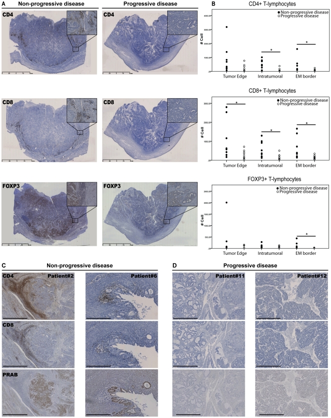
Background: Every year approximately 74,000 women die of endometrial cancer, mainly due to recurrent or metastatic disease. The presence of tumor infiltrating lymphocytes (TILs) as well as progesterone receptor (PR) positivity has been correlated with improved prognosis. This study describes two mechanisms by which progesterone inhibits metastatic spread of endometrial cancer: by stimulating T-cell infiltration and by inhibiting epithelial-to-mesenchymal cell transition (EMT).
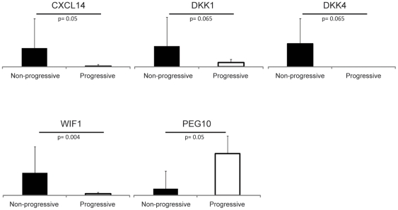
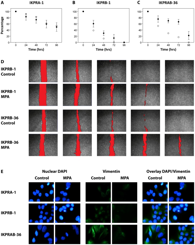
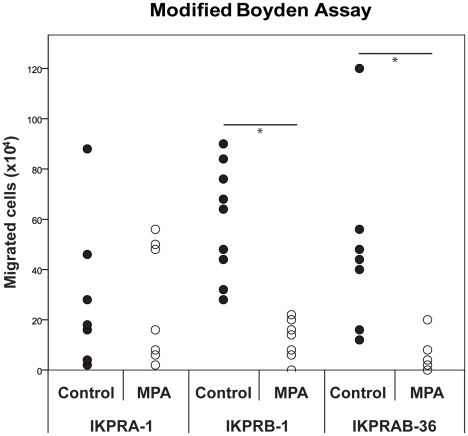
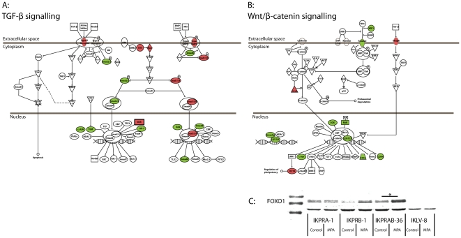
Methodology and principal findings: Paraffin sections from patients with (n = 9) or without (n = 9) progressive endometrial cancer (recurrent or metastatic disease) were assessed for the presence of CD4+ (helper), CD8+ (cytotoxic) and Foxp3+ (regulatory) T-lymphocytes and PR expression. Progressive disease was observed to be associated with significant loss of TILs and loss of PR expression. Frozen tumor samples, used for genome-wide expression analysis, showed significant regulation of pathways involved in immunesurveillance, EMT and metastasis. For a number of genes, such as CXCL14, DKK1, DKK4, PEG10 and WIF1, quantitive RT-PCR was performed to verify up- or downregulation in progressive disease. To corroborate the role of progesterone in regulating invasion, Ishikawa (IK) endometrial cancer cell lines stably transfected with PRA (IKPRA), PRB (IKPRB) and PRA+PRB (IKPRAB) were cultured in presence/absence of progesterone (MPA) and used for genome-wide expression analysis, Boyden- and wound healing migration assays, and IHC for known EMT markers. IKPRB and IKPRAB cell lines showed MPA induced inhibition of migration and loss of the mesenchymal marker vimentin at the invasive front of the wound healing assay. Furthermore, pathway analysis of significantly MPA regulated genes showed significant down regulation of important pathways involved in EMT, immunesuppression and metastasis: such as IL6-, TGF-β and Wnt/β-catenin signaling.
Conclusion: Intact progesterone signaling in non-progressive endometrial cancer seems to be an important factor stimulating immunosurveilance and inhibiting transition from an epithelial to a more mesenchymal, more invasive phenotype.
Conflict of interest statement
Figures
Similar articles
-
Fluorene-9-bisphenol inhibits epithelial-mesenchymal transition of human endometrial cancer Ishikawa cells by repressing TGF-β signaling pathway.Environ Sci Pollut Res Int. 2019 Sep;26(26):27407-27413. doi: 10.1007/s11356-019-05184-0. Epub 2019 Jul 20. Environ Sci Pollut Res Int. 2019. PMID: 31327139
-
Estrogen-related receptor alpha induces epithelial-mesenchymal transition through cancer-stromal interactions in endometrial cancer.Sci Rep. 2019 Apr 30;9(1):6697. doi: 10.1038/s41598-019-43261-z. Sci Rep. 2019. PMID: 31040369 Free PMC article.
-
Metformin Increases E-cadherin in Tumors of Diabetic Patients With Endometrial Cancer and Suppresses Epithelial-Mesenchymal Transition in Endometrial Cancer Cell Lines.Int J Gynecol Cancer. 2016 Sep;26(7):1213-21. doi: 10.1097/IGC.0000000000000761. Int J Gynecol Cancer. 2016. PMID: 27643646
-
Progesterone: the ultimate endometrial tumor suppressor.Trends Endocrinol Metab. 2011 Apr;22(4):145-52. doi: 10.1016/j.tem.2011.01.005. Epub 2011 Feb 25. Trends Endocrinol Metab. 2011. PMID: 21353793 Free PMC article. Review.
-
Epithelial to Mesenchymal Transition and Cell Biology of Molecular Regulation in Endometrial Carcinogenesis.J Clin Med. 2019 Mar 30;8(4):439. doi: 10.3390/jcm8040439. J Clin Med. 2019. PMID: 30935077 Free PMC article. Review.
Cited by
-
Construction of the systemic anticancer immune environment in tumour-bearing humanized mouse by using liposome-encapsulated anti-programmed death ligand 1 antibody-conjugated progesterone.Front Immunol. 2023 Jul 10;14:1173728. doi: 10.3389/fimmu.2023.1173728. eCollection 2023. Front Immunol. 2023. PMID: 37492571 Free PMC article.
-
S100 expression in dendritic cells is inversely correlated with tumor grade in endometrial carcinoma.Obstet Gynecol Sci. 2014 May;57(3):201-7. doi: 10.5468/ogs.2014.57.3.201. Epub 2014 May 15. Obstet Gynecol Sci. 2014. PMID: 24883291 Free PMC article.
-
Epithelial-to-Mesenchymal Transition in the Female Reproductive Tract: From Normal Functioning to Disease Pathology.Front Oncol. 2017 Jul 5;7:145. doi: 10.3389/fonc.2017.00145. eCollection 2017. Front Oncol. 2017. PMID: 28725636 Free PMC article. Review.
-
The migratory capacity of human trophoblastic BeWo cells: effects of aldosterone and the epithelial sodium channel.J Membr Biol. 2013 Mar;246(3):243-55. doi: 10.1007/s00232-013-9526-y. Epub 2013 Jan 26. J Membr Biol. 2013. PMID: 23354843
-
Inhibition of AKT with the orally active allosteric AKT inhibitor, MK-2206, sensitizes endometrial cancer cells to progestin.PLoS One. 2012;7(7):e41593. doi: 10.1371/journal.pone.0041593. Epub 2012 Jul 24. PLoS One. 2012. PMID: 22911820 Free PMC article.
References
-
- Ferlay J, Shin H-R, Bray F, Forman D, Mathers C, et al. Estimates of worldwide burden of cancer in 2008: GLOBOCAN 2008. International Journal of Cancer. 2010;127:2893–2917. - PubMed
-
- Amant F, Moerman P, Neven P, Timmerman D, Van Limbergen E, et al. Endometrial cancer. Lancet. 2005;366:491–505. - PubMed
-
- Jemal A, Siegel R, Xu J, Ward E. Cancer Statistics, 2010. CA Cancer J Clin. 2010;60:277–300. - PubMed
-
- Cohn DE, Horowitz NS, Mutch DG, Kim S-M, Manolitsas T, et al. Should the Presence of Lymphvascular Space Involvement Be Used to Assign Patients to Adjuvant Therapy Following Hysterectomy for Unstaged Endometrial Cancer? Gynecologic Oncology. 2002;87:243–246. - PubMed
-
- Jolly S, Vargas CE, Kumar T, Weiner SA, Brabbins DS, et al. The impact of age on long-term outcome in patients with endometrial cancer treated with postoperative radiation. Gynecologic Oncology. 2006;103:87–93. - PubMed
Publication types
MeSH terms
Substances
LinkOut - more resources
Full Text Sources
Research Materials
Miscellaneous
