

Co-infection with the friend retrovirus and mouse scrapie does not alter prion disease pathogenesis in susceptible mice
- PMID: 22295118
- PMCID: PMC3266293
- DOI: 10.1371/journal.pone.0030872
Co-infection with the friend retrovirus and mouse scrapie does not alter prion disease pathogenesis in susceptible mice
Erratum in
- PLoS One. 2012;7(6). doi:10.1371/annotation/c6b9e78a-451b-4a34-bcd3-24a820bfa32f. Priola, Sue [corrected to Priola, Suzette A]
Abstract
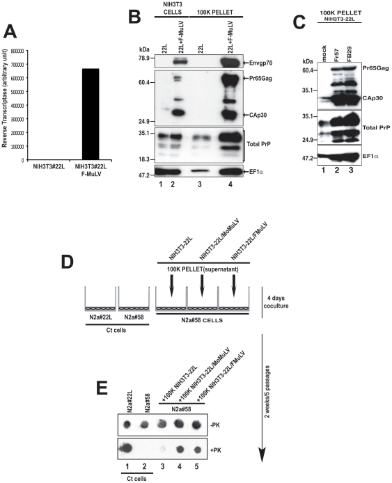
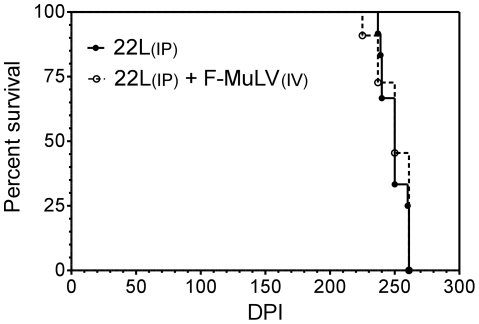
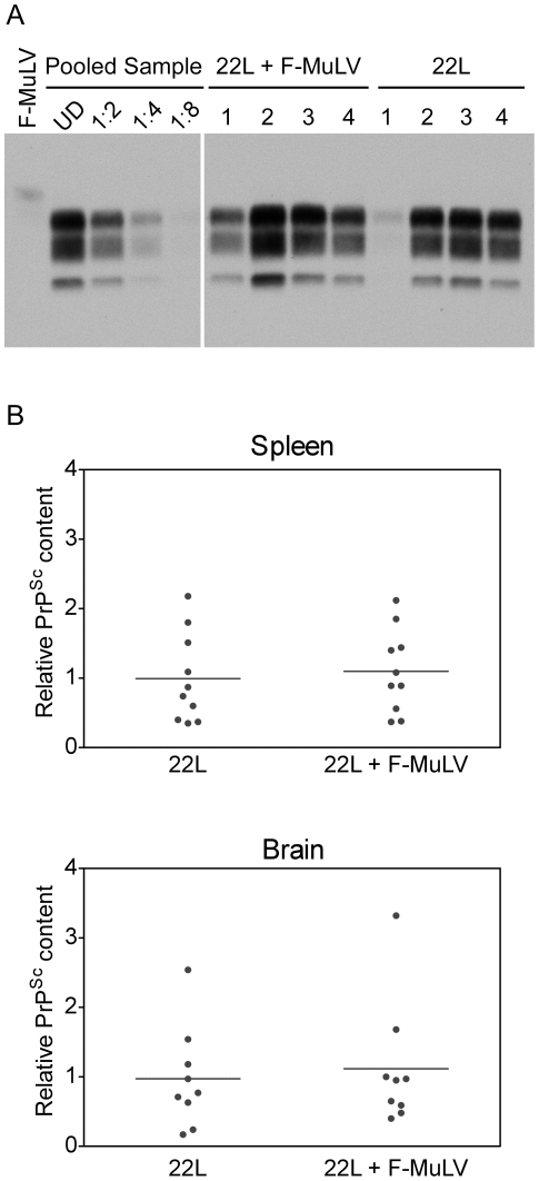
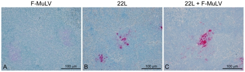
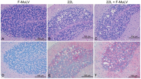
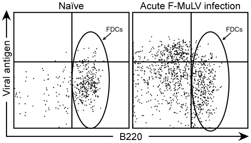
Prion diseases are fatal, transmissible neurodegenerative diseases of the central nervous system. An abnormally protease-resistant and insoluble form (PrP(Sc)) of the normally soluble protease-sensitive host prion protein (PrP(C)) is the major component of the infectious prion. During the course of prion disease, PrP(Sc) accumulates primarily in the lymphoreticular and central nervous systems. Recent studies have shown that co-infection of prion-infected fibroblast cells with the Moloney murine leukemia virus (Mo-MuLV) strongly enhanced the release and spread of scrapie infectivity in cell culture, suggesting that retroviral coinfection might significantly influence prion spread and disease incubation times in vivo. We now show that another retrovirus, the murine leukemia virus Friend (F-MuLV), also enhanced the release and spread of scrapie infectivity in cell culture. However, peripheral co-infection of mice with both Friend virus and the mouse scrapie strain 22L did not alter scrapie disease incubation times, the levels of PrP(Sc) in the brain or spleen, or the distribution of pathological lesions in the brain. Thus, retroviral co-infection does not necessarily alter prion disease pathogenesis in vivo, most likely because of different cell-specific sites of replication for scrapie and F-MuLV.
Conflict of interest statement
Figures
Similar articles
-
Retrovirus infection strongly enhances scrapie infectivity release in cell culture.EMBO J. 2006 Jun 21;25(12):2674-85. doi: 10.1038/sj.emboj.7601162. Epub 2006 May 25. EMBO J. 2006. PMID: 16724107 Free PMC article.
-
Mouse neuroblastoma cells release prion infectivity associated with exosomal vesicles.Biol Cell. 2008 Oct;100(10):603-15. doi: 10.1042/BC20080025. Biol Cell. 2008. PMID: 18422484
-
Acute formation of protease-resistant prion protein does not always lead to persistent scrapie infection in vitro.J Biol Chem. 2004 Jul 9;279(28):29218-25. doi: 10.1074/jbc.M402576200. Epub 2004 May 7. J Biol Chem. 2004. PMID: 15133048
-
Follicular dendritic cells in scrapie pathogenesis.Arch Virol Suppl. 2000;(16):13-21. doi: 10.1007/978-3-7091-6308-5_2. Arch Virol Suppl. 2000. PMID: 11214915 Review.
-
Genetic and infectious prion diseases.Arch Neurol. 1993 Nov;50(11):1129-53. doi: 10.1001/archneur.1993.00540110011002. Arch Neurol. 1993. PMID: 8105771 Review.
Cited by
-
Prion strains depend on different endocytic routes for productive infection.Sci Rep. 2017 Jul 31;7(1):6923. doi: 10.1038/s41598-017-07260-2. Sci Rep. 2017. PMID: 28761068 Free PMC article.
-
Propagation and Dissemination Strategies of Transmissible Spongiform Encephalopathy Agents in Mammalian Cells.Int J Mol Sci. 2022 Mar 8;23(6):2909. doi: 10.3390/ijms23062909. Int J Mol Sci. 2022. PMID: 35328330 Free PMC article. Review.
-
Cellular mechanisms responsible for cell-to-cell spreading of prions.Cell Mol Life Sci. 2018 Jul;75(14):2557-2574. doi: 10.1007/s00018-018-2823-y. Epub 2018 May 14. Cell Mol Life Sci. 2018. PMID: 29761205 Free PMC article. Review.
-
Recombinant prion protein refolded with lipid and RNA has the biochemical hallmarks of a prion but lacks in vivo infectivity.PLoS One. 2013 Jul 30;8(7):e71081. doi: 10.1371/journal.pone.0071081. Print 2013. PLoS One. 2013. PMID: 23936256 Free PMC article.
-
Reactivated endogenous retroviruses promote protein aggregate spreading.Nat Commun. 2023 Aug 18;14(1):5034. doi: 10.1038/s41467-023-40632-z. Nat Commun. 2023. PMID: 37596282 Free PMC article.
References
-
- Castilla J, Saa P, Hetz C, Soto C. In vitro generation of infectious scrapie prions. Cell. 2005;121:195–206. - PubMed
-
- Priola SA. Prion protein diversity and disease in the transmissible spongiform encephalopathies. In: Caughey B, editor. Prion Proteins. San Diego: Academic Press; 2001. pp. 1–27. - PubMed
-
- Kimberlin RH, Walker CA. Pathogenesis of mouse scrapie: dynamics of agent replication in spleen, spinal cord and brain after infection by different routes. J Comp Pathol. 1979;89:551–562. - PubMed
-
- Weissmann C, Bueler H, Fischer M, Sailer A, Aguzzi A, et al. PrP-deficient mice are resistant to scrapie. Ann N Y Acad Sci. 1994;724:235–240. - PubMed
Publication types
MeSH terms
Substances
Grants and funding
LinkOut - more resources
Full Text Sources
Research Materials
