

Spindle cell carcinoma progressed from transitional cell carcinoma of the urinary bladder
- PMID: 22295151
- PMCID: PMC3267490
Spindle cell carcinoma progressed from transitional cell carcinoma of the urinary bladder
Abstract
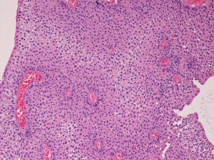
The author reports a very rare case of spindle cell carcinoma (SpCC) of the urinary bladder progressed from ordinary papillary transitional cell carcinoma (TCC). A 63-year-old man complained of hematuria. A transurethral endoscopic examination revealed a papillary tumor, and transuthetral resection of bladder tumor (TUR-BT) was performed and was diagnosed as ordinary papillary urothelial TCC. Since then, he was treated with TUR-BT eight times. Chemotherapy, radiation, radical cystectomy and lymph nodes dissection were performed 16 years after the first TUR-BT. However, he developed rectal mucosal metastasis. He is now alive 17 years after the first presentation. All the TUR-BT specimens were ordinary papillary TCCs without invasion (pTa). Immunohistochemically, the TUR-BT specimens were positive for pancytokeratin, high molecular weight cytokeratin (CK), CK 5/6, CK 7, CK 18, CK 19, CK 20, p53, p63, Ki-67 (10%), and negative for other antigens examined including vimentin. The cystectomy bladder specimens show broad ulcers and polypoid lesions, and malignant spindle cells (SpCC) invading into muscular layer were present. No TCC elements were recognized. The tumor cells were positive strongly for vimentin, and less strongly for pancytokeratin, high molecular weight cytokeratin, CK 5/6, CK 14, CK 18, p53, p63 and Ki-67 (95%), and negative for other antigens examined. The rectal metastatic lesion showed SpCC without TCC elements, and were strongly positive for vimentin, and weakly positive for pancytokeratin, S100 protein, p53, p63, Ki-67 (90%), neuron-specific enolase, CD56, KIT and PDGFRA. It was negative for other antigen examined. It is strongly suggested that the present SpCC were progressed from ordinary TCC.
Keywords: Spindle cell carcinoma; immunohistochemistry; urinary bladder.
Figures


Similar articles
-
Urinary bladder urothelial carcinoma with expression of KIT and PDGFRA and showing diverse differentiations into plasmacytoid, clear cell, acantholytic, nested, and spindle variants, and into adenocarcinoma, signet-ring cell carcinoma, small cell carcinoma, large cell carcinoma, and pleomorphic carcinoma.Int J Clin Exp Pathol. 2013 May 15;6(6):1150-6. Print 2013. Int J Clin Exp Pathol. 2013. PMID: 23696935 Free PMC article.
-
[Spindle cell carcinoma of the urinary bladder: a case report].Hinyokika Kiyo. 1993 Oct;39(10):947-51. Hinyokika Kiyo. 1993. PMID: 8266862 Japanese.
-
Synchronous squamous cell carcinoma of the kidney, squamous cell carcinoma of the ureter, and sarcomatoid carcinoma of the urinary bladder: a case report.Pathol Res Pract. 2010 Jun 15;206(6):379-83. doi: 10.1016/j.prp.2009.07.021. Epub 2009 Sep 18. Pathol Res Pract. 2010. PMID: 19766408
-
[Transitional cell carcinoma of bladder occurring during pregnancy: report of two patients].Nihon Hinyokika Gakkai Zasshi. 1994 Nov;85(11):1683-6. doi: 10.5980/jpnjurol1989.85.1683. Nihon Hinyokika Gakkai Zasshi. 1994. PMID: 7807778 Review. Japanese.
-
[Metastatic small intestinal tumor associated with transitional cell carcinoma: a report of 2 cases and review of cases in Japan].Hinyokika Kiyo. 2005 Jan;51(1):41-4. Hinyokika Kiyo. 2005. PMID: 15732341 Review. Japanese.
Cited by
-
Cutaneous pseudolymphoma: a case report with an immunohistochemical study.Int J Clin Exp Pathol. 2013 Apr 15;6(5):966-72. Print 2013. Int J Clin Exp Pathol. 2013. PMID: 23638232 Free PMC article.
-
Urinary bladder urothelial carcinoma with expression of KIT and PDGFRA and showing diverse differentiations into plasmacytoid, clear cell, acantholytic, nested, and spindle variants, and into adenocarcinoma, signet-ring cell carcinoma, small cell carcinoma, large cell carcinoma, and pleomorphic carcinoma.Int J Clin Exp Pathol. 2013 May 15;6(6):1150-6. Print 2013. Int J Clin Exp Pathol. 2013. PMID: 23696935 Free PMC article.
References
-
- Honda N, Ohshita H, Fukatsu H, Segawa A. Spindle cell carcinoma of the urinary bladder: a case report. Kikyoukika Kiyo. 1993;39:947–951. - PubMed
-
- Serio G, Zampatti C, Ceppi M. Spindle and giant cell carcinoma of the urinary bladder: a clinico-pathologic, light microscopic and immunohistochemical study. Br J Urol. 1995;75:167–172. - PubMed
-
- Barber NJ, Kane TP, Conroy B, Britton JP. Condyloma acuminatum-like lesion of the urinary bladder progressed to invasive spindle cell carcinoma. Scand J Urol Nephrol. 2003;37:512–514. - PubMed
-
- Gold RP, Saitas V, Pellman C. Congenital uteropelvic junction with calcified renal pelvis and superimposed spindle cell urothelial carcinoma. Urol Radiol. 1990;12:15–17. - PubMed
-
- Terada T, Kawaguchi M. Primary clear cell ade-nocarcinoma of the peritoneum. Tohoku J Exp Med. 2005;206:271–275. - PubMed
Publication types
MeSH terms
LinkOut - more resources
Full Text Sources
Medical
Research Materials
Miscellaneous