

Malignant intraductal oncocytic papillary neoplasm of the common bile duct
- PMID: 22295153
- PMCID: PMC3267492
Malignant intraductal oncocytic papillary neoplasm of the common bile duct
Abstract
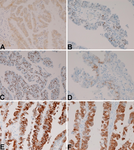
Recently, several cases of intraductal oncocytic papillary neoplasm (IOPN) of the liver and hepatic bile ducts have been reported. The author herein reports the first case of IOPN of the common bile duct (CBD). A 78-year-old man was admitted to our hospital because of jaundice. Imaging modalities including US, CT, MRI revealed an intraductal tumor of the middle CBD and biliary dilation distal to the tumor. A partial resection of the CBD was performed. Grossly, a papillary tumor measuring 20 × 15 mm was found within the CBD. Mucus is absent. Histologically, the papillary tumor was composed of atypical oncocytes. The atypia was enough to be diagnosed as adenocarcinoma. No invasive features were noted. Immunohistochemically, the tumor cells were positive for pancytokeratins (CK), CK 7, CK 18, CK19, EMA, CA19-9, CEA, mitochondria, p53 protein, C-erbB2, Ki-67 (labeling = 80%), MUC2, MUC5AC and MUC-6,. The tumor cells were negative for CK8, CK20, chromogranin, synaptophysin, neuron-specific enolase, S100 protein, CD56, MUC1, CD10 and CDX2. These immunohistochemical findings were compatible with IOPN. The patient died of other non-tumorous disease 7 year after the operation. In summary, the author presented the first case of IOPN of the CBD.
Keywords: Common bile duct; histopathology; immunohistochemistry; intraductal oncocytic papillary neoplasm.
Figures


Similar articles
-
Non-invasive intraductal papillary neoplasms of the common bile duct: a clinicopathologic study of six cases.Int J Clin Exp Pathol. 2012;5(7):690-7. Epub 2012 Sep 5. Int J Clin Exp Pathol. 2012. PMID: 22977666 Free PMC article.
-
Spindle Cell Carcinoma of the Common Bile Duct: Case Report with Immunohistochemical Analysis.Case Rep Gastroenterol. 2010 Sep 18;4(3):374-380. doi: 10.1159/000320674. Case Rep Gastroenterol. 2010. PMID: 21060703 Free PMC article.
-
Intraductal tubular neoplasms of the bile ducts.Am J Surg Pathol. 2012 Nov;36(11):1647-55. doi: 10.1097/PAS.0b013e3182684d4f. Am J Surg Pathol. 2012. PMID: 23073323
-
Intraductal papillary neoplasm of the bile ducts: A case report and literature review.World J Gastroenterol. 2015 Nov 21;21(43):12498-504. doi: 10.3748/wjg.v21.i43.12498. World J Gastroenterol. 2015. PMID: 26604656 Free PMC article. Review.
-
Undifferentiated carcinoma of the common bile duct with intraductal tumor thrombi: report of a case.Surg Today. 2011 Apr;41(4):579-84. doi: 10.1007/s00595-009-4304-2. Epub 2011 Mar 23. Surg Today. 2011. PMID: 21431499 Review.
Cited by
-
Noninvasive Intracystic Oncocytic Papillary Neoplasm of the Gall Bladder.J Gastrointest Cancer. 2016 Sep;47(3):318-23. doi: 10.1007/s12029-015-9741-0. J Gastrointest Cancer. 2016. PMID: 26073617 No abstract available.
References
-
- Tearda T, Mitsui T, Nakanuma Y, Miura S, Toya D. Intrahepatic biliary papillomatosis arising in non-obstructive intrahepatic biliary dilations confined to the hepatic left lobe. Am J Gastroenterol. 1991;86:1523–1526. - PubMed
-
- Sudo Y, Harada K, Tsuneyama K, Katayanagi K, Zen Y, Nakanuma Y. Oncocytic biliary cystademnoma is a form of intraductal oncocytic papillary neoplasm of the liver. Mod Pathol. 2001;14:1304–1309. - PubMed
-
- Martin RC, Klimstra DS, Schwarz L, Yilmaz A, Blumgard LH, Jarnagin W. Hepatic intraductal oncocytic papillary carcinoma. Cancer. 2002;95:2180–2187. - PubMed
-
- Terada T, Taniguchi M. Intraductal oncocytic papillary neoplasm of the liver. Pathol Int. 2004;54:116–123. - PubMed
-
- Rouzbahman M, Serra S, Adsay NV, Bejarano PA, Nakanuma Y, Chetty R. Oncocytic papillary neoplasms of the biliary tract: a clinicopathological, mucin core and Wnt pathway protein analysis of four cases. Pathology. 2007;39:413–418. - PubMed
Publication types
MeSH terms
Substances
LinkOut - more resources
Full Text Sources
Research Materials
Miscellaneous