. 2012 Feb;4(1):30-9.
doi: 10.3978/j.issn.2072-1439.2010.08.04.

Involvement of Rho kinase (ROCK) in sepsis-induced acute lung injury

Affiliations

Involvement of Rho kinase (ROCK) in sepsis-induced acute lung injury

Ismail Cinel et al. J Thorac Dis. 2012 Feb.

Abstract

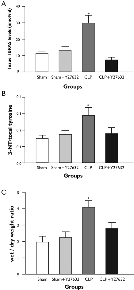
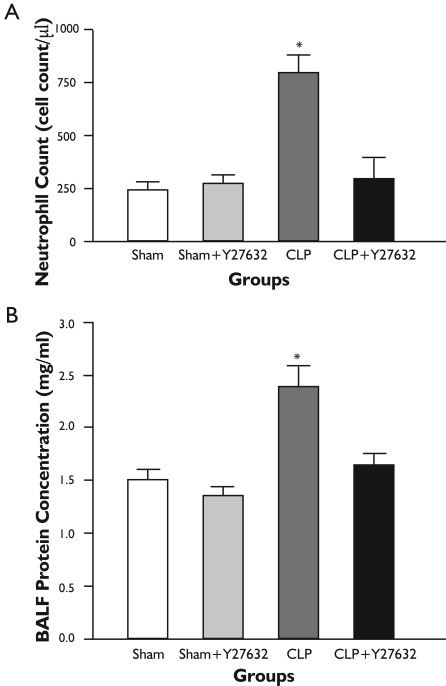
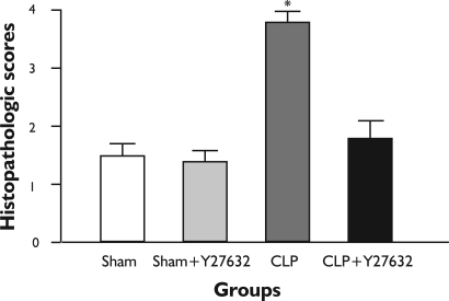
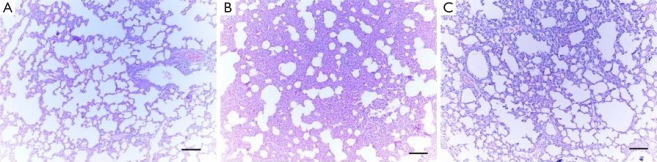
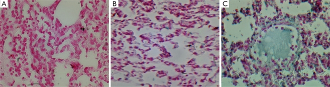
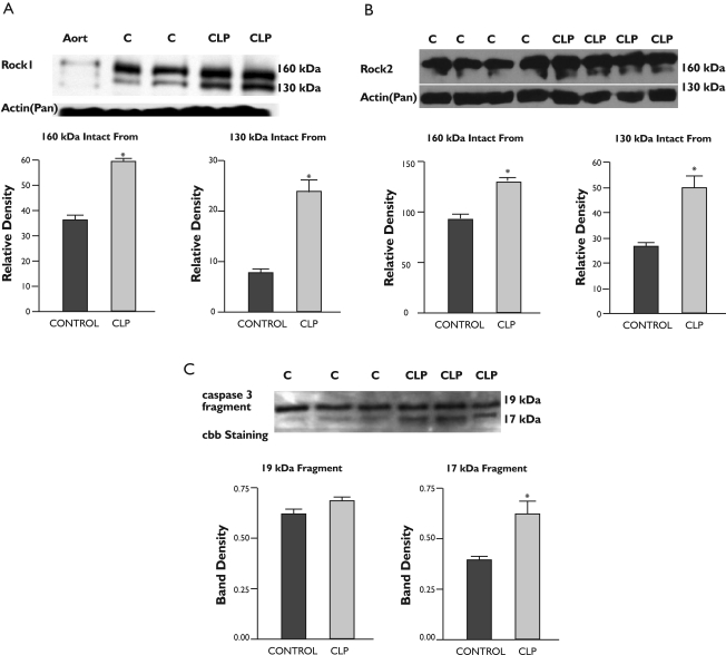
Indirect acute lung injury is associated with high morbidity and mortality. We investigated the link between Rho kinase (ROCK) activation and apoptotic cell death in sepsis induced acute lung injury. This hypothesis was tested by administering a specific, selective inhibitor of ROCK (Y-27632) to rats subjected to cecal ligation and puncture (CLP). Rats were randomly divided into 4 groups as; sham-operated, sham + Y-27632, CLP and CLP + Y-27632. Twenty-four hours later, each experiment was terminated and lungs analyzed. Histopathology was assessed by hematoxylin-eosin staining and the presence of apoptosis was evaluated through the TUNEL assay. Pulmonary activity of caspase 3 and ROCK 1 & 2 were measured by western blot. Interstitial edema, severely damaged pulmonary architecture with massive infiltration of the inflammatory cells and an increase in lung tissue TBARS levels as well as 3-NT to total tyrosine ratios were observed in untreated CLP animals. Pretreatment of animals with Y-27632, reduced lung injury in the CLP induced septic rats in each of these parameters of lung injury (p<0.05). Western immunoblot revealed active caspase cleavage and increased expression of active fragment of ROCK 1 & 2 in the CLP group. TUNEL assay showed an increase in percentage of apoptotic cells when comparing the CLP group with the CLP + Y-27632 group. These results suggest an important role of Rho kinase in sepsis induced lung injury by a mechanism that might be related to oxidative and/or nitrosative stress mediated caspase cleavage leading to apoptosis.

Keywords: 3-Nitrotyrosine; ROCK; Rho kinase; Sepsis; acute lung injury; apoptosis; nitric oxide; peroxynitrite; reactive oxygen species.


Conflict of interest statement

No potential conflict of interest.

Figures

Figure 1.
Figure 1.. Lung tissue TBARS levels, 3-Nitrotyrosine/Total Tyrosine Ratio and Wet to Dry Weight Ratio (W:D).
A: CLP resulted in increased lung TBARS levels compared with the sham operated animals. The CLP-induced increase was reduced by Y-27632 treatment; B: CLP increased 3-nitrotyrosine/Total Tyrosine ratios and Y-27632 prevented these increases; C: Lung tissue W/D weight ratios were significantly increased in CLP group in comparison to sham-operated group. Y-27632 treatment caused significant decrease in the W/D weight ratio in comparison to CLP group. All data represent mean±S.E.M.; For comparison analysis of variance (ANOVA) followed by Tukey post hoc test was used. *P<0.05 compared with the other groups.
Figure 2.
Figure 2.. Number of Inflammatory Cells and Protein Concentrations in Bronchoalveolar Lavage Fluid.
A: In the CLP group, cellular infiltration in the BALF was found to be increased when compared with sham-operated group, whereas Y-27632 treatment in CLP group caused decrease in cellular infiltration in the BALF compared to the CLP group; B: In the CLP group, protein concentrations in the BALF was found to be increased and Y-27632 prevented these increases. All data represent mean±S.E.M.; For comparison analysis of variance (ANOVA) followed by Tukey post hoc test was used. *P<0.05 compared with the other groups.
Figure 3.
Figure 3.. Histopathological scores of the Lung Tissues.
Histopathological scores of the lung tissue. CLP resulted in increased lung histopathologic scores compared with sham-operated animals. The CLP induced increase was reduced by Y-27632 treatment. For comparison Kruskall-Wallis variance analysis followed by Dunn test was used. *P<0.05 compared with other groups.
Figure 4.
Figure 4.. Photomicrographs of Lung Tissues (Hematoxylin & Eosin X100).
A: Normal pulmonary histology was observed in sham group (grade 1); B: CLP group revealed severe interstitial infiltration of neutrophils and destructed pulmonary architecture (grade 3); C: In the CLP + Y-27632 group, there was improvement of the deranged histopathology observed in CLP Group (grade 2).
Figure 5.
Figure 5.. Photomicrographs of Lung Tissues (Tunel Assay X400).
A: Sham group 5 µm section stained with Tacs Blue and Nuclear Fast Red; B: CLP + Y-27632 group 5 µm section stained with Tacs Blue and Nuclear Fast Red; C: CLP group 5 µm section stained with Tacs Blue and Nuclear Fast Red.]
Figure 6.
Figure 6.. Western Blot findings in Lung Homogenates from Sham operated and CLP rats.
A: The homogenates were subjected to SDS gel electrophoresis and transferred to PVDF membranes, which were incubated with specific antibody against ROCK 1; B: The same as A. except that ROCK 2 antibody was used; C: The homogenates were subjected to SDS gel electrophoresis and transferred to PVDF membranes, which were incubated with specific antibody against 19/17 kDa fragment of caspase 3. Sham-operated lung homogenate, CLP: CLP lung homogenate, Cbb: Coomassie brillant blue. See Methods section for details.

References

    1. Angus DC, Wax RS. Epidemiology of sepsis: an update. Crit Care Med. 2001;29:S109–16. - PubMed
    1. Cinel I, Dellinger RP. Advances in pathogenesis and management of sepsis. Curr Opin Infect Dis. 2007;20:345–52. - PubMed
    1. Cinel I, Opal SM. Molecular biology of inflammation and sepsis: a primer. Crit Care Med. 2009;37:291–304. - PubMed
    1. Hotchkiss RS, Swanson PE, Freeman BD, Tinsley KW, Cobb JP, Matuschak GM, et al. Apoptotic cell death in patients with sepsis, shock, and multiple organ dysfunction. Crit Care Med. 1999;27:1230–51. - PubMed
    1. Cinel I, Buyukafsar K, Cinel L, Polat A, Atici S, Tamer L, et al. The role of poly(ADP-ribose) synthetase inhibition in preventing endotoxemia-induced intestinal epithelial apoptosis. Pharmacol Res. 2002;46:119–27. - PubMed