

Psoas muscle metastases in non-small cell lung cancer
- PMID: 22295171
- PMCID: PMC3256543
- DOI: 10.3978/j.issn.2072-1439.2011.04.05
Psoas muscle metastases in non-small cell lung cancer
Abstract
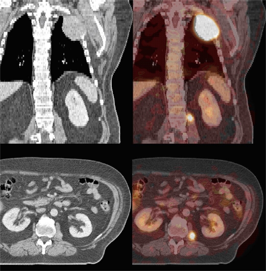
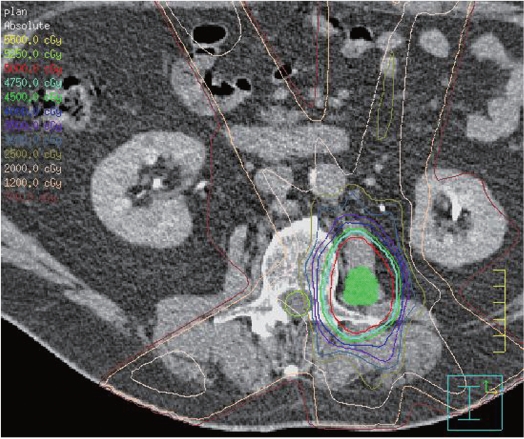
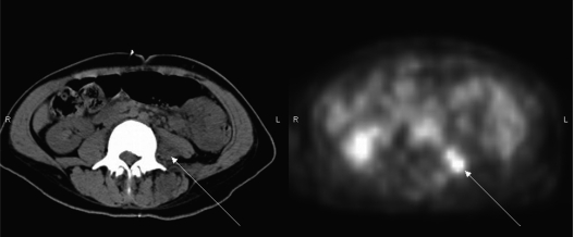
Lung cancer is the leading cause of cancer-related death in the U.S. and often spreads via lymphatics or through hematogenous metastasis to the brain, bone and adrenal glands. Isolated metastases to skeletal muscle, including the psoas muscles, are very uncommon. The present report is a case series of three patients with psoas metastases from non-small cell lung cancer (NSCLC) and a review of the relevant literature. Three patients presented with psoas muscle metastases from NSCLC detected on diagnostic imaging. All patients were treated with radiotherapy to the psoas muscle, and two patients were treated with curative intent on an oligometastatic paradigm. Radiotherapy to the psoas muscle was effective and well tolerated.
Keywords: carcinoma; lung; metastases; psoas muscle.
Conflict of interest statement
No potential conflict of interest.
Figures
References
-
- American Cancer Society [Internet] Facts & Figures 2007. Atlanta: American Cancer Society;2007. Available from: http://www.mitsubishi-motors.com/en/corporate/ir/library/pdf/fact_2007.pdf.
-
- Perez CA, Pajak TF, Rubin P, Simpson JR, Mohiuddin M, Brady LW, et al. Long-term observations of the patterns of failure in patients with unresectable non-oat cell carcinoma of the lung treated with definitive radiotherapy. Report by the Radiation Therapy Oncology Group. Cancer. 1987;59:1874–81. - PubMed
-
- Hu C, Chang EL, Hassenbusch SJ, 3rd, Allen PK, Woo SY, Mahajan A, et al. Nonsmall cell lung cancer presenting with synchronous solitary brain metastasis. Cancer. 2006;106:1998–2004. - PubMed
-
- Flexner S. Exhibition of specimens from a case of carcinoma of the pancreas with multiple carcinosis. Bull Johns Hopkins Hosp. 1894;5:16–17.
-
- Ampil FL, Lall C, Datta R. Palliative management of metastatic tumors involving the psoas muscle: case reports and review of the literature. Am J Clin Oncol. 2001;24:313–4. - PubMed
Publication types
LinkOut - more resources
Full Text Sources