. 2011:2011:867152.
doi: 10.4061/2011/867152. Epub 2011 Oct 19.

Lymphangiogenesis and Axillary Lymph Node Metastases Correlated with VEGF-C Expression in Two Immunocompetent Mouse Mammary Carcinoma Models

Affiliations

Lymphangiogenesis and Axillary Lymph Node Metastases Correlated with VEGF-C Expression in Two Immunocompetent Mouse Mammary Carcinoma Models

Yuko Ito et al. Int J Breast Cancer. 2011.

Abstract

Lymphangiogenesis and the expression of vascular endothelial cell growth factor C (VEGF-C) in tumors have been considered to be causally promoting lymphatic metastasis. There are only a few studies on lymphatic metastasis in immunocompetent allograft mouse models. To study the relationship between VEGF-C-mediated lymphangiogenesis and axillary lymph node metastasis, we used two mouse mammary carcinoma cell lines; the BJMC338 has a low metastatic propensity, whereas the BJMC3879 has a high metastatic propensity although it originated from the former cell line. Each cell line was injected separately into two groups of female BALB/c mice creating in vivo mammary cancer models. The expression level of VEGF-C in BJMC3879 was higher than BJMC338. As the parent cell line, BJMC3879-derived tumors showed higher expression of VEGF-C compared to BJMC338-derived tumors. This higher expression of VEGF-C in BJMC3879-derived tumors was associated with marked increase in infiltrating macrophages and enhanced expression of lymphatic vessel endothelial hyaluronan receptor-1 (LYVE-1) reflecting increased tumoral lymphatic density and subsequent induction of axillary lymph node metastasis. Our mouse mammary carcinoma models are allotransplanted tumors showing the same axillary lymph node metastatic spectrum as human breast cancers. Therefore, our mouse models are ideal for exploring the various molecular mechanisms of cancer metastasis.


Figures

Figure 1
Figure 1
TEM micrographs (a and b), immunofluorecent staining (c and d), Western blot analysis (e), and RT-PCR analysis (f) of VEGF-C in the two-mouse mammary carcinoma cell lines. Both BJMC338 cells (a) and BJMC3879 cells (b) have the similar ultrastructure. BJMC338 cells (c) have minimally any activity of VEGF-C, whereas BJMC3879 cells (d) have moderate expression. Western blot analysis (e) demonstrates the same activity of VEGF-C as in (c and d). VEGF-C mRNA expression in BJMC3879 cells is higher than that of BJMC338 cells (f). Green fluorescence (FITC) indicates activity of VEGF-C, and red fluorescence (PI) shows nuclei of cells in (c and d).
Figure 2
Figure 2
Histopathology of the inoculated tumors at 10 weeks postinoculation. H&E staining (a and b) show viable region (white ∗) and necrotic region (black ∗) in BJMC338 tumor (a) and BJMC3879 tumor (b). TEM micrographs (c and d) indicate tumor cells in the viable region of BJMC338 tumor (c) and BJMC3879 tumor (d).
Figure 3
Figure 3
Histopathology by H&E staining of axially lymph nodes (a and b) and lungs (c and d) in mice at 10 weeks postinoculation. Axially lymph node and lung in BJMC338 tumor (a and c) show normal appearance, whereas those of BJMC3879 tumor (b and d) demonstrate metastatic foci.
Figure 4
Figure 4
Immunohistochemistry of LYVE-1 in BJMC338 tumors (a, c, e, and g) and BJMC3879 tumors (b, d, f, and h) at 4 weeks (a and b), 6 weeks (c and d), 8 weeks (e and f), and 10 weeks postinoculation (g and h). Note the upregulation of LYVE-1 in BJMC3879 tumors.
Figure 5
Figure 5
Density of lymphatic vessels (LVD) in tumors. The LVD in BJMC3879 tumors (black bars) is always significantly higher than that of BJMC338 tumors (white bars) (*P < 0.01, **P < 0.001).
Figure 6
Figure 6
TEM micrographs of lymphatic vessels in BJMC3879 tumors at 10 weeks postinoculation (a) and 4 weeks postinoculation (b). The boxed areas in (b) are observed with high-power view (c and d). (a) A leukocyte (∗) is seen between endothelial cells (arrows), whereas tumor cells (arrowheads) are observed in the lumen. (b) Lymphatic capillary (arrow) and blood capillaries (arrowheads) are detected in the connective tissue surrounding tumors. Casein-like droplet (arrow) is located in the opening junction (c), and characteristic over-lapping junction (arrow) is showed (d).
Figure 7
Figure 7
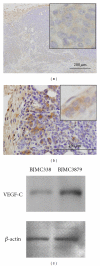
Immunohistochemistry (a and b) and Western blot analysis (c) of VEGF-C in BJMC338 tumor (a and c) and BJMC3879 tumor (b and c) at 8 weeks postinoculation.
Figure 8
Figure 8
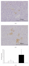
Immunohistochemistry of CD68 in BJMC338 tumor (a) and BJMC3879 tumor (b) at 4 weeks postinoculation. The density of macrophages in BJMC3879 tumors was significantly higher than that in BJMC338 tumors (*P < 0.001) (c).

Similar articles

Cited by

References

    1. Schoppmann SF, Birner P, Studer P, Breiteneder-Geleff S. Lymphatic microvessel density and lymphovascular invasion assessed by anti-podoplanin immunostaining in human breast cancer. Anticancer Research. 2001;21(4):2351–2355. - PubMed
    1. Bono P, Wasenius VM, Heikkilä P, Lundin J, Jackson DG, Joensuu H. High LYVE-1-positive lymphatic vessel numbers are associated with poor outcome in breast cancer. Clinical Cancer Research. 2004;10(21):7144–7149. - PubMed
    1. Nakamura Y, Yasuoka H, Tsujimoto M, et al. Lymph vessel density correlates with nodal status, VEGF-C expression, and prognosis in breast cancer. Breast Cancer Research and Treatment. 2005;91(2):125–132. - PubMed
    1. Van der Auwera I, Cao Y, Tille JC, et al. First international consensus on the methodology of lymphangiogenesis quantification in solid human tumours. British Journal of Cancer. 2006;95(12):1611–1625. - PMC - PubMed
    1. Mohammed RAA, Green A, El-Shikh S, Paish EC, Ellis IO, Martin SG. Prognostic significance of vascular endothelial cell growth factors -A, -C and -D in breast cancer and their relationship with angio- and lymphangiogenesis. British Journal of Cancer. 2007;96(7):1092–1100. - PMC - PubMed