. 2012 Mar 7;134(9):4163-8.
doi: 10.1021/ja2095766. Epub 2012 Feb 28.

Hydrophobic core flexibility modulates enzyme activity in HIV-1 protease

Affiliations

Hydrophobic core flexibility modulates enzyme activity in HIV-1 protease

Seema Mittal et al. J Am Chem Soc. .

Abstract

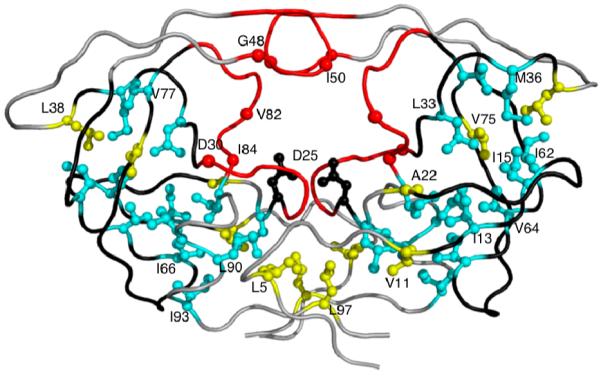
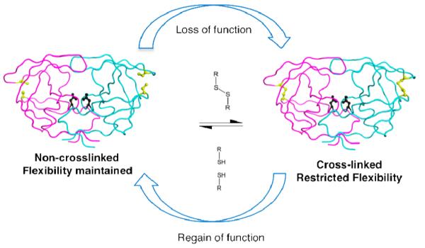
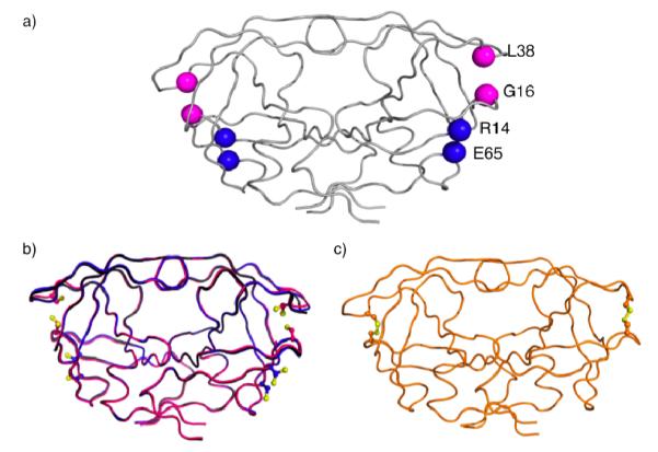
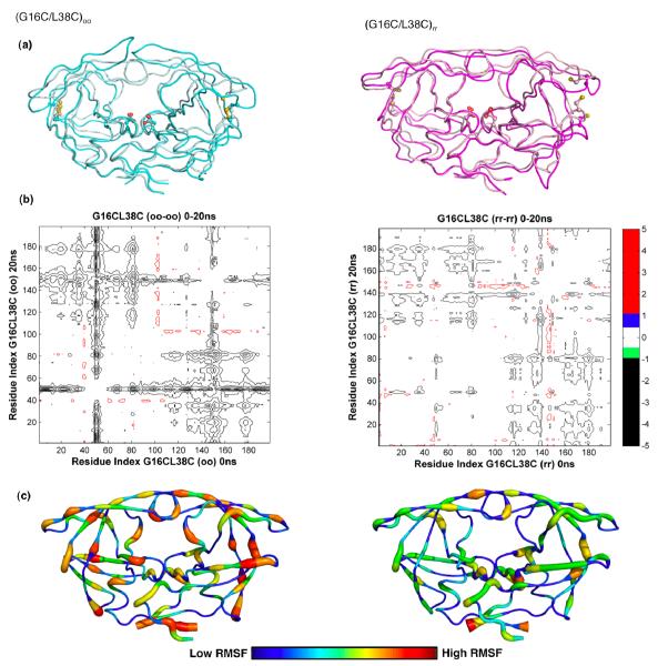
Human immunodeficiency virus Type-1 (HIV-1) protease is crucial for viral maturation and infectivity. Studies of protease dynamics suggest that the rearrangement of the hydrophobic core is essential for enzyme activity. Many mutations in the hydrophobic core are also associated with drug resistance and may modulate the core flexibility. To test the role of flexibility in protease activity, pairs of cysteines were introduced at the interfaces of flexible regions remote from the active site. Disulfide bond formation was confirmed by crystal structures and by alkylation of free cysteines and mass spectrometry. Oxidized and reduced crystal structures of these variants show the overall structure of the protease is retained. However, cross-linking the cysteines led to drastic loss in enzyme activity, which was regained upon reducing the disulfide cross-links. Molecular dynamics simulations showed that altered dynamics propagated throughout the enzyme from the engineered disulfide. Thus, altered flexibility within the hydrophobic core can modulate HIV-1 protease activity, supporting the hypothesis that drug resistant mutations distal from the active site can alter the balance between substrate turnover and inhibitor binding by modulating enzyme activity.


Figures

Figure 1
Figure 1
Drug resistance mutations in HIV-1 protease. Active site and the primary active site residues causing drug resistance (D30N, G48V,I50L/V, V82A/F/T, I84V) are colored in red. Hydrophobic core residues associated with drug resistance (I13V, I15V, L24I, L33F, M36I, I62V, I64V, I66F, V77I, I85V, L89M, L90M, I93L) are colored cyan. Remaining hydrophobic core residues (L5, V11, A22, L38, V75, L97) and rest of the protease are in yellow and gray, respectively. Catalytic Asps and the loops containing residues 11-22, 31-38 and 58-78 are displayed in black.
Figure 2
Figure 2
Schematic of cysteine redox chemistry used in this study. The cysteine substituted and non-cross-linked protease is flexible and as active as WT enzyme. Upon oxidation of cysteines, protease gets cross-linked via disulfide bond. Resulting loss of hydrophobic core flexibility is accompanied by loss of catalytic activity that is reversible upon reduction of the disulfide bond.
Figure 3
Figure 3
Ribbon diagrams of crystal structures of protease with engineered cysteines (a) Sites for engineering cysteines in HIV-1 protease are shown with backbone Cα (G16C/L38C) in red and (R14C/E65C) in blue. (b) The backbone structural superposition of DRV complexes of (G16C/L38C)rr and (R14C/E65C)rr on WT in black. Under reducing conditions, no disulfide bonds were observed. Alternate conformations for cysteine side chains were, however, seen for 50% of substituted cysteines in each pair analyzed. (c) apo(G16C/L38C)oo with disulfide bonds on both sides of the dimer shown in orange color.
Figure 4
Figure 4
Molecular dynamics simulation analyses. (a) Backbone superposition of (G16C/L38C)oo from 0ns (cyan) and at 20ns (pale cyan) and (G16C/L38C)rr from 0ns (light pink) to 20ns (magenta) in MD simulations. The side chains of active site aspartic acids and the engineered cysteines are displayed. (b) Differences in internal Cα-Cα distances between the 0ns and 20ns snapshots of the cross-linked (oo) and non-crosslinked (rr) forms of (G16C/L38C) variant are shown in the double difference plots. Each contour line represents a deviation by 0.5Å. Black, green, blue and red distinguish the contour ranges −1.0 Å and below, −1.0 to −0.5 Å, 0.5-1.0 Å and 1 Å and above, respectively. (c) Average RMSF of protease residues in (G16C/L38C)oo and (G16C/L38C)rr proteases from 20ns MD simulation trajectories. Protease molecules from 5, 10, 15 and 20ns simulations were superposed on to the 0ns crystal structure using the most invariant residues, 24 to 26 and 85 to 95. The average Cα RMSFs were calculated and mapped on to a representative protease molecule with the most variable regions depicted in red and the most invariant regions depicted in blue.

References

    1. Kohl NE, Emini EA, Schleif WA, Davis LJ, Heimbach JC, Dixon RA, Scolnick EM, Sigal IS. Proc Natl Acad Sci U S A. 1988;85:4686–4690. - PMC - PubMed
    1. Kramer RA, Schaber MD, Skalka AM, Ganguly K, Wong-Staal F, Reddy EP. Science. 1986;231:1580–4. - PubMed
    1. Muzammil S, Ross P, Freire E. Biochemistry. 2003;42:631–8. - PubMed
    1. Ohtaka H, Schon A, Freire E. Biochemistry. 2003;42:13659–66. - PubMed
    1. Clemente JC, Moose RE, Hemrajani R, Whitford LR, Govindasamy L, Reutzel R, McKenna R, Agbandje-McKenna M, Goodenow MM, Dunn BM. Biochemistry. 2004;43:12141–12151. - PubMed

Publication types

Substances