

Review
doi: 10.1021/cr2001178.
Epub 2012 Feb 2.
Gold nanoparticles in chemical and biological sensing
Affiliations
- PMID: 22295941
- PMCID: PMC4102386
- DOI: 10.1021/cr2001178
Item in Clipboard
Review
Gold nanoparticles in chemical and biological sensing
Chem Rev.
.
No abstract available
Figures
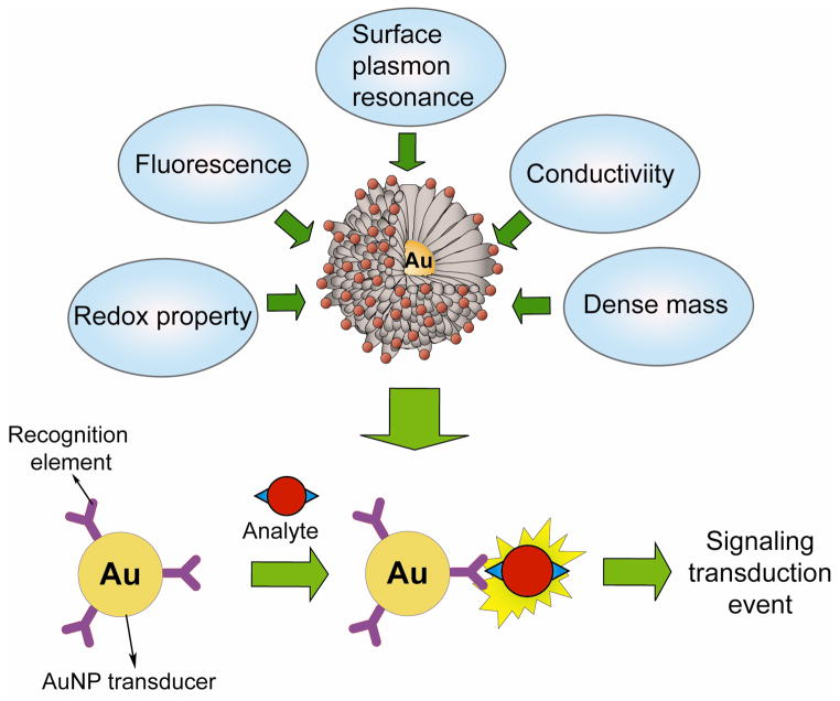
Physical properties of AuNPs and schematic illustration of an AuNP-based detection system.
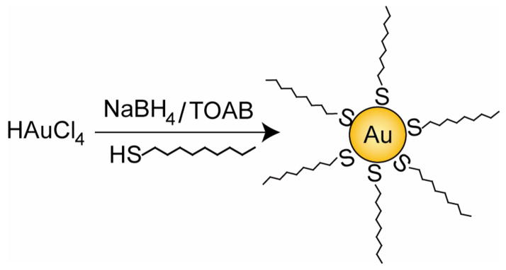
Brust-Schiffrin method for two-phase synthesis of AuNPs by reduction of gold salts in presence of external thiol ligands.
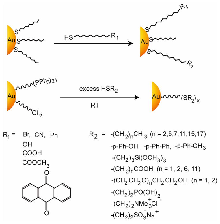
General scheme for place exchange reaction for alkanethiol-protected AuNPs using functionalized thiols, giving examples of particles that can be rapidly generated.
Schematic depiction of red-to-blue color colorimetric sensing of metal ions using chelating ligands.
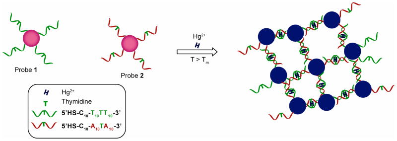
Colorimetric detection of Hg2+ using DNA functionalized AuNPs exploiting thymidine- Hg2+-thymidine coordination chemistry.
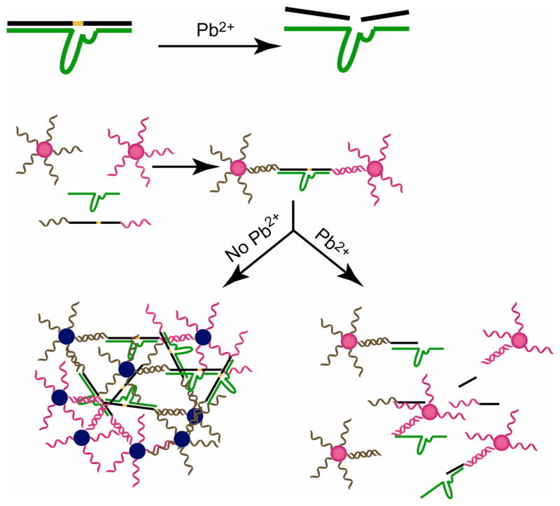
Pb2+ directed assembly of AuNPs by the DNAzymes resulting in detection.
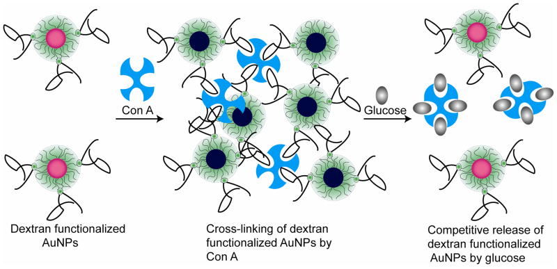
Colorimetric glucose sensing via the dissociation of Con A-aggregated dextran coated AuNPs. The presence of Con A crosslinks dextran-coated AuNPs with concomitant blue-shift in SPR. The addition of glucose diminishes the Con A-AuNPs interaction releasing the individual dextran-coated AuNPs.
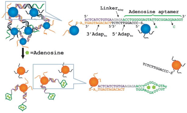
Schematic representation of colorimetric sensing of adenosine using aptamer linked AuNPs (left panel). The mutated linker with two mutations denoted by short black arrows was used as a control. Reprinted with permission from Angew. Chem. Int. Ed. (Ref 293). Copyright 2006 Wiley-VCH Verlag & Co. KGaA, Weinheim.
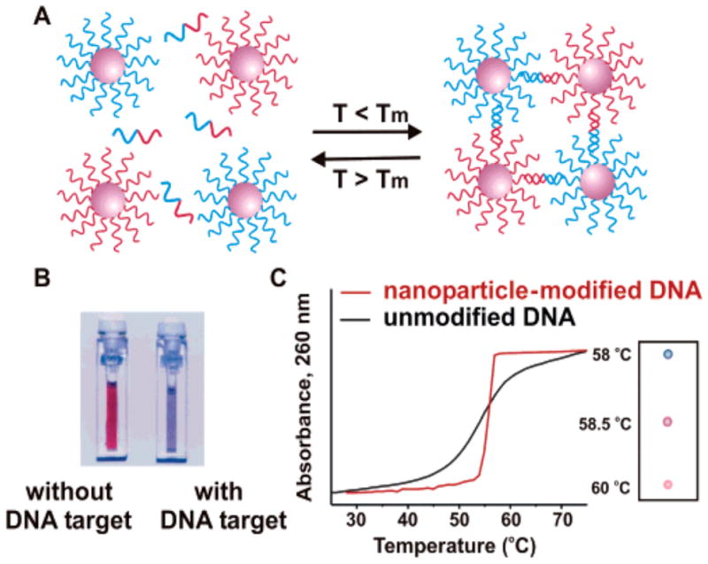
Aggregation of oligonucleotide AuNPs in presence of complementary target DNA (A), leading to change in color of solution from red to blue (B). Reprinted with permission from Science and Chem. Rev. (Ref and Ref 16). Copyright 1997 American Association for the Advancement of Science and 2005 American Chemical Society.
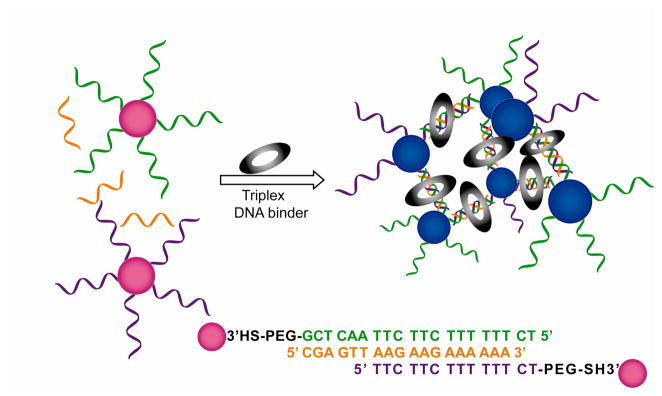
Schematic illustration of structure and color change of DNA functionalized AuNPs in presence of triplex DNA binders.
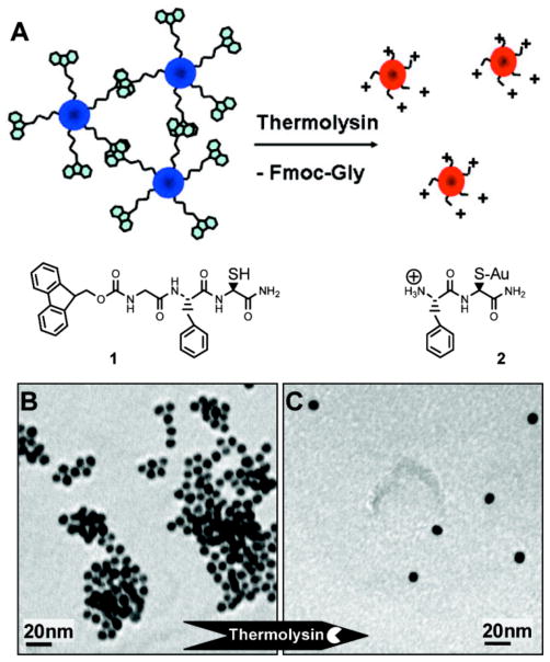
Schematic illustration of thermolysin triggered dispersion of AuNP assemblies (A), TEM images of AuNPs (functionalized with 1) before (B) and after (C) addition of thermolysin and generation of 2. Reprinted with permission from J. Am. Chem. Soc. (Ref 358). Copyright 2007 American Chemical Society.
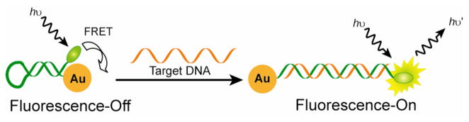
Schematic illustration of DNA detection, showing the conformational changes of dye-oligonucleotide-AuNP conjugates before and after hybridization with the target DNA.
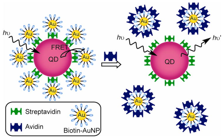
Competitive inhibition assay for the detection of avidin using QD-AuNP conjugates.
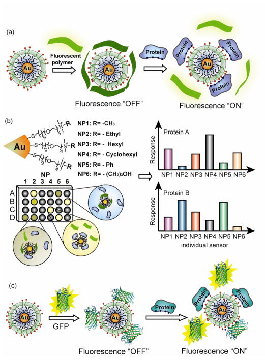
Schematic illustration of ‘chemical nose’ sensor array based on AuNP-fluorescent polymer/GFP conjugates. (a) The competitive binding between protein and quenched polymer-AuNP complexes leads to the restoration of fluorescence (b) The combination of an array of sensors generates fingerprint response patterns for individual proteins. (c) The competitive binding between protein and nanoparticle-GFP complexes leads to fluorescence light-up.
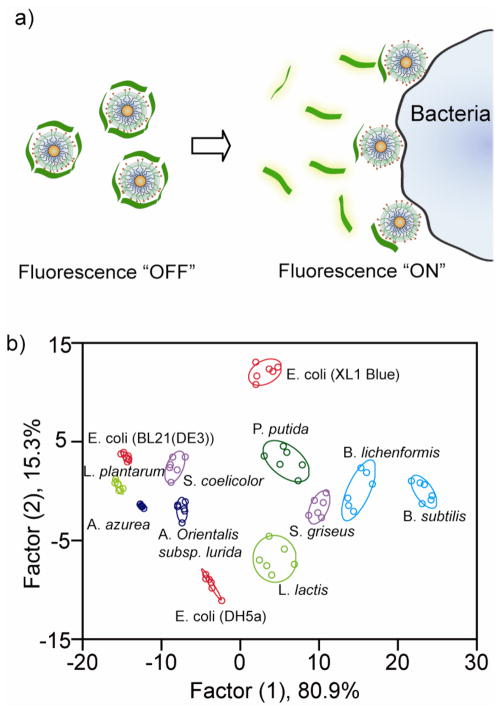
Array-based sensing of bacteria. a) Displacement assay between bacteria and the AuNP-PPE complex. b) Canonical score plot for the first two factors of simplified fluorescence response patterns obtained with NP-PPE assembly arrays against bacteria (95% confidence ellipses shown). Reprinted with permission from Angew. Chem. Int. Ed. (Ref ). Copyright 2008 Wiley-VCH Verlag & Co. KGaA.
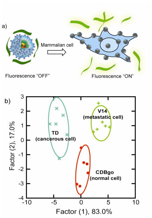
a) Schematic depiction of the fluorophore-displacement cell detection array. Displacement of quenched fluorescent polymer by a cell with consequent restoration of polymer fluorescence. b) Canonical score plot for the first two factors of simplified fluorescence response patterns obtained with AuNP-conjugated polymer assembly arrays against different mammalian cell types. Reprinted with permission with permission from Proc. Natl. Acad. Sci. U.S.A. (Ref 398). Copyright 2009 National Academy of Sciences.
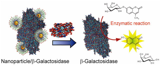
A schematic representation of the sensor system comprised of β-galactosidase (β-Gal) and cationic AuNPs. β-Gal is displaced from the β-Gal/AuNP complex by protein analytes, restoring the catalytic activity of β-Gal towards the fluorogenic substrate 4-methylumbelliferyl-β-D-galactopyranoside, resulting in an amplified signal for detection. Reprinted with permission from J. Am. Chem. Soc. (Ref 401). Copyright 2010 American Chemical Society.
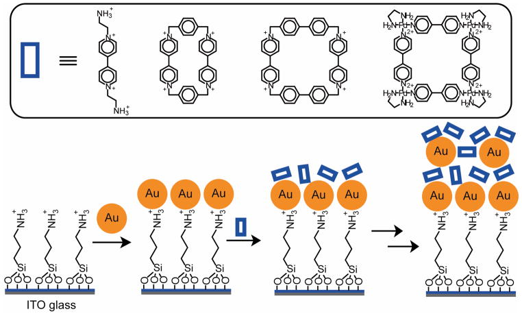
Electroactive multilayers formed by the simultaneous deposition of anionic AuNPs and oligocationic cyclophanes. The AuNPs provide excellent conductive surfaces, while the macrocycles serve as π-acceptors to bind with π-donor analytes (e.g. hydroquinone), generating electrochemical responses.
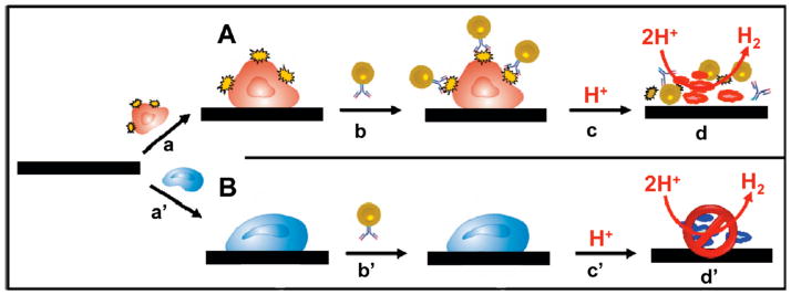
Specific identification of (A) Tumoral Cell Line (HMy2) expressing surface HLA-DR molecules compared to a (B) PC-3 Cell Line that is negative to this marker using AuNP-conjugated antibody coupled with an electrochemical sensor. The specific binding of AuNPs with tumor cells catalyses hydrogen evolution in the acidic environment, generating electrochemical responses. Reprinted with permission from Anal. Chem. (Ref 574). Copyright 2009 Americal Chemical Society.
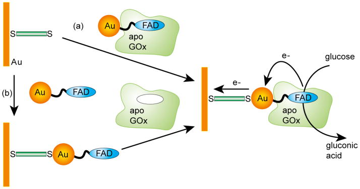
Fabrication of GOx electrode by the reconstitution of GOx on a FAD-functionalized AuNP : (a) The adsorption of AuNP-reconstituted of apo-GOx to a dithiol monolayer assembled on a gold electrode and (b) the adsorption of FAD-AuNPs to a dithiol-modified gold electrode followed by the reconstitution of apo-GOx onto functional AuNPs. Reprinted with permission from Science (Ref 608). Copyright 2003 American Association for the Advancement of Science.
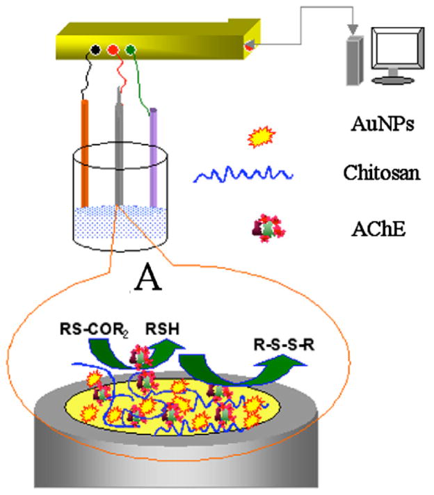
Acetylcholinesterase enzyme biosensor constructs using AuNPs/chitosan hydrogel matrices for the detection of pestisides. A) Megascopic interface of AChE/Chitosan–AuNP modified gold electrode. Reprinted with permission from J. Electroanal. Chem. (Ref 689). Copyright 2007 Elsevier B.V.
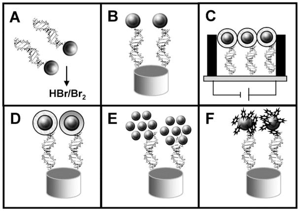
Schematic procedure of the different strategies used for the integration of AuNPs into DNA sensing systems: (A) previous dissolving of AuNPs by using HBr/Br2 mixture followed by Au(III) ions detection, (B) direct detection of AuNPs anchored onto the surface of the genosensor, (C) conductometric detection, (D) enhancement with silver or gold followed by detection, (E) AuNPs as carriers of other AuNPs, (F) AuNPs as carriers of other electroactive labels. Reprinted with permission from Electroanalysis (Ref 691). Copyright 2007 Wiley-VCH Verlag GmbH & Co. KGaA, Weinheim.
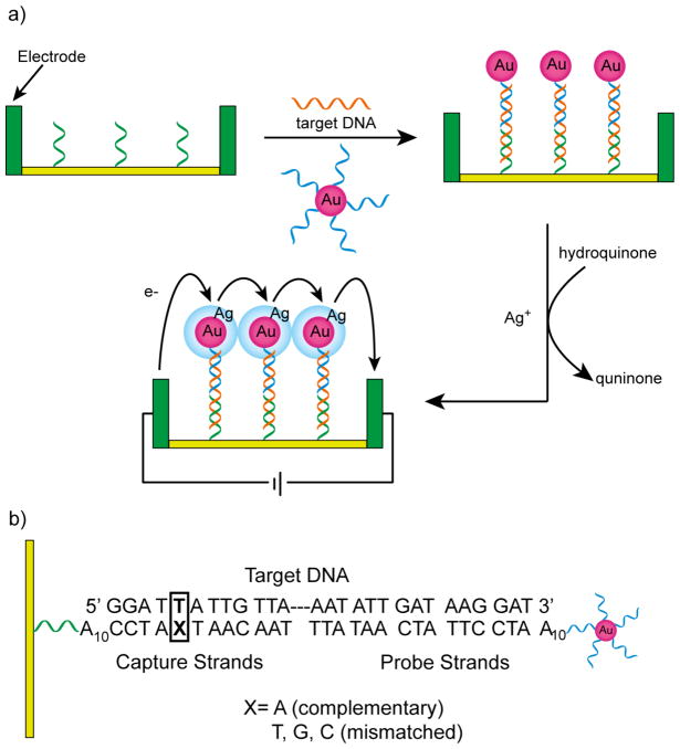
a) Schematic illustration of electrical detection of DNA based on “sandwich” hybridization with DNA functionalized AuNPs followed by silver enhancement. b) Sequences of capture, target, and probe DNA strands. Reproduced with permission from Science (Ref 732). Copyright 2002 American Association for the Advancement of Science.
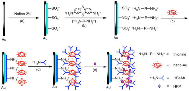
Schematic illustration of the stepwise immunosensor fabrication process to detect HBsAg: (a) formation of nafion monolayer; (b) adsorption of thionine; (c) formation of AuNP-monolayer; (d) HBsAb loading; (e) blocking with HRP. Reprinted with permission from Anal. Chim. Acta (Ref 752). Copyright 2005 Elsevier B.V.
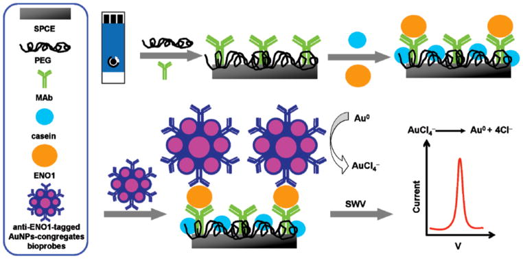
Schematic representation of the operation of the electrochemical immunosensor for the detection of ENO1. Reprinted with permission from Anal. Chem. (Ref 838). Copyright 2010 American Chemical Society.
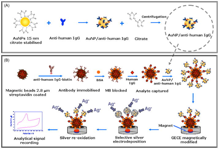
(A) AuNP conjugation with anti-human IgG; (B) analytical procedure for the sandwich type assay and the obtaining of the analytical signal based on the catalytic effect of AuNPs on the silver electrodeposition. Reprinted with permission from Biosens. Bioelectron. (Ref ). Copyright 2009 Elsevier B.V
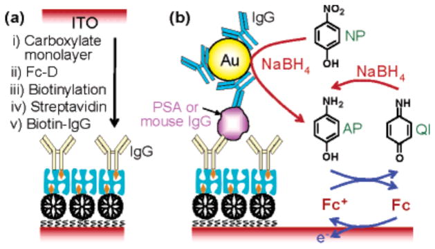
(a) Schematic representation of the preparation of an immunosensing layer. (b) Schematic view of electrochemical detection of mouse IgG or PSA. Reprinted with permission from J. Am. Chem. Soc. (Ref 868). Copyright 2006 American Chemical Society.
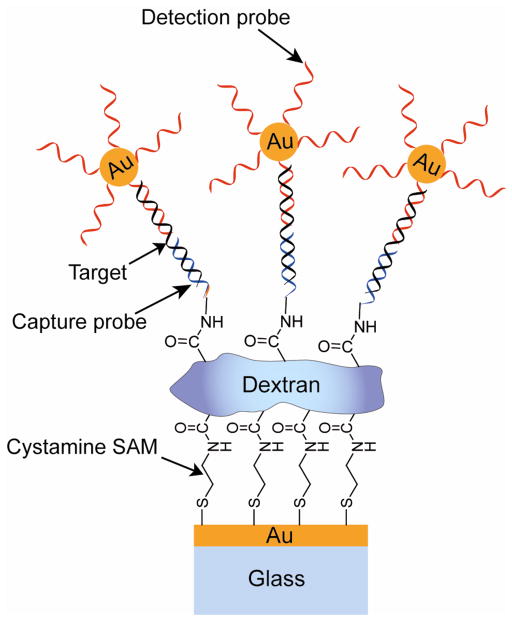
Schematic representation of sandwich DNA detection assay via AuNP-mediated SPR signal amplification. The SPR measurements were carried out by injecting the oligonucleotide functionalized AuNPs into the flow cell housing sensors covered with various duplexes or capture probes. The intermediate dextran layer reduces the nonspecific adsorption of AuNPs, improving detection sensitivity. Adapted from Ref .
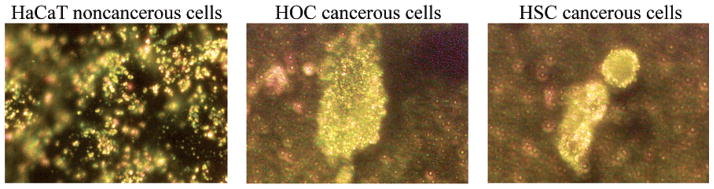
SPR light scattering images to distinguish between normal cells (left panel, HaCaT) and cancerous cells (middle & right panel, HOC and HSC) after incubation with anti-EGFR conjugated AuNPs. The anti-EGFR conjugated AuNPs bind specifically to the surface of cancer cells resulting in a sharper SPR band with a red shifted maxima. Reproduced with permission from Nano Lett. (Ref 969). Copyright 2005 American Chemical Society.
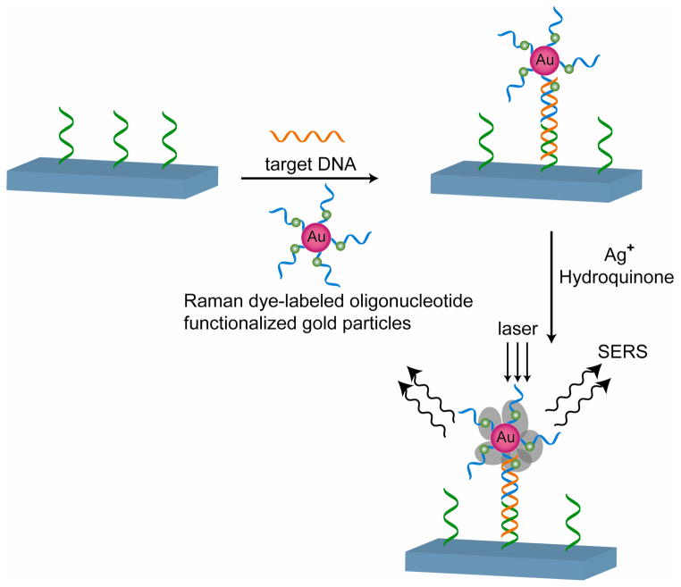
Schematic representation of three component sandwich assay for SERS-based oligonucleotide detection. Reproduced with permission from Science (Ref 987). Copyright 2002 American Association for the Advancement of Science.
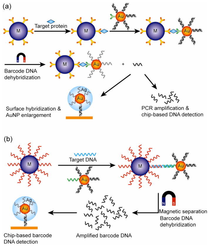
AuNP-based bio-barcode assay of (a) proteins and (b) DNA
References
-
- Diamond D. Principles of Chemical and Biological Sensors. John Wiley & Sons, Inc; New York, NY: 1998.
-
- Sadik OA, Land WH, Wang J. Electroanalysis. 2003;15:1149–1159.
-
- Sapsford KE, Bradburne C, Detehanty JB, Medintz IL. Mater Today. 2008;11:38–49.
-
- Burnworth M, Rowan SJ, Weder C. Chem Eur J. 2007;13:7828–7836. - PubMed
Publication types
MeSH terms
Substances
Grants and funding
LinkOut - more resources
Full Text Sources
Other Literature Sources
