. 2012 Apr;220(4):336-49.
doi: 10.1111/j.1469-7580.2012.01474.x. Epub 2012 Feb 1.

Three-dimensional and molecular analysis of the arterial pole of the developing human heart

Affiliations

Three-dimensional and molecular analysis of the arterial pole of the developing human heart

Aleksander Sizarov et al. J Anat. 2012 Apr.

Abstract

Labeling experiments in chicken and mouse embryos have revealed important roles for different cell lineages in the development of the cardiac arterial pole. These data can only fully be exploited when integrated into the continuously changing morphological context and compared with the patterns of gene expression. As yet, studies on the formation of separate ventricular outlets and arterial trunks in the human heart are exclusively based on histologically stained sections. So as to expand these studies, we performed immunohistochemical analyses of serially sectioned human embryos, along with three-dimensional reconstructions. The development of the cardiac arterial pole involves several parallel and independent processes of formation and fusion of outflow tract cushions, remodeling of the aortic sac and closure of an initial aortopulmonary foramen through formation of a transient aortopulmonary septum. Expression patterns of the transcription factors ISL1, SOX9 and AP2α show that, in addition to fusion of the SOX9-positive endocardial cushions, intrapericardial protrusion of pharyngeal mesenchyme derived from the neural crest contributes to the separation of the developing ascending aorta from the pulmonary trunk. The non-adjacent walls of the intrapericardial arterial trunks are formed through addition of ISL1-positive cells to the distal outflow tract, while the facing parts of the walls form from the protruding mesenchyme. The morphogenetic steps, along with the gene expression patterns reported in this study, are comparable to those observed in the mouse. They confirm the involvement of mesenchymal tissues derived from endocardium, mesoderm and migrating neural crest cells in the process of initial septation of the distal part of the outflow tract, and its subsequent separation into discrete intrapericardial arterial trunks.


Figures

Fig. 1
Fig. 1
The contours of the developing heart (red and blue lines) superimposed on the silhouettes of embryos at consecutives stages to illustrate the ambiguity of terms ‘dorsal/ventral’ and ‘cranial/caudal’ in describing the morphogenesis of the curved and changing pharyngeal region. (A) The contour of the stage 13 embryo, the reconstructed heart of which is shown in Fig. 2. (B) Stage 15 embryo (see Fig. 3). (C) Stage 16 embryo (see Fig. 6).
Fig. 2
Fig. 2
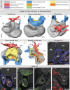
Three-dimensional (3D) and molecular analysis of the cardiac outflow tract at stage 13. (A–E) At this stage the myocardial outflow tract (OFT), connecting the right ventricle with the aortic sac (as), is tubular and its lumen is lined by connexin40-positive endocardium (F,G). The 3rd, 4th and 6th aortic arches originate directly from the aortic sac. The dotted line in (D) refers to the distance between the origins of tiny right and left pulmonary arteries. Note the canal-like part of the looping heart tube interposed between the ballooning left and right ventricles (dotted line in B). (G–J) Sections through the distal outflow tract, which were incubated with antibodies as indicated. The asterisks in (G) and (H) point to the appearance of mesenchymal tissue within the distal outflow tract taking the form of columns at later stages (see text). The small arrows in (J) point to contiguity between neural crest-derived cells of outflow tract and pharyngeal mesenchyme. Scale bars: 200 μm. Abbreviations: AVC, atrioventricular canal; e/tr, (undivided) developing esophagus and trachea; LA/RA, left/right atrium; LSCV, left superior cardinal vein; lsh, left sinus horn; LV/RV, left/right ventricle; SV, sinus venosus.
Fig. 3
Fig. 3
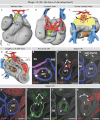
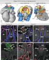
Three-dimensional (3D) and molecular analysis of the cardiac outflow tract at stage 15. (A–D) The intrapericardial mesenchymal tissue forming the column-like structures at ventral and dorsal aspects of the distal outflow tract (asterisks in A and B). Note the appearance of the longitudinal arterial channel, the future ascending aorta (AAo) giving rise to the 3rd and 4th aortic arches. The 6th aortic arches originate separately from the unseptated distal outflow tract (E) as the result of the remodeling of the aortic sac. The dotted line in (C) points to the decreasing distance between the origins of the left and right pulmonary arterial branches. The arrows in (A) and (D) point to the characteristic bend of the myocardial outflow tract, dividing it into proximal and distal parts. Dotted lines in (D) refer to the spirally oriented channels in the proximal outflow tract connecting the unseptated distal outflow tract with the right ventricle (white line) and the primary interventricular foramen (green line). (E) Section through the proximal outflow tract showing the unfused endocardial cushions producing aortic and pulmonary arterial channels (green and white arrows, respectively). (F–J) Sections through the distal outflow tract, which were incubated with antibodies, as indicated. The star in (F) points to the mesenchyme located between the 4th and 6th and between the right and left aortic arches. Asterisks in (F)–(K) point to the ventrally and dorsally located columns of mesenchymal tissue (see text). Scale bars: 200 μm. Abbreviations: pv, pulmonary vein; tr, trachea; for other abbreviations, see Fig. 1.
Fig. 4
Fig. 4
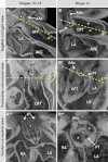
Three-dimensional (3D) morphology of the distal outflow tract, as assessed by high-resolution episcopic microscopy. (A–A**) Different views of the unseptated distal outflow tract at stages 14–15 with the dorsal wall between the 4th and 6th aortic arches slightly protruding intrapericardially (star), which leads to the appearance of the future extrapericardial ascending aorta (AAo). (B–B**) Different views through the septated distal outflow tract at stage 16. Note that progressive protrusion of the dorsal wall of the outflow tract intrapericardially leads to the formation of the so-called aortopulmonary septum (#, outlined by the dotted line in B, B*) separating the developing intrapericardial aortic channel from the future pulmonary trunk (PT). Yellow asterisks indicate endocardial cushions. See text for further description.
Fig. 5
Fig. 5
The images show the appearance and development of the arterial valves. (A, E, I) Ventral views of the near frontal cuts through the high-resolution episcopic microscopy (HREM) datasets of the embryos at consecutive stages taken through the middle portion of the intrapericardial outflow tract. Note the considerable rotation of the line between aortic (Ao) and pulmonary (Pu) arterial channels. (B–D) Sections of stages 14–15 embryos through the intrapericardial extension of the non-myocardial components derived from the pharyngeal mesenchyme (asterisks, outlined by dotted lines), which were incubated with antibodies as indicated. Note that these extensions are positive for SOX9 and ISL1, while being negative for αSMA. The arrows point to the cellular condensations within one of the outflow tract cushions (see text). (F–H) Sections of the stage 16 embryo, showing the appearance of the intercalated cushions (asterisks, outlined by dotted lines) within the middle portion of the outflow tract, which were incubated with antibodies as indicated. The intercalated cushions only weakly express ISL1, while beginning to express SOX9 and remaining negative for αSMA. (J,J*,K,K*) Sections through the developing pulmonary (PuV) and aortic (AoV) valves in the stage 18 embryonic heart, which were incubated with antibodies as indicated. The developing leaflets of the valves scarcely express αSMA, but are strongly positive for SOX9. The tissue between the discrete and separate walls of the arterial trunks (#) is now negative for αSMA. Scale bars: 200 μm.
Fig. 6
Fig. 6
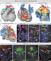
Three-dimensional (3D) and molecular analysis of the cardiac outflow tract at stage 16. (A–C) The muscular outflow tract has further shortened, and the mass of the right ventricle is recognisable ventrally. The extrapericardial portions of the perpendicularly oriented ascending aorta and pulmonary trunk are separated from each other by mesenchymal tissue (# in B,C). The dotted line in (C) points to the decreasing distance between the origin of the pulmonary arterial branches. (D) The relations between developing ascending aorta and pulmonary trunk (white and green dotted lines, respectively) now resemble the situation seen in the formed heart. (E) Section through the proximal outflow tract showing the still unfused endocardial cushions producing aortic and pulmonary arterial channels (green and white arrows, respectively). (F–K) Sections through the distal outflow tract, which were incubated with antibodies as indicated. The protrusion from the dorsal wall of the aortic sac now extends into the pericardial cavity (#), and interposes between the ascending aorta and the pulmonary trunk. The contact between the protrusion and the distal ends of the outflow tract cushions expressing SOX9 (yellow asterisks) is marked by a dashed line. The yellow arrows in (J) point to the sharp border of the AP2α expression domain. Scale bars: 200 μm. For abbreviations, see previous figures.
Fig. 7
Fig. 7
Three-dimensional (3D) and molecular analysis of the cardiac outflow tract at stage 18. (A–C) The myocardial outflow tract has further shortened, along with formation of the right ventricular infundibulum (RVOT). The intrapericardial ascending aorta and pulmonary trunk now possess their own discrete non-myocardial walls. The dotted line in (B) points to the proximal parts of the 6th arches, which now form the bifurcation of the pulmonary trunk. The left-sided 6th arch is persisting to become the arterial duct, while the right-sided 6th arch distal to the origin of the right pulmonary artery is regressing. Both 4th arches are of roughly equal size, while the third arches are no longer recognisable. (D) Section through the left ventricular outflow tract (LVOT) and developing aortic valve (AoV). The asterisks in (C) and (D) refer to the myocardialising mesenchyme between the ventricular outflow tracts. (E–J) Sections through the right ventricular outflow tract, developing pulmonary valve (PuV), pulmonary trunk and ascending aorta were incubated with antibodies as indicated. The tissue between great arteries (#) is ISL1-negative and expresses AP2α, which is also expressed in the facing walls of the arterial trunks. In contrast to the wall of the ascending aorta, the pulmonary truncal wall expressed transcription factor NKX2-5, reflecting the cardiogenic potential of these mesenchymal cells. Scale bars: 200 μm. For abbreviations, see previous figures.
Fig. 8
Fig. 8
Schematic summary of the processes leading to septation of the distal outflow tract. Note that this is a highly simplified 2D view of the morphologically complex 3D structure. (A) At stages 14 and 15, the aortopulmonary foramen within the distal outflow tract is bordered dorsally by ISL1-positive mesenchyme, which gives rise to the extrapericardial 6th aortic arches and the future ascending aorta. At both sides of this ISL1-positive mesenchyme, neural crest-derived cells are migrating ventrally along and cranial to the 6th aortic arches. (B) At stage 16, the endocardial cushions have been formed and fused in the outflow tract to separate the developing aortic and pulmonary channels. Progressive protrusion of the neural crest-derived mesenchyme results initially in the formation of an obliquely oriented aortopulmonary septum (#), and subsequent closure of the aortopulmonary foramen. (C) At stage 18, the continuous addition of ISL1-positive cells at the arterial pole results in the formation of the intrapericardial arterial trunks, the facing walls of which are formed from neural crest-derived mesenchyme. The stars in (A–C) point to the ISL1-positive mesenchyme at the level of the sixth aortic arches, which remains interposed between them. Abbreviations: L/R, left/right; RVOT, right ventricular infundibulum.

Similar articles

Cited by

References

    1. Akiyama H, Chaboisser M-C, Behringer RR, et al. Essential role of Sox9 in the pathway that controls formation of cardiac valves and septa. Proc Natl Acad Sci USA. 2004;101:6502–6507. - PMC - PubMed
    1. Anderson RH, Webb S, Brown NA, et al. Formation of the ventricular outflow tracts, arterial valves, and intrapericardial arterial trunks. Heart. 2003;89:1110–1118. - PMC - PubMed
    1. Beall AC, Rosenquist TH. Smooth muscle cells of neural crest origin form the aorticopulmonary septum in the avian embryo. Anat Rec. 1990;226:360–366. - PubMed
    1. Bergwerff M, Verberne ME, DeRuiter MC, et al. Neural crest cell contribution to the developing circulatory system: implications for vascular morphology. Circ Res. 1998;82:221–231. - PubMed
    1. Betters E, Liu Y, Kjaeldgaard A, et al. Analysis of early human neural crest development. Dev Biol. 2010;344:578–592. - PMC - PubMed

Publication types

Substances