. 2012 Feb 2:9:3.
doi: 10.1186/1743-8977-9-3.

Amorphous silica nanoparticles size-dependently aggravate atopic dermatitis-like skin lesions following an intradermal injection

Affiliations

Amorphous silica nanoparticles size-dependently aggravate atopic dermatitis-like skin lesions following an intradermal injection

Toshiro Hirai et al. Part Fibre Toxicol. .

Abstract

Background: Due to the rising use of nanomaterials (NMs), there is concern that NMs induce undesirable biological effects because of their unique physicochemical properties. Recently, we reported that amorphous silica nanoparticles (nSPs), which are one of the most widely used NMs, can penetrate the skin barrier and induce various biological effects, including an immune-modulating effect. Thus, it should be clarified whether nSPs can be a risk factor for the aggravation of skin immune diseases. Thus, in this study, we investigated the relationship between the size of SPs and adjuvant activity using a model for atopic dermatitis.

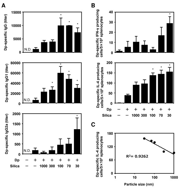
Results: We investigated the effects of nSPs on the AD induced by intradermaly injected-mite antigen Dermatophagoides pteronyssinus (Dp) in NC/Nga mice. Ear thickness measurements and histopathological analysis revealed that a combined injection of amorphous silica particles (SPs) and Dp induced aggravation of AD in an SP size-dependent manner compared to that of Dp alone. In particular, aggravation was observed remarkably in nSP-injected groups. Furthermore, these effects were correlated with the excessive induction of total IgE and a stronger systemic Th2 response. We demonstrated that these results are associated with the induction of IL-18 and thymic stromal lymphopoietin (TSLP) in the skin lesions.

Conclusions: A particle size reduction in silica particles enhanced IL-18 and TSLP production, which leads to systemic Th2 response and aggravation of AD-like skin lesions as induced by Dp antigen treatment. We believe that appropriate regulation of nanoparticle physicochemical properties, including sizes, is a critical determinant for the design of safer forms of NMs.


Figures

Figure 1
Figure 1
Effects of amorphous silica nanoparticles (nSPs) on atopic dermatitis (AD)-like skin lesions induced by Dermatophagoides pteronyssinus (Dp). NC/Nga mice were intradermally injected on the ventral side of both ears with Dp alone or Dp plus SPs in PBS on days 1, 3, 5, 7, 9, 11, 13, 16, and 18. Ear thickness was measured on days 0 and 19 (A). The ears were removed 24 h after the last intradermal injection. Sections were prepared, and the ears were stained with H&E to assess representative symptoms of AD (B and C) or with toluidine blue to assess mast cell infiltration (D and E). Representative histological findings of the PBS, Dp, Dp + mSP1000, and Dp + nSP30 groups are shown. Several representative symptoms of AD were scored in the H&E sections (C). Infiltration of mast cells was evaluated as the number of cells in toluidine blue sections in a high power field (E). Data are presented as mean ± SE of 4-5 animals per group. The results are representative of two separate experiments. *P < 0.05 vs. Dp group. Scale bars: 100 μm (B) and 50 μm (D).
Figure 2
Figure 2
Effects of amorphous silica particles (SPs) on total and Dermatophagoides pteronyssinus (Dp)-specific IgE production in plasma. NC/Nga mice were intradermally injected on the ventral side of both ears with Dp alone or Dp plus SPs in PBS on days 1, 3, 5, 7, 9, 11, 13, 16, and 18. To evaluate AD severity, plasma was collected 24 h after the last intradermal injection and analyzed by ELISA for total and Dp-specific IgE (A and C). A regression analysis of total IgE was also performed (B). Data are presented as mean ± SE of 4-5 animals per group. The results are representative of two separate experiments. *P < 0.05 vs. Dp group.
Figure 3
Figure 3
Analysis of the local immune response induced by Dermatophagoides pteronyssinus (Dp) + amorphous silica particles (SPs). NC/Nga mice were intradermally injected on the ventral side of both ears with Dp alone or Dp plus SPs in PBS on days 1, 3, 5, 7, 9, 11, 13, 16, and 18. At 24 h after the last intradermal injection, the ears were removed and homogenized. The homogenates were centrifuged at 10,000 rpm for 5 min, and the supernatants were collected. The levels of interleukin (IL)-4, IL-5, IL-13, interferon (IFN)-γ, IL-18, and thymic stromal lymphopoietin (TSLP) in the supernatants were examined by ELISA (A, B, and D). A regression analysis of IL-18 was also performed (C). Data are presented as mean ± SE of 4-5 animals per group. The results are representative of two separate experiments. *P < 0.05 vs. Dp group.
Figure 4
Figure 4
Analysis of the systematic immune response induced by Dermatophagoides pteronyssinus (Dp) + amorphous silica particles (SPs). NC/Nga mice were intradermally injected on the ventral side of both ears with Dp alone or Dp plus SPs in PBS on days 1, 3, 5, 7, 9, 11, 13, 16, and 18. At 24 h after the last intradermal injection, splenocytes and plasma were collected from each group. Dp-specific IgG and its subclasses (A) in plasma were analyzed by ELISA. Splenocytes from each group were cultured with 100 μg ml-1 Dp. The levels of Dp-specific interferon (IFN)- γ and interleukin (IL)-4-producing cells were examined by individual cytokine-specific ELISPOT assay (B). A regression analysis of Dp-specific IL-4-producing cells was also performed (C). Data are presented as mean ± SE of 4-5 animals per group. The results are representative of two separate experiments. *P < 0.05 vs. Dp group.

References

    1. Bowman DM, van Calster G, Friedrichs S. Nanomaterials and regulation of cosmetics. Nat Nanotechnol. 2010;5:92. doi: 10.1038/nnano.2010.12. - DOI - PubMed
    1. Liu D, Gu N. Nanomaterials for fresh-keeping and sterilization in food preservation. Recent Pat Food Nutr Agric. 2009;1:149–154. - PubMed
    1. Salata O. Applications of nanoparticles in biology and medicine. J Nanobiotechnology. 2004;2:3. doi: 10.1186/1477-3155-2-3. - DOI - PMC - PubMed
    1. Choksi AN, Poonawalla T, Wilkerson MG. Nanoparticles: a closer look at their dermal effects. J Drugs Dermatol. 2010;9:475–481. - PubMed
    1. Nel A, Xia T, Madler L, Li N. Toxic potential of materials at the nanolevel. Science. 2006;311:622–627. doi: 10.1126/science.1114397. - DOI - PubMed

Publication types

LinkOut - more resources