Review
. 2012 Mar 1;4(3):a006049.
doi: 10.1101/cshperspect.a006049.

Signaling in innate immunity and inflammation

Affiliations
Review

Signaling in innate immunity and inflammation

Kim Newton et al. Cold Spring Harb Perspect Biol. .

Abstract

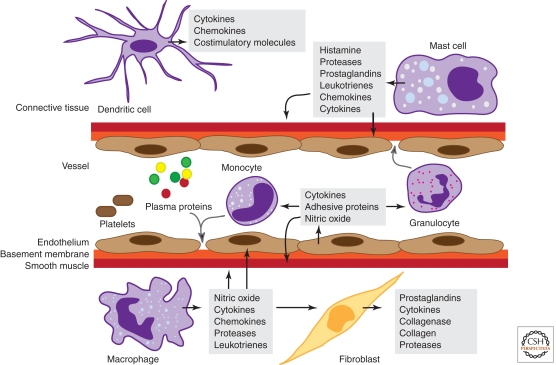
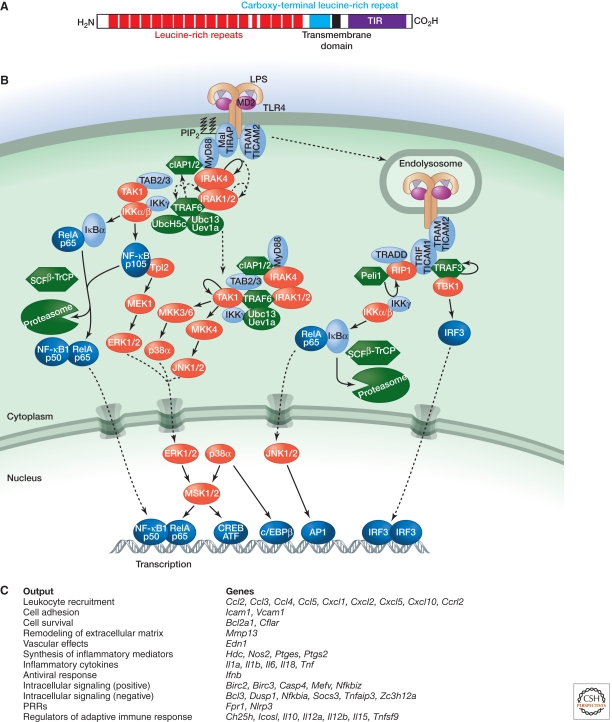
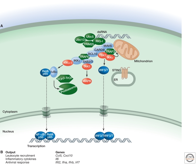
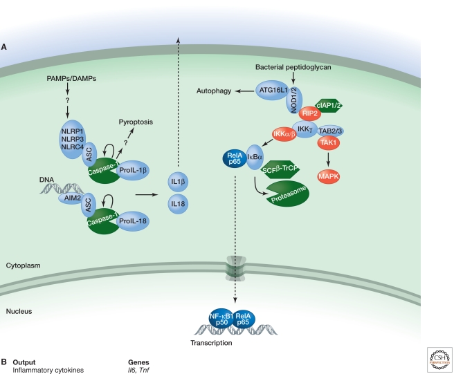
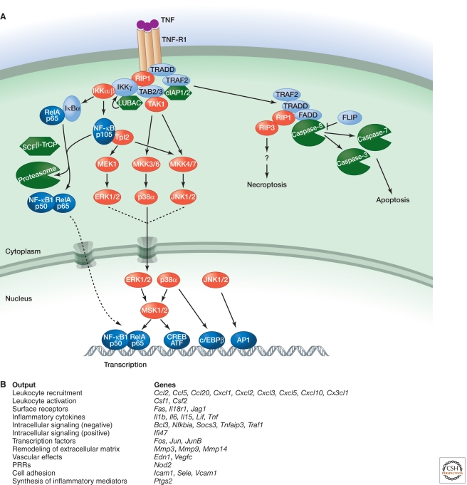
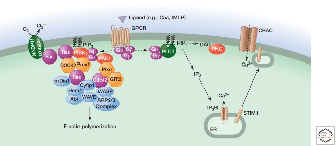
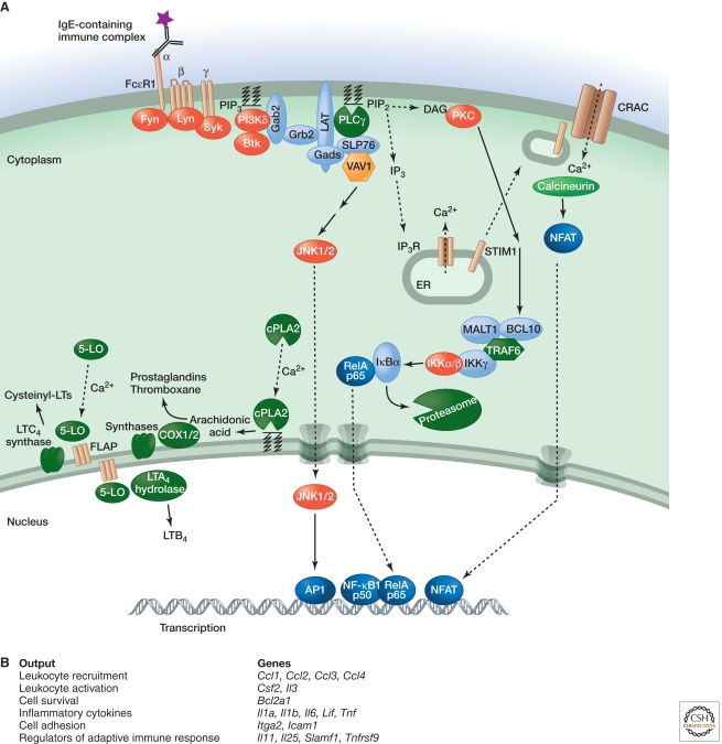
Inflammation is triggered when innate immune cells detect infection or tissue injury. Surveillance mechanisms involve pattern recognition receptors (PRRs) on the cell surface and in the cytoplasm. Most PRRs respond to pathogen-associated molecular patterns (PAMPs) or host-derived damage-associated molecular patterns (DAMPs) by triggering activation of NF-κB, AP1, CREB, c/EBP, and IRF transcription factors. Induction of genes encoding enzymes, chemokines, cytokines, adhesion molecules, and regulators of the extracellular matrix promotes the recruitment and activation of leukocytes, which are critical for eliminating foreign particles and host debris. A subset of PRRs activates the protease caspase-1, which causes maturation of the cytokines IL1β and IL18. Cell adhesion molecules and chemokines facilitate leukocyte extravasation from the circulation to the affected site, the chemokines stimulating G-protein-coupled receptors (GPCRs). Binding initiates signals that regulate leukocyte motility and effector functions. Other triggers of inflammation include allergens, which form antibody complexes that stimulate Fc receptors on mast cells. Although the role of inflammation is to resolve infection and injury, increasing evidence indicates that chronic inflammation is a risk factor for cancer.


Figures

Figure 1.
Figure 1.
Cells and mediators of the inflammatory response. Molecules derived from plasma proteins and cells in response to tissue damage or pathogens mediate inflammation by stimulating vascular changes, plus leukocyte migration and activation. Granulocytes include neutrophils, basophils, and eosinophils.
Figure 2.
Figure 2.
Signaling by TLR4. (A) Domain structure of human TLR4. (B) Binding of LPS to TLR4 and the coreceptor MD2 triggers interactions between the cytoplasmic TIR domain of TLR4 and TIR-containing adaptor proteins (Mal, MyD88, and TRAM). MyD88 binds IRAK4, which requires its kinase activity to bind the kinases IRAK1 and IRAK2 sequentially. The MyD88–IRAK complex also engages the ubiquitin ligase TRAF6 to make polyubiquitin chains that activate the IKK complex for NF-κB- and ERK-dependent gene transcription. Ubiquitin ligases cIAP1 and cIAP2 recruited to the TLR4 signaling complex regulate translocation of a subset of signaling components to the cytoplasm, where TAK1 activation initiates a MAPK cascade that stimulates gene expression. TLR4 activated at the plasma membrane is endocytosed but can signal within the endosomal compartment via the adaptors TRAM and TRIF. The kinase and ubiquitin ligase combination of RIP1 and Peli1 interacts with TRIF to signal NF-κB activation, whereas TBK1 and TRAF3 stimulate IRF3-dependent transcription. (C) Functional outputs of some of the genes upregulated by TLR4 signaling.
Figure 3.
Figure 3.
Signaling by RIG-I. (A) RIG-I binding to dsRNA that has a 5′ triphosphate and polyubiquitin, the latter generated by the ubiquitin ligase TRIM25 and E2 ubiquitin-conjugating enzymes Ubc5 and Ubc13, promotes RIG-I binding to mitochondrial MAVS. Subsequently, a larger complex containing the adaptor proteins CARD9 and BCL10 is assembled for MAPK and NF-κB activation. TRAF3, the kinases TBK1 and IKKε, and ER-resident protein STING are required for activation of transcription factors IRF3 and IRF7. (B) Functional outputs of some of the genes upregulated by MAVS signaling.
Figure 4.
Figure 4.
Signaling by NLRs. (A) NLRP1, NLRP3, and NLRC4 respond to diverse PAMPS and DAMPs by engaging the caspase-1 adaptor protein ASC, whereas AIM2 binds ASC in response to cytoplasmic dsDNA. Activation of caspase-1 within each inflammasome complex results in processing of pro-IL1β and pro-IL18 and secretion of their biologically active forms. Caspase-1 activation also triggers a rapid form of cell death termed pyroptosis. NOD1 and NOD2 sense different components of bacterial peptidoglycan and stimulate either the autophagy machinery or gene transcription via NF-κB and MAPK activation. The latter outcome requires interaction of NOD1 or NOD2 with the kinase RIP2, which may be ubiquitylated by cIAPs in order to recruit TAB2/3 and IKKγ for TAK1 and IKK activation. (B) Functional outputs of some of the genes upregulated by NOD1 or NOD2 signaling.
Figure 5.
Figure 5.
Signaling by TNF-R1. (A) Binding of TNF to TNF-R1 causes the cytoplasmic death domain (DD) in TNF-R1 to bind the DD-containing proteins TRADD and RIP1. TRADD also binds TRAF2, which serves as an adaptor for the ubiquitin ligases cIAP1 and cIAP2. Ubiquitylation of RIP1, and potentially other components of the complex, recruits IKKγ and TAK1 for NF-κB and MAPK activation. Recruitment of LUBAC for linear ubiquitylation of IKKγ may stabilize the signaling complex. Translocation of TRADD, TRAF2, and RIP1 to the cytoplasm nucleates a second complex that contains the adaptor protein FADD and caspase-8. If c-FLIP levels are low, activation of caspase-8 and its substrates caspase-3 and caspase-7 causes apoptotic cell death. Inhibition of protein synthesis and caspases, as might occur in a virus-infected cell, promotes necroptotic cell death that is dependent on the kinase activities of RIP1 and RIP3. (B) Functional outputs of some of the genes upregulated by TNF signaling.
Figure 6.
Figure 6.
Signaling by GPCRs activated by chemoattractants C5a and fMLP. C5a or fMLP binding to their respective GPCRs triggers dissociation of Gαi-GTP from Gβγ, the latter interacting with PLCβ, the p101 regulatory subunit of PI3Kγ, and PAK1. Activated PLCβ generates the second messengers IP3 and DAG to elevate intracellular calcium and activate PKC, respectively. These outcomes regulate JNK activation, vesicle exocytosis, and superoxide production by the NADPH oxidase. PIP3 generated by PI3Kγ, whose activation also involves the GTPase Ras, stimulates GEFs (DOCK2 and Prex1) that activate Rac GTPases. PAK1 interacts with the GEF PIXα for activation of another Rho family GTPase called Cdc42. Rac1, Rac2, and Cdc42 together regulate chemotaxis by coordinating alterations to the actin cytoskeleton via mDia1 and the ARP2/3 complex. Rac2 is also an essential component of the NADPH oxidase. The signaling components regulating gene transcription are less defined.
Figure 7.
Figure 7.
Signaling by FcεRI. (A) Binding of the Fc region of antigen-bound IgE to FcεRI activates the Src family kinases Lyn and Fyn. Tyrosine phosphorylation of the FcRγ ITAM recruits the tyrosine kinase Syk, which is required for phosphorylation of LAT transmembrane adaptor proteins. Phosphorylated LAT1 binds PLCγ and the adaptors Gads and Grb2. Gads recruits the adaptor SLP76, which regulates activation of PLCγ and the GEF Vav1. Grb2 binds Gab2, which is phosphorylated by Fyn and binds the p85 regulatory subunit of PI3Kδ. PIP3 generated by PI3Kδ retains signaling components such as Gab2, PLCγ, and Btk at the plasma membrane. IP3 generated by PLCγ depletes ER calcium stores, which causes a STIM1-dependent influx of calcium that promotes mast cell degranulation. Elevated intracellular calcium also activates the phosphatase calcineurin, stimulates NFAT-dependent gene expression, and triggers the translocation of cPLA2 and 5-lipoxygenase (5-LO) to the nuclear envelope, cytoplasmic lipid bodies, or ER. cPLA2 releases arachidonic acid from membrane phospholipids. COX enzymes and downstream synthases metabolize arachidonic acid into prostaglandins and thromboxane, whereas leukotriene (LT) synthesis from arachidonic acid involves five-lipoxygenase-activating protein (FLAP), 5-LO, and downstream LTC4 synthase or LTA4 hydrolase. DAG generated by PLCγ activates PKC, which is important for IKK activation via MALT1, BCl10, and TRAF6, as well as subsequent NF-κB-dependent gene transcription. IKKβ has also been implicated in mast cell degranulation independent of NF-κB activation. (B) Functional outputs of some of the genes upregulated by FcεRI signaling.

References

    1. Albrecht EA, Chinnaiyan AM, Varambally S, Kumar-Sinha C, Barrette TR, Sarma JV, Ward PA 2004. C5a-induced gene expression in human umbilical vein endothelial cells. Am J Pathol 164: 849–859 - PMC - PubMed
    1. Alvarez SE, Harikumar KB, Hait NC, Allegood J, Strub GM, Kim EY, Maceyka M, Jiang H, Luo C, Kordula T, et al. 2010. Sphingosine-1-phosphate is a missing cofactor for the E3 ubiquitin ligase TRAF2. Nature 465: 1084–1088 - PMC - PubMed
    1. Alvarez-Errico D, Lessmann E, Rivera J 2009. Adapters in the organization of mast cell signaling. Immunol Rev 232: 195–217 - PMC - PubMed
    1. Andreu P, Johansson M, Affara NI, Pucci F, Tan T, Junankar S, Korets L, Lam J, Tawfik D, DeNardo DG, et al. 2010. FcRγ activation regulates inflammation-associated squamous carcinogenesis. Cancer Cell 17: 121–134 - PMC - PubMed
    1. Baba Y, Nishida K, Fujii Y, Hirano T, Hikida M, Kurosaki T 2008. Essential function for the calcium sensor STIM1 in mast cell activation and anaphylactic responses. Nat Immunol 9: 81–88 - PubMed

MeSH terms

LinkOut - more resources