. 2012 Mar;139(6):1164-74.
doi: 10.1242/dev.077362. Epub 2012 Feb 1.

Molecular mechanism underlying the regulatory specificity of a Drosophila homeodomain protein that specifies myoblast identity

Affiliations

Molecular mechanism underlying the regulatory specificity of a Drosophila homeodomain protein that specifies myoblast identity

Brian W Busser et al. Development. 2012 Mar.

Abstract

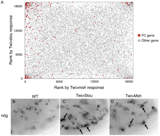
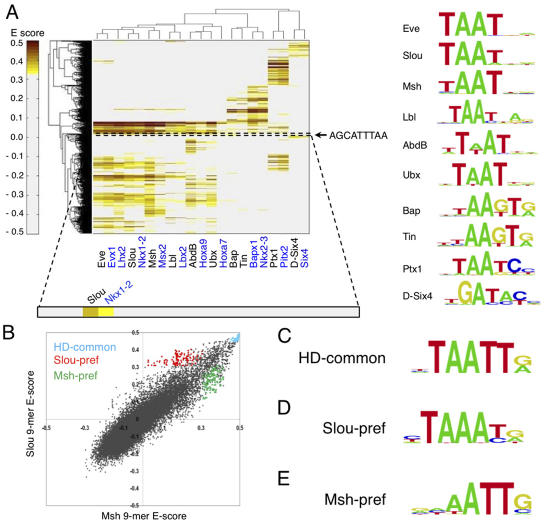
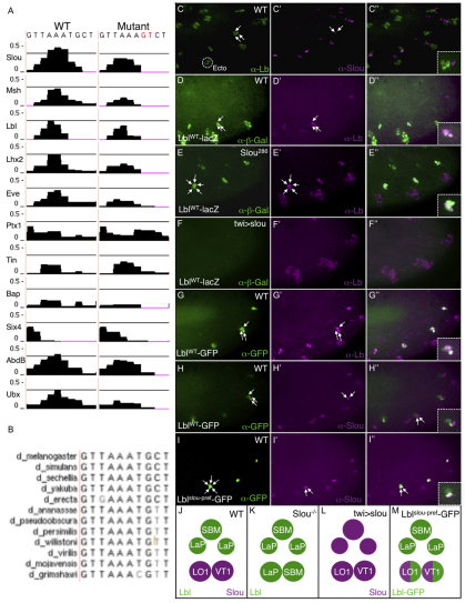
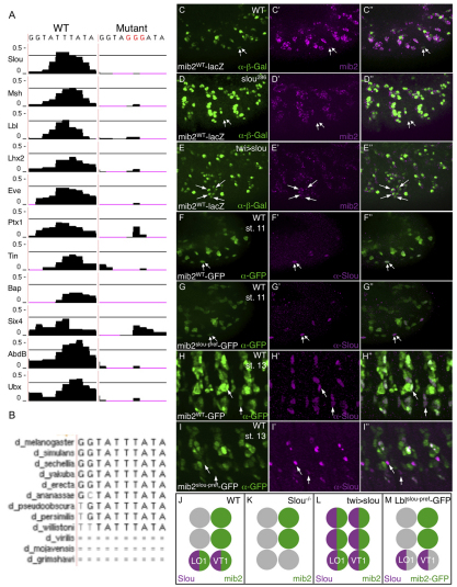
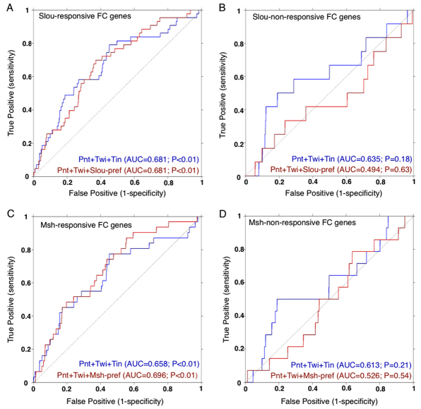
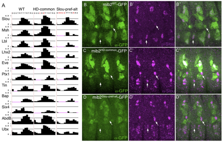
A subfamily of Drosophila homeodomain (HD) transcription factors (TFs) controls the identities of individual muscle founder cells (FCs). However, the molecular mechanisms by which these TFs generate unique FC genetic programs remain unknown. To investigate this problem, we first applied genome-wide mRNA expression profiling to identify genes that are activated or repressed by the muscle HD TFs Slouch (Slou) and Muscle segment homeobox (Msh). Next, we used protein-binding microarrays to define the sequences that are bound by Slou, Msh and other HD TFs that have mesodermal expression. These studies revealed that a large class of HDs, including Slou and Msh, predominantly recognize TAAT core sequences but that each HD also binds to unique sites that deviate from this canonical motif. To understand better the regulatory specificity of an individual FC identity HD, we evaluated the functions of atypical binding sites that are preferentially bound by Slou relative to other HDs within muscle enhancers that are either activated or repressed by this TF. These studies showed that Slou regulates the activities of particular myoblast enhancers through Slou-preferred sequences, whereas swapping these sequences for sites that are capable of binding to multiple HD family members does not support the normal regulatory functions of Slou. Moreover, atypical Slou-binding sites are overrepresented in putative enhancers associated with additional Slou-responsive FC genes. Collectively, these studies provide new insights into the roles of individual HD TFs in determining cellular identity, and suggest that the diversity of HD binding preferences can confer regulatory specificity.


Figures

Fig. 1.
Fig. 1.
Differential responsiveness of individual FC genes to Slou or Msh overexpression. (A) mRNA expression profiles of mesodermal cells overexpressing Slou or Msh under control of Twist (Twi)-Gal4 (Twi>Slou and Twi>Msh, respectively) compared with wild type. On each axis, genes are ranked by Bayesian t-statistic (Choe et al., 2005) from the most likely upregulated relative to wild type (lower left corner) to the most likely downregulated. Responses of previously documented FC (red) and other (gray) genes are shown, ranked from the most likely upregulated (lower left) to the most likely downregulated (upper right). ‘Other genes’ include genes known to be not expressed in FCs and genes not tested for expression in FCs. (B-D) Expression of Nidogen (Ndg) (B-D) mRNA in wild-type (B), Slou overexpressing (C) and Msh-overexpressing (D) stage 12 embryos. Arrows indicate groups of cells that have increased expression of Ndg, which occurs in different somatic mesodermal cells in the Twi>Slou and Twi>Msh embryos. See also supplementary material Table S1.
Fig. 2.
Fig. 2.
Identification of Slou- and Msh-preferred binding sites. (A) Hierarchical agglomerative clustering analysis of E-scores for 9816 ungapped 9-mers with E-score>0.31 (y-axis) against HD TFs (x-axis). Drosophila HDs and their mouse orthologs are shown with black and blue labels, respectively. Proteins are clustered according to their 9-mer binding profiles. The color bar indicates 9-mer E-scores. Logos are shown for all Drosophila HD TFs, as determined by the PRIORITY algorithm (Gordân et al., 2010). The location of the nucleotide sequence of the Slou-preferred 9-mer (AGCATTTAA) that was mutated in the lbl FC enhancer (Fig. 3) is indicated by a dotted box on the heatmap and is shown to the right of the heatmap. (B) Scatter plot comparing the PBM-derived binding preferences of Slou and Msh. Cyan dots represent 9-mers common to all examined Drosophila HD TFs; red dots and green dots represent 9-mers preferentially bound by Slou or Msh, respectively; black dots represent all other 9-mers. (C-E) Motif logos for: all 9-mers bound by all HD TFs examined [‘HD-common’ (C)]; 9-mers preferentially bound by Slou [‘Slou-pref’ (D)] or Msh [‘Msh-pref’ (E)].
Fig. 3.
Fig. 3.
A Slou-preferred binding site in the lbl FC enhancer mediates the repressive effect of Slou on Lbl-expressing FCs. (A) E-score (y-axis) binding profiles of the indicated HD TFs for a Slou-preferred binding site in the wild-type lbl FC enhancer and a version of the enhancer in which this site is mutated. The horizontal black line represents a threshold binding E-score of 0.31 (see Materials and methods for details). The low level binding of AbdB is biologically irrelevant as AbdB is not expressed in the anterior abdominal hemisegments of the Drosophila embryo where effects of the HD-binding site mutation are observed. In addition, whereas there are three overlapping Slou-preferred 9-mers with enrichment scores much higher than the 0.31 threshold, only one of these 9-mers binds to AbdB at this same cutoff, and this sequence has a lower score than any of the Slou-preferred 9-mers. (B) Conservation of the Slou-preferred binding site in the lbl enhancer. (C-I″) The images enclosed by the dotted boxes represent zoomed-in views of the cells indicated by the arrows in the main part of each panel. (C-C″) Lbl (green), which is expressed in three cells in the somatic mesoderm, and Slou (magenta), which is expressed in two adjacent cells, are not co-expressed. (D-D″) β-Gal (green) driven by the lblWT-lacZ transgene is expressed in the three Lbl-positive somatic mesodermal cells (magenta). (E-E″) When crossed into the slou286 loss-of-function mutant, both the lblWT-lacZ reporter (green) and endogenous Lbl (magenta) are derepressed into five adjacent mesodermal cells. (F-F″) Ectopic mesodermal expression of Slou in embryos containing the lblWT-lacZ transgene extinguishes both reporter β-gal (green) and endogenous Lbl (magenta) expression. (G-G″) GFP (green) driven by the wild-type lbl muscle enhancer (lblWT-GFP) is expressed in three Lbl-positive (magenta) cells. (H-H″) GFP (green) driven by the lblWT-GFP construct does not co-express with the two Slou-positive (magenta) cells. (I-I″) GFP (green) driven by the lbl muscle enhancer containing a mutant Slou-preferred binding site (lblslou-pref-GFP) is derepressed into the two Slou-positive (magenta) FCs. (J-M) Schematic depiction of the effects of cis and trans manipulations of Slou on activity of the lbl gene and its muscle enhancer. Lbl protein- or enhancer-expressing (green) and Slou-expressing (magenta) cells are shown.
Fig. 4.
Fig. 4.
A Slou-preferred binding site in the mib2 FC enhancer mediates the activating function of Slou in two FCs that co-express Slou and Mib2. (A) E-score (y-axis) binding profiles of the indicated HD TFs for a Slou-preferred binding site in the wild-type mib2 FC enhancer and a version of the enhancer in which this site is mutated. Note that the binding of the two Hox TFs, Ubx and AbdB, immediately adjacent to the center of the Slou-pref site in the mib2 enhancer is unlikely to account for the targeted loss of activity of the mutant enhancer because Hox TFs globally influence muscle segmental patterning, whereas the Slou-preferred site mutant exerts a cell-specific effect. Indeed, no FCs other than LO1 and VT1 show altered GFP reporter expression in embryos containing the mib2Slou-pref-GFP transgene (G-I). (B) The Slou-preferred binding site in the mib2 enhancer is highly conserved. (C-C″) β-Gal (green) driven by the mib2WT-lacZ transgene is co-expressed with endogenous mib2 mRNA (magenta). Arrows indicate the same two Slou-expressing cells as shown in Fig. 3 (the FCs of muscles LO1 and VT1). (D-D″) Loss of mib2 mRNA (magenta) and mib2 FC enhancer-driven β-gal (green) from the same two Slou-positive cells in slou286 mutant embryos. (E-E″) Ectopic expression of mib2 mRNA (magenta) and β-gal (green) activated by the mib2 FC enhancer in response to overexpression of Slou (twi>slou). Arrows indicate cells that do not express mib2 or β-gal in wild-type embryos (compare with C). (F-F″,H-H″) Co-expression of GFP (green) and Slou (magenta) in Slou-positive LO1 and VT1 FCs at stage 11 (F) and the corresponding myotubes at stage 13 (H); in both cases, embryos contain the mib2WT-GFP transgene. (G-G″,I-I″) Attenuation of GFP (green) driven by the mib2 FC enhancer containing a mutant Slou-preferred binding site (mib2slou-pref-GFP) in Slou (magenta)-expressing LO1 and VT1 FCs at stage 11 (G-G″) and myotubes at stage 13 (I-I″). The loss of reporter expression increases over time. (J-M) Schematic depiction of the effects of cis and trans manipulations of Slou on activity of the mib2 gene and its enhancer. mib2 gene- or enhancer-expressing (green), Slou-expressing (magenta) and non-expressing cells (gray) are shown.
Fig. 5.
Fig. 5.
Enrichment of Slou-preferred and Msh-preferred binding sites located within putative CRMs in the noncoding sequences of Slou- and Msh-responsive FC genes. (A-D) Receiver operating characteristic (ROC) curves showing the discrimination of Slou-responsive (A), Slou-nonresponsive (B), Msh-responsive (C) and Msh-non-responsive (D) FC genes by the indicated AND combinations of Pnt, Twi, Tin, Slou-preferred and Msh-preferred binding motifs (see supplementary material Table S5 for the entire set of Lever results). The area under the ROC curve (AUC) for each gene set and motif combination is shown. Foreground gene sets are listed in supplementary material Table S5 and the background was generated as described in the Materials and methods. Slou-preferred and Msh-preferred sites are over-represented together with the known FC regulators Pnt and Twi, in the noncoding regions of Slou-responsive or Msh-responsive FC genes, respectively. This effect does not occur with FC genes that are known not to be Slou or Msh responsive.
Fig. 6.
Fig. 6.
The specific nucleotides of a Slou-preferred binding site in a Slou-responsive FC enhancer are crucial for enhancer activity. (A) E-score (y-axis) binding profiles of the indicated HD TFs for a Slou-preferred binding site in the wild-type mib2 FC enhancer, a version in which this site is changed to one that binds all FCI-HD TFs (HD-common) and a version in which this site is changed to a different Slou-preferred binding sequence (Slou-pref-alt). (B-B″) Co-expression of Slou (magenta) with GFP (green) in Slou-expressing myotubes in stage 13 embryos containing the mib2WT-GFP transgene. (C-D″) Attenuation of GFP (green) driven by the mib2 FC enhancer containing a Slou-preferred site that has been exchanged for a HD-common site (C, mib2HD-common-GFP) or a Slou-preferred site that has been exchanged for another Slou-preferred site (D, mib2Slou-pref-alt-GFP) in Slou-expressing LO1 and VT1 myotubes in stage 13 embryos. Arrows indicate myotubes LO1 and VT1.

References

    1. Azpiazu N., Frasch M. (1993). tinman and bagpipe - two homeobox genes that determine cell fates in the dorsal mesoderm of Drosophila. Genes Dev. 7, 1325–1340 - PubMed
    1. Barolo S., Carver L. A., Posakony J. W. (2000). GFP and beta-galactosidase transformation vectors for promoter/enhancer analysis in Drosophila. Biotechniques 29, 726–732 - PubMed
    1. Baylies M. K., Arias A. M., Bate M. (1995). wingless is required for the formation of a subset of muscle founder cells during Drosophila embryogenesis. Development 121, 3829–3837 - PubMed
    1. Baylies M. K., Bate M., Ruiz Gomez M. (1998). Myogenesis: a view from Drosophila. Cell 93, 921–927 - PubMed
    1. Berger M. F., Bulyk M. L. (2009). Universal protein-binding microarrays for the comprehensive characterization of the DNA-binding specificities of transcription factors. Nat. Protoc. 4, 393–411 - PMC - PubMed

Publication types

MeSH terms