Review
. 2012 Feb 1;14(1):13.
doi: 10.1186/1532-429X-14-13.

Clinical utility of cardiovascular magnetic resonance in hypertrophic cardiomyopathy

Affiliations
Review

Clinical utility of cardiovascular magnetic resonance in hypertrophic cardiomyopathy

Martin S Maron. J Cardiovasc Magn Reson. .

Abstract

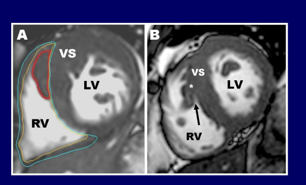
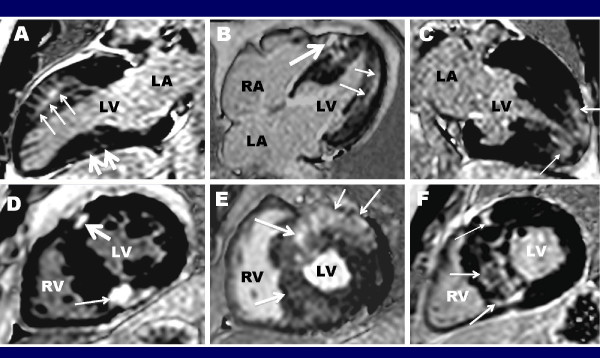
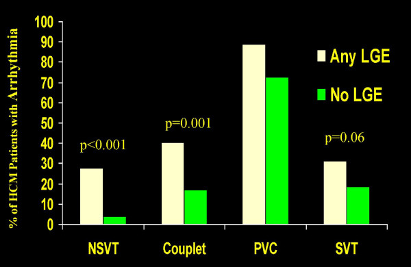
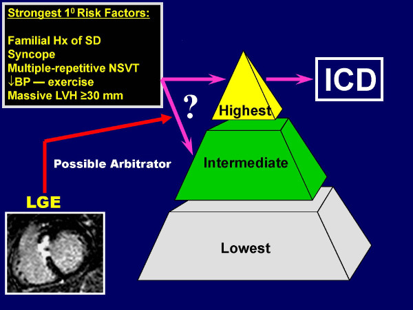
Hypertrophic cardiomyopathy (HCM) is characterized by substantial genetic and phenotypic heterogeneity, leading to considerable diversity in clinical course including the most common cause of sudden death in young people and a determinant of heart failure symptoms in patients of any age. Traditionally, two-dimensional echocardiography has been the most reliable method for establishing a clinical diagnosis of HCM. However, cardiovascular magnetic resonance (CMR), with its high spatial resolution and tomographic imaging capability, has emerged as a technique particularly well suited to characterize the diverse phenotypic expression of this complex disease. For example, CMR is often superior to echocardiography for HCM diagnosis, by identifying areas of segmental hypertrophy (ie., anterolateral wall or apex) not reliably visualized by echocardiography (or underestimated in terms of extent). High-risk HCM patient subgroups identified with CMR include those with thin-walled scarred LV apical aneurysms (which prior to CMR imaging in HCM remained largely undetected), end-stage systolic dysfunction, and massive LV hypertrophy. CMR observations also suggest that the cardiomyopathic process in HCM is more diffuse than previously regarded, extending beyond the LV myocardium to include thickening of the right ventricular wall as well as substantial morphologic diversity with regard to papillary muscles and mitral valve. These findings have implications for management strategies in patients undergoing invasive septal reduction therapy. Among HCM family members, CMR has identified unique phenotypic markers of affected genetic status in the absence of LV hypertrophy including: myocardial crypts, elongated mitral valve leaflets and late gadolinium enhancement. The unique capability of contrast-enhanced CMR with late gadolinium enhancement to identify myocardial fibrosis has raised the expectation that this may represent a novel marker, which may enhance risk stratification. At this time, late gadolinium enhancement appears to be an important determinant of adverse LV remodeling associated with systolic dysfunction. However, the predictive significance of LGE for sudden death is incompletely resolved and ultimately future large prospective studies may provide greater insights into this issue. These observations underscore an important role for CMR in the contemporary assessment of patients with HCM, providing important information impacting diagnosis and clinical management strategies.


Figures

Figure 1
Figure 1
Cardiac sarcomere showing the location of known disease-causing genes for HCM. Adapted from Wheeler et al.[20] Not shown are genes previously linked to HCM, but with lesser degrees of evidence for disease causing: titin, vinculin, muscle LIM protein, telethonin, cardiac ankyrin repeat protein, calreticulin 3, calsequestrin 2, phospholamban, ryanodine receptor 2.
Figure 2
Figure 2
CMR end-diastolic images demonstrating diverse patterns of LVH in HCM. (A) involving ventricular septum (VS), but sparing the LV free wall (FW); (B) hypertrophy of the basal anterior free wall and a portion of the contiguous anterior septum, representing the most common area of LV wall thickening in HCM; (C) massive hypertrophy (wall thickness, 33 mm) limited to basal posterior ventricular septum (asterisk); (D) focal area sharply confined to basal anterior septum (arrows); (E) localized to LV apex (asterisks); (F) segmental LV hypertrophy of the basal anterior septum and anterolateral wall (asterisks), separated by regions of normal LV thickness (arrows). Adapted with permission, from Maron MS et al.[29] FW = free wall; LA = left atrium; LV = left ventricle; RA = right atrium; RV = right ventricle; VS = ventricular septum.
Figure 3
Figure 3
CMR can identify segmental LV hypertrophy that may not be reliably visualized by two-dimensional echocardiography. (A) normal 2-D echocardiogram in a patient with a family history of HCM; (B) this same patient then underwent CMR, which reveals an area of segmental hypertrophy in the anterolateral LV wall (asterisk) consistent with a diagnosis of HCM. Reproduced with permission of American Heart Association; from Rickers C et al.;[35] (C) two-dimensional echocardiographic end-diastolic basal short-axis view demonstrates a maximal LV wall thickness of 18 mm in the anterolateral free wall consistent with the diagnosis of HCM; (D) in the same patient, an end-diastolic short-axis CMR at the same level of LV shows a focal area of massive LV hypertrophy (35 mm) in the same region of the LV wall reported to be 18 mm by echocardiography. The finding of massive hypertrophy by CMR, characterized this patient as high risk, and prompted recommendation for ICD therapy for primary prevention of sudden death. Reproduced with permission, from Maron MS et al.;[42](E) echocardiography was considered non-diagnostic; (F) in the same patient, CMR clearly demonstrates segmental hypertrophy confined to the LV apex, consistent with a diagnosis of apical HCM. Reproduced with permission, from Moon et al.[43] LV = left ventricle; RV = right ventricle; VS = ventricular septum.
Figure 4
Figure 4
CMR end-diastolic images demonstrating diversity of the phenotypic expression within HCM. (A) increased wall thickness in the superior segment (thin arrow) and extreme hypertrophy of the inferior segment (thick arrow) of the RV wall; Reproduced with permission, from Maron MS et al.[29](B) medium-sized LV apical aneurysm (arrowheads) and maximal LV wall thickening at mid-ventricular level with muscular apposition of septum and LV free wall producing distinct proximal (P) and distal chambers; Reproduced with permission, from Maron MS et al.[50](C) anomalous insertion of papillary muscle (thin arrows) directly into the anterior leaflet of the mitral valve (thick arrow) (in the absence of chordae tendinae) producing obstruction to blood flow from the apposition of the papillary muscle and basal ventricular septum (asterisk); (D) extraordinarily long anterior mitral valve leaflet measuring 33 mm; PML is of normal length (although not well visualized in this frame); Reproduced with permission, from Maron MS et al.[53](E) multiple accessory papillary muscles, 4 in number (arrows); Reproduced with permission from Harrigan C et al.[54](F) 7-year-old asymptomatic genotype positive/phenotype negative HCM girl with 3 deep myocardial crypts in the basal (posterior) inferior LV free wall. Ao = aorta; RV = right ventricle; LA = left atrium; LV = left ventricle; VS = ventricular septum
Figure 5
Figure 5
Right ventricular crista supraventricularis in HCM. When determining. where to measure the maximal LV wall thickness, it is important to be aware that HCM patients often have prominent and hypertrophied right ventricular muscular structures, the most common of which is the crista supraventricularis. In some HCM patients, the crista supraventricularis is not only significantly hypertrophied (panel A, outlined in red) but not uncommonly inserts from its origin in the RV cavity to directly adjacent to the ventricular septum. As a result, this RV muscle structure may be inappropriately included in the measurement of septal thickness---resulting in an overestimation of the maximal LV wall thickness; Reproduced with permission, from Maron MS et al.[47]. In order to avoid including the crista supraventricularis as part of the septum, close inspection of the CMR short-axis cine images can help clarify this issue; (B) in a different HCM patient than panel A, the crista supraventicularis muscle is noted to move away from the septum toward the RV cavity with a small area of blood pool noted between the crista and septum (arrow), allowing for a more accurate delineation of the epicardial border of the septum (asterisk).
Figure 6
Figure 6
Contrast-enhanced CMR images in 6 different HCM patients demonstrating the diverse pattern and extent of late gadolinium enhancement in this disease. (A) extensive transmural LGE in the anterior wall (small arrows) with smaller focal area in the inferior wall (small arrows); (B) mid-myocardial LGE in the lateral wall (small arrows) and diffuse LGE in the ventricular septum which extends into the RV wall (large arrows) in a 26 year-old man with "end-stage" phase of HCM with an ejection fraction of 40%; (C) LGE confined to the LV apex (arrows); (D) LGE localized to the insertion area of the RV wall into the anterior (large arrow) and posterior ventricular septum (small arrow); (E) transmural LGE involving the majority of the ventricular septum (large arrow) and lateral wall (small arrow). (F) Basal short-axis image with transmural LGE located predominantly in the ventricular septum (arrows). RA = right atrium; RV = right ventricle; LA = left atrium; LV = left ventricle
Figure 7
Figure 7
Prevalence of arrhythmia on 24-hour Holter ECG with respect to presence of late gadolinium enhancement in patients with HCM. Adapted with permission, from Adabag et al.[93]. ECG = electrocardiogram; HCM = hypertrophic cardiomyopathy; LGE = late gadolinium enhancement; NSVT = nonsustained ventricular tachycardia; PVC = premature ventricular contraction; SVT = supraventricular tachychardia
Figure 8
Figure 8
Role of CMR in Sudden Death Risk Stratification. Results of contrast-enhanced CMR with late gadolinium enhancement could be used as a potential arbitrator to arrive at a decision regarding ICD therapy for primary prevention of sudden death in HCM patients in whom risk still remains ambiguous after assessment with current conventional risk factors.
Figure 9
Figure 9
Quantification of LGE. (A) Identical LV short-axis contrast-enhanced cardiovascular MR images show LGE depicted with gray-scale thresholding techniques at 2, 4, and 6 SDs above mean signal intensity of normal remote myocardium, with visual assessment, and with 2 SDs above mean of external region of interest (ROI). LV endocardium and epicardium are outlined in red and green, respectively. Solid black areas in LV myocardium represent areas of delayed enhancement at corresponding semiautomated threshold; (B) Graph illustrates volumes of delayed enhancement (horizontal axis values) assessed by using various gray-scale thresholding techniques (2, 4, and 6 SDs above mean), visual assessment, and an external region of interest. Reproduced with permission, from Harrigan C et al.[113]

References

    1. Maron BJ. Hypertrophic cardiomyopathy: a systematic review. JAMA. 2002;287:1308–20. - PubMed
    1. Wigle ED, Rakowski H, Kimball BP, Williams WG. Hypertrophic cardiomyopathy. Clinical spectrum and treatment. Circulation. 1995;92:1680–92. - PubMed
    1. Maron BJ, McKenna WJ, Danielson GK, Kappenberger LJ, Kuhn HJ, Seidman CE, Shah PM, Spencer WH, Spirito P, Ten Cate FJ, Wigle ED. American College of Cardiology/European Society of Cardiology clinical expert consensus document on hypertrophic cardiomyopathy. A report of the American College of Cardiology Foundation Task Force on Clinical Expert Consensus Documents and the European Society of Cardiology Committee for Practice Guidelines. J Am Coll Cardiol. 2003;42:1687–713. - PubMed
    1. Alcalai R, Seidman JG, Seidman CE. Genetic basis of hypertrophic cardiomyopathy: from bench to the clinics. J Cardiovasc Electrophysiol. 2008;19:104–10. - PubMed
    1. Seidman JG, Seidman C. The genetic basis for cardiomyopathy: from mutation identification to mechanistic paradigms. Cell. 2001;104:557–67. - PubMed

MeSH terms