. 2011 Oct 19;55(4):e36.
doi: 10.4081/ejh.2011.e36.

Autophagic degradation of farnesylated prelamin A as a therapeutic approach to lamin-linked progeria

Affiliations

Autophagic degradation of farnesylated prelamin A as a therapeutic approach to lamin-linked progeria

V Cenni et al. Eur J Histochem. .

Erratum in

  • Eur J Histochem. 2013;57(4):e42

Abstract

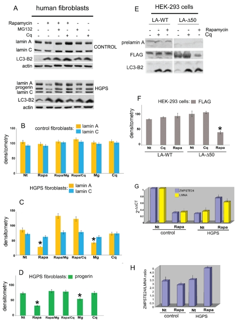
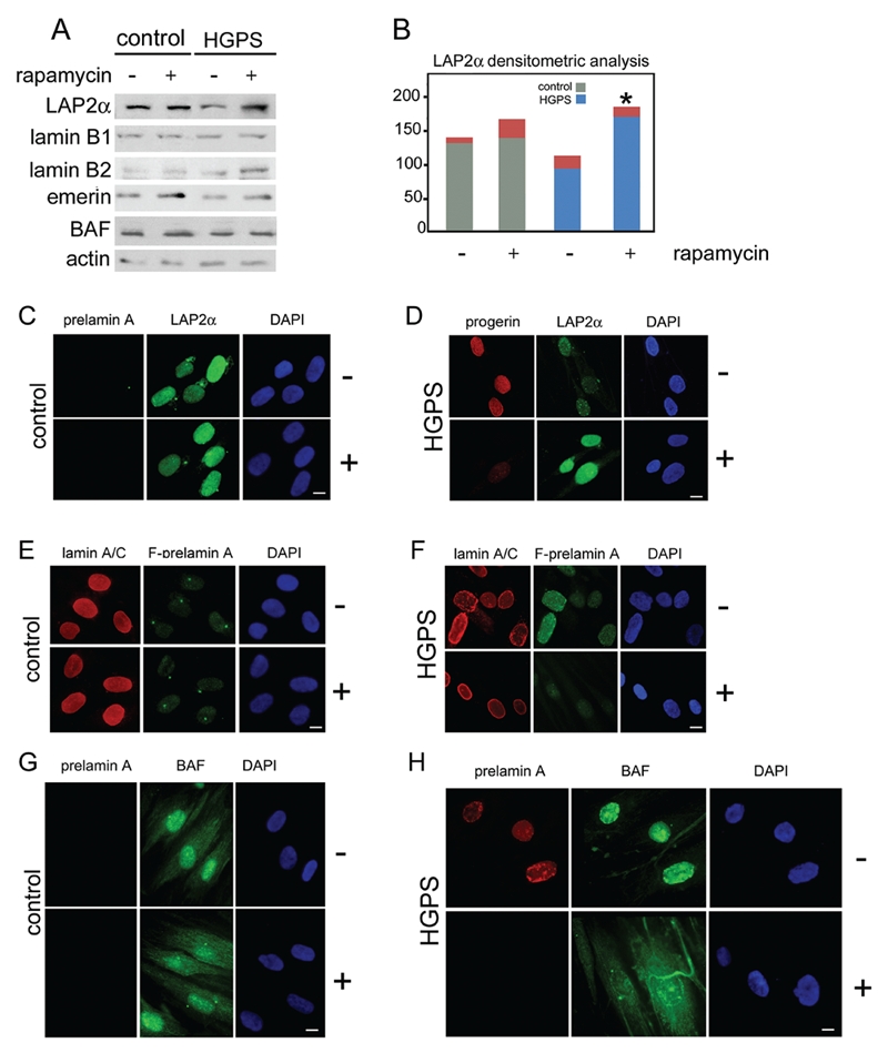
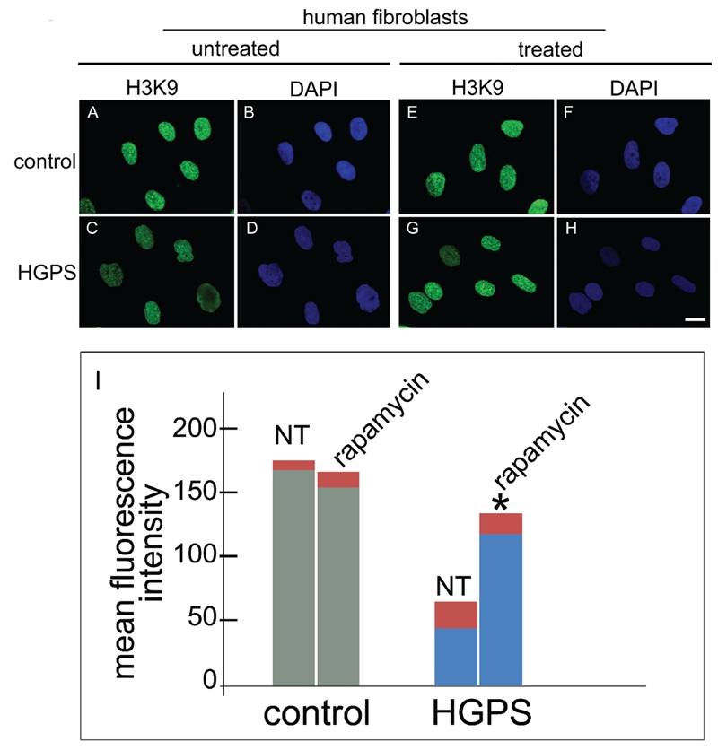
Farnesylated prelamin A is a processing intermediate produced in the lamin A maturation pathway. Accumulation of a truncated farnesylated prelamin A form, called progerin, is a hallmark of the severe premature ageing syndrome, Hutchinson-Gilford progeria. Progerin elicits toxic effects in cells, leading to chromatin damage and cellular senescence and ultimately causes skin and endothelial defects, bone resorption, lipodystrophy and accelerated ageing. Knowledge of the mechanism underlying prelamin A turnover is critical for the development of clinically effective protein inhibitors that can avoid accumulation to toxic levels without impairing lamin A/C expression, which is essential for normal biological functions. Little is known about specific molecules that may target farnesylated prelamin A to elicit protein degradation. Here, we report the discovery of rapamycin as a novel inhibitor of progerin, which dramatically and selectively decreases protein levels through a mechanism involving autophagic degradation. Rapamycin treatment of progeria cells lowers progerin, as well as wild-type prelamin A levels, and rescues the chromatin phenotype of cultured fibroblasts, including histone methylation status and BAF and LAP2alpha distribution patterns. Importantly, rapamycin treatment does not affect lamin C protein levels, but increases the relative expression of the prelamin A endoprotease ZMPSTE24. Thus, rapamycin, an antibiotic belonging to the class of macrolides, previously found to increase longevity in mouse models, can serve as a therapeutic tool, to eliminate progerin, avoid farnesylated prelamin A accumulation, and restore chromatin dynamics in progeroid laminopathies.


Figures

Figure 1
Figure 1
Rapamycin reduces progerin level in HGPS cells. A) Western blotting evaluation of lamin A/C and progerin in control (CONTROL) and Hutchinson Gilford progeria cells (HGPS). Whole lysates from control and HGPS cells untreated (−) or treated (+) with rapamycin (Rapamycin), MG132 or chloroquine-diphosphate (Cq) were subjected to lamin A/C, LC3-B2 and actin antibodies detection; B) densitometric analysis of lamin A and C immunolabeled bands detected in control fibroblasts; C) densitometric analysis of lamin A and C immunolabeled bands detected in HGPS fibroblasts; P=0.0358 for lamin A (rapa), P=0.0298 for lamin A (Mg); D) densitometric analysis of progerin immunoblotted bands; P=0.0390 for progerin (rapa); P=0.0458 for progerin (Mg); E) prelamin A, FLAG and LC3-B2 protein levels evaluation in HEK-293 cells expressing FLAG-tagged wild type prelamin A (LA-WT) or progerin (LA-Δ50). Immunolabeled bands observed in untreated (−) or rapamycin (Rapamycin) and chloroquine-diphosphate (Cq) treated (+) cells are shown; F) densitometric of FLAG immunoblotted bands; G) RT-PCR analysis of ZMPSTE24 and LMNA mRNA expression in untreated (Nt) and rapamycintreated HGPS cells (Rapa) and control (control); 2 −ΔΔCT values are reported relative to untreated control samples. P=0.0236 for LA-Δ50 (rapa); H) ratio between ZMPSTE24 and LMNA mRNA expression. Values are means of duplicate experiments ± S.D. In B, C, D and F densitometric analysis of triplicate experiments was performed, and the mean values ± S.D. are reported; asterisk indicates statistically significant difference with respect to lamin A or progerin densitometry in untreated samples; statistical significance was calculated by the Mann-Whitney test vs untreated HGPS samples, or cells expressing LMNA constructs.
Figure 2
Figure 2
Nuclear envelope/lamina proteins in rapamycin treated cells. A) Western Blotting analysis of LAP2α (LAP2 ), lamin B1 (lamin B1), lamin B2 (lamin B2), emerin (emerin) and Barrier-to-autointegration factor (BAF) in control (control) and HGPS cells (HGPS) untreated (−) or treated (+) with rapamycin. Actin was detected as protein loading control. Immunolabeled bands are shown; B) densitometric analysis of LAP2α immunolabeled bands detected in Western blotting analysis performed in control and HGPS untreated (−) or rapamycin treated cells (+). Asterisk indicates statistically significant difference, P=0.0319 for HGPS + rapamycin (+) vs untreated HGPS (−); C) prelamin A and LAP2α immunofluorescence labeling performed in untreated (−) or rapamycin treated (+) control cells. Prelamin A was evaluated using a goat-polyclonal antibody visualized by TRITC-conjugated secondary antibody (red). LAP2α distribution was evaluated using a rabbitpolyclonal antibody visualized by FITC-conjugated secondary antibody (green); D) progerin and LAP2α immunolabeling detection performed in untreated (−) or rapamycin treated (+) HGPS cells. Progerin (progerin) detection was performed using a mouse-monoclonal antibody visualized by Cy3-conjugated secondary antibody (red). LAP2α distribution was evaluated using a rabbit-polyclonal antibody visualized by FITC-conjugated secondary antibody (green). In untreated HGPS cells progerin staining was observed, while LAP2α labeling was decreased. Rapamycin treatment dramatically reduced progerin labeling and restored LAP2α staining levels; E) Lamin A/C and farnesylated-prelamin A detection performed in untreated (−) or rapamycin treated (+) control cells. Lamin A/C (lamin A/C) was evaluated using a goat-polyclonal antibody visualized by TRITC-conjugated secondary antibody (red). Farnesylated-prelamin A (F-prelamin A) staining was performed using a rabbit-polyclonal antibody visualized by FITC-conjugated secondary antibody (green). Lamin A/C was detected in untreated or treated cells at the same levels. F-prelamin A was undetected in both samples; F) Lamin A/C and farnesylated-prelamin A detection performed in untreated (−) or rapamycin treated (+) HGPS cells. Lamin A/C (lamin A/C) was evaluated using a goat-polyclonal antibody visualized by TRITC-conjugated secondary antibody (red). Farnesylated-prelamin A (F-prelamin A) staining was performed using a rabbit-polyclonal antibody visualized by FITC-conjugated secondary antibody (green). Lamin A/C staining was observed at the nuclear lamina of both untreated and treated cells. F-prelamin A staining observed in untreated cells was strongly reduced by rapamycin treatment; G) prelamin A BAF detection performed in untreated (−) or rapamycin treated (+) control cells. Prelamin A was stained by a goat-polyclonal antibody visualized by TRITC-conjugated secondary antibody (red). BAF immunolabeling detection was performed by a rabbit-polyclonal antibody visualized by FITC-conjugated secondary antibody (green). Prelamin A was undetectable in untreated or rapamycin treated cells. BAF showed a normal cellular distribution in both samples; H) prelamin A and Barrier-to-autointegration (BAF) detection was performed in untreated (−) or rapamycin treated (+) HGPS cells. Prelamin A was stained by a goat-polyclonal antibody visualized by TRITC-conjugated secondary antibody (red). BAF was labeled by rabbit-polyclonal antibody and visualized by FITC-conjugated secondary antibody (green). Prelamin A staining was present at the nuclear lamina of untreated cells while in rapamycin treated cells lamin A precursor was undetectable. BAF nuclear localization was observed in HGPS untreated cells. The normal BAF nucleo-cytoplasmic cellular distribution was recovered by rapamycin treatment. In panel C, D, E, F,G and H nuclei were counterstained with DAPI. Scale bar, 10 µm.
Figure 3
Figure 3
Rapamycin treatment rescues the heterochromatin marker trimethyl-H3K9 in HGPS cells. Immunofluorescence labeling of trimethyl-H3K9 (H3K9) in control and HGPS cells untreated (untreated) or treated (treated) with rapamycin using a rabbit-polyclonal antibody visualized by FITC-conjugated secondary antibody (green). In control untreated cells (A) a proper trimethyl-H3K9 staining was observed, while a clearly detectable decrease of immunolabeling was observed in untreated HGPS cells (C). Rapamycin treatment of control cells did not affect trimethyl-H3K9 staining (E), while rescued trimethyl-H3K9 distribution in HGPS cells (G). Nuclei in B, D, F and H were counterstained with DAPI. Scale bar = 10 µm; I) The mean fluorescence intensity of 300 nuclei per sample stained for trimethyl-H3K9 was measured by the NIS software and plotted for untreated (NT) or rapamycin-treated (treated) control and HGPS nuclei. Asterisk indicates statistically significant difference, P=0.0331 for HGPS + rapamycin vs untreated HGPS.

References

    1. Hennekam RC. Hutchinson-Gilford progeria syndrome: review of the phenotype. Am J Med Genet A. 2006;140:2603–24. - PubMed
    1. Dominguez-Gerpe L, Araujo-Vilar D. Prematurely aged children: molecular alterations leading to Hutchinson-Gilford progeria and Werner syndromes. Curr Aging Sci. 2008;1:202–12. - PubMed
    1. Eriksson M, Brown WT, Gordon LB, Glynn MW, Singer J, Scott L, et al. Recurrent de novo point mutations in lamin A cause Hutchinson-Gilford progeria syndrome. Nature. 2003;423:293–8. - PMC - PubMed
    1. Csoka AB, Cao H, Sammak PJ, Constantinescu D, Schatten GP, Hegele RA. Novel lamin A/C gene (LMNA) mutations in atypical progeroid syndromes. J Med Genet. 2004;41:304–8. - PMC - PubMed
    1. Columbaro M, Capanni C, Mattioli E, Novelli G, Parnaik VK, Squarzoni S, et al. Rescue of heterochromatin organization in Hutchinson-Gilford progeria by drug treatment. Cell Mol Life Sci. 2005;62:2669–78. - PMC - PubMed

Publication types