

Silencing of Fem1cR3 gene expression in the DBA/2J mouse precedes retinal ganglion cell death and is associated with histone deacetylase activity
- PMID: 22297488
- PMCID: PMC3339913
- DOI: 10.1167/iovs.11-8872
Silencing of Fem1cR3 gene expression in the DBA/2J mouse precedes retinal ganglion cell death and is associated with histone deacetylase activity
Abstract
Purpose: Downregulation of normal gene expression in dying retinal ganglion cells has been documented in both acute and chronic models of optic nerve disease. The authors examined the mechanism and timing of this phenomenon in DBA/2J mice, using genetically modified substrains of this inbred line.
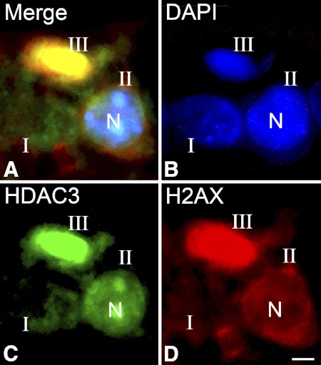
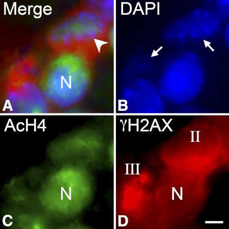
Methods: DBA/2J mice, doubly congenic for the Bax mutant allele and the ganglion cell reporter gene Fem1c(Rosa3) (R3), were evaluated to elucidate the timing of loss of normal gene expression during the apoptotic process. The localization of histone deacetylase 3 (HDAC3) and nuclear histone H4 acetylation were examined by immunofluorescence in dying cells. The role of HDACs in gene silencing during glaucoma was interrogated using the global HDAC inhibitor trichostatin A (TSA).
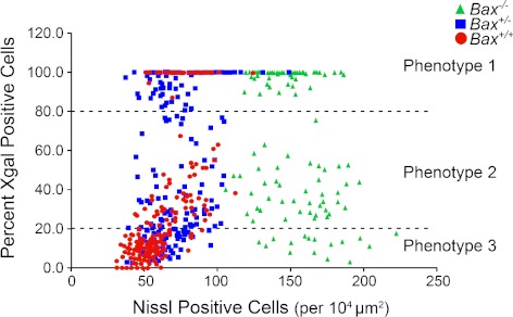
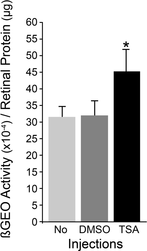
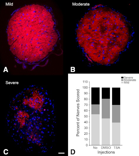
Results: Silencing of the R3 allele occurred in Bax(-/-) ganglion cells, indicating that this process preceded the committed step of the intrinsic apoptotic pathway. Weekly TSA treatment, between the ages of 6 and 10 months, was able to attenuate the loss of R3 expression in the retina, but had no effect on optic nerve degeneration. Dying cells in aging DBA/2J mice exhibited nuclear localization of HDAC3 and a decrease in the level of H4 acetylation.
Conclusions: Retinal ganglion cells exhibit a loss of normal gene expression as an early (pre-BAX involvement) part of their apoptotic program during glaucomatous degeneration. This process can be ameliorated, but not completely blocked, using HDAC inhibitors. Epigenetic changes to active chromatin, such as deacetylation, may be mediated by HDAC3 in dying neurons.
Figures
Similar articles
-
Role of HDACs in optic nerve damage-induced nuclear atrophy of retinal ganglion cells.Neurosci Lett. 2016 Jun 20;625:11-5. doi: 10.1016/j.neulet.2015.12.012. Epub 2015 Dec 28. Neurosci Lett. 2016. PMID: 26733303 Free PMC article. Review.
-
Histone H4 deacetylation plays a critical role in early gene silencing during neuronal apoptosis.BMC Neurosci. 2010 May 26;11:62. doi: 10.1186/1471-2202-11-62. BMC Neurosci. 2010. PMID: 20504333 Free PMC article.
-
Targeting HDAC3 in the DBA/2J spontaneous mouse model of glaucoma.Exp Eye Res. 2020 Nov;200:108244. doi: 10.1016/j.exer.2020.108244. Epub 2020 Sep 21. Exp Eye Res. 2020. PMID: 32971093 Free PMC article.
-
Histone deacetylase 3 (HDAC3) plays an important role in retinal ganglion cell death after acute optic nerve injury.Mol Neurodegener. 2014 Sep 28;9:39. doi: 10.1186/1750-1326-9-39. Mol Neurodegener. 2014. PMID: 25261965 Free PMC article.
-
BAX to basics: How the BCL2 gene family controls the death of retinal ganglion cells.Prog Retin Eye Res. 2017 Mar;57:1-25. doi: 10.1016/j.preteyeres.2017.01.002. Epub 2017 Jan 4. Prog Retin Eye Res. 2017. PMID: 28064040 Free PMC article. Review.
Cited by
-
Roles of Histone Acetyltransferases and Deacetylases in the Retinal Development and Diseases.Mol Neurobiol. 2023 Apr;60(4):2330-2354. doi: 10.1007/s12035-023-03213-1. Epub 2023 Jan 13. Mol Neurobiol. 2023. PMID: 36637745 Review.
-
Acetylation preserves retinal ganglion cell structure and function in a chronic model of ocular hypertension.Invest Ophthalmol Vis Sci. 2014 Oct 30;55(11):7486-93. doi: 10.1167/iovs.14-14792. Invest Ophthalmol Vis Sci. 2014. PMID: 25358731 Free PMC article.
-
Spink2 modulates apoptotic susceptibility and is a candidate gene in the Rgcs1 QTL that affects retinal ganglion cell death after optic nerve damage.PLoS One. 2014 Apr 3;9(4):e93564. doi: 10.1371/journal.pone.0093564. eCollection 2014. PLoS One. 2014. PMID: 24699552 Free PMC article.
-
Role of HDACs in optic nerve damage-induced nuclear atrophy of retinal ganglion cells.Neurosci Lett. 2016 Jun 20;625:11-5. doi: 10.1016/j.neulet.2015.12.012. Epub 2015 Dec 28. Neurosci Lett. 2016. PMID: 26733303 Free PMC article. Review.
-
Intravitreal AAV2 gene delivery to feline retinal ganglion cells.Vision Res. 2025 Jan;226:108519. doi: 10.1016/j.visres.2024.108519. Epub 2024 Nov 16. Vision Res. 2025. PMID: 39549467 Free PMC article.
References
-
- Nickells RW. From ocular hypertension to ganglion cell death: a theoretical sequence of events leading to glaucoma. Can J Ophthalmol. 2007;42:278–287 - PubMed
-
- Whitmore AV, Libby RT, John SWM. Glaucoma: thinking in new ways—a role for autonomous axonal self-destruction and compartmentalised processes? Prog Retin Eye Res. 2005;24:639–662 - PubMed
-
- Schlamp CL, Johnson EC, Li Y, Morrison JC, Nickells RW. Changes in Thy1 gene expression associated with damaged retinal ganglion cells. Mol Vis. 2001;7:192–201 - PubMed
-
- Huang W, Fileta J, Guo Y, Grosskreutz CL. Downregulation of Thy1 in retinal ganglion cells in experimental glaucoma. Curr Eye Res. 2006;31:265–271 - PubMed
Publication types
MeSH terms
Substances
Grants and funding
LinkOut - more resources
Full Text Sources
Molecular Biology Databases
Research Materials
