Review
. 2012 Jan;92(1):193-235.
doi: 10.1152/physrev.00043.2010.

Fast synaptic inhibition in spinal sensory processing and pain control

Affiliations
Review

Fast synaptic inhibition in spinal sensory processing and pain control

Hanns Ulrich Zeilhofer et al. Physiol Rev. 2012 Jan.

Abstract

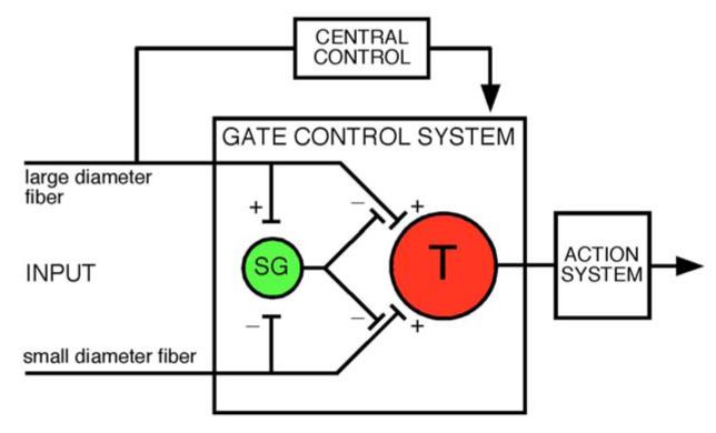
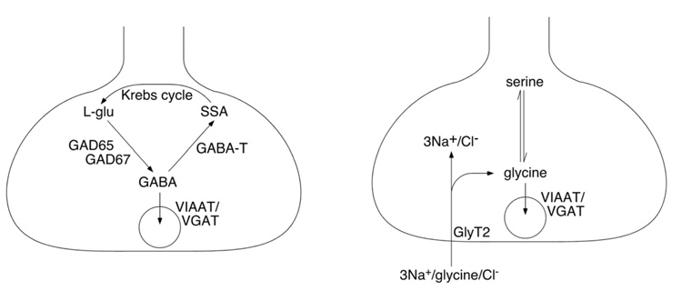
The two amino acids GABA and glycine mediate fast inhibitory neurotransmission in different CNS areas and serve pivotal roles in the spinal sensory processing. Under healthy conditions, they limit the excitability of spinal terminals of primary sensory nerve fibers and of intrinsic dorsal horn neurons through pre- and postsynaptic mechanisms, and thereby facilitate the spatial and temporal discrimination of sensory stimuli. Removal of fast inhibition not only reduces the fidelity of normal sensory processing but also provokes symptoms very much reminiscent of pathological and chronic pain syndromes. This review summarizes our knowledge of the molecular bases of spinal inhibitory neurotransmission and its organization in dorsal horn sensory circuits. Particular emphasis is placed on the role and mechanisms of spinal inhibitory malfunction in inflammatory and neuropathic chronic pain syndromes.


Figures

Figure 1
Figure 1
Gate control theory of pain (modified from ref. 248). This model proposed that inhibitory interneurons (yellow) located in the substantia gelatinosa (SG) would determine whether nociceptive input from the periphery would be relayed through the spinal transmission system (red, T) to higher CNS areas where pain would be consciously perceived.
Figure 2
Figure 2
Membrane topology of cys loop ion channels as proposed by Karlin and Akabas (186)
Figure 3
Figure 3
GABAA receptor subunits and ligands (A) Dendrogram of mammalian GABAA receptors (modified from ref. 30). (B) wheel arrangement of the five subunits of a typical GABAA receptor containing α, β and γ subunits seen from the extracellular side. Data based on (32, 33). (C) Chemical structures of GABA and of the GABAA receptor agonist muscimol. (D) Chemical structures of GABAA receptor blockers.
Figure 4
Figure 4
Inhibitory (strychnine-sensitive) glycine receptor subunits and ligands. (A) Dendrogram of mammalian inhibitory glycine receptors. (B) Chemical structures of glycine and of other putative endogenous glycine receptor agonists β-alanine and taurine. (C) chemical structure of the glycine receptor antagonist strychnine.
Figure 5
Figure 5
Key elements of GABAergic (A) and glycinergic (B) presynaptic terminals. Abbreviations: GABA-T, GABA transaminase; SSA, succinic semialdehyde.
Figure 6
Figure 6
Laminar organization of the spinal cord and distribution of inhibitory neurotransmitter receptors. (A) Spinal laminae illustrated in a coronal section of the lumbar spinal cord taken from a mouse whose sciatic nerve has been injected with cholera toxin B subunit in order to label axons and terminals of myelinated sensory nerve fibers and motoneurons (curtsey of Dres. Jolly Paul and Jean-Marc Fritschy). (B) Distribution of GABAA receptor subunits is shown as pseudocolor images. Highest density, yellow, low density, blue (modified from ref. (417). (C) Distribution of glycine receptor subunits GlyRα1 and GlyRα3 in the spinal dorsal horn (modified from ref. 140). Counterstaining against calcitonin gene related peptide, which marks lamina II outer.
Figure 7
Figure 7
Distribution of GABAergic and glycinergic neurons in the dorsal horn. (A) Dorsal horn laminae (same as figure 6A). (B) Distribution of GABAergic neurons visualized as EGFP expression driven by the GAD67 promoter, and (C) distribution of glycinergic neurons visualized through EGFP expression driven by the GlyT2 promoter.
Figure 8
Figure 8
Subtypes of dorsal horn interneurons defined by the morphology of their dendritic trees (A) and their firing patterns (B). An islet cell-like morphology and tonic action potential firing are good predictors of an inhibitory (GABAergic or glycinergic) phenotype.
Figure 9
Figure 9
Generation of spinal interneuron diversity (A) The neural tube is patterned by morphogen gradients secreted from the floor and the roof plate (FP and RP, respectively). Morphogen activity, such as sonic hedgehog (Shh) activity from the FP or Wnt and Bone morphogenic protein (BMP) activity from the RP, lead to the concentration dependent activation or repression of various transcription factors, and thereby to the generation of distinct progenitor domains. Within the ventricular zone (VZ) of the ventral neural tube five distinct progenitor domains are formed. Neurons which arise from the VZ populate the mantle zone (MZ). Each progenitor domain gives rise to a different type of ventral neuron. Therefore five types of neurons are generated in the ventral spinal cord (V3, Mn, V2, V1, and V0). In the dorsal spinal cord six types of interneurons (dI1-6) are generated from six different progenitor domains. Only the three dorsal most populations (dI1-3) are dependent on morphogen signals from the RP, like BMPs or Wnts. The three ventral most interneuron populations (dI4-dI6) are also generated in the absence of a dorsal signaling center. (B and C) A transcription factor code for dorsal spinal interneuron specification. (B) During the early phase of neurogenesis six types of dorsal interneurones (dI1-6) arise from six distinct progenitor domains (P1-6). Individual progenitor domains (P1-P6) express a unique combination of transcription factors thereby establishing the identity of the respective interneuron population. Newborn dorsal interneurons also express a unique set of transcription factors required for the further specification of their identity. (C) During the late phase of neurogenesis mainly two types of late born interneurones (dILA and dILB) arise from a broad progenitor domain (PdL) expressing a seemingly uniform transcription factor code (e.g. Mash1 and Gsh1/2). This suggests the involvement of additional mechanisms than combinatorial expression of transcription factors to generate neuronal diversity. The two late born neuron populations are distinguished by the expression of a different set of transcription factors subsequently determining their identity.
Figure 10
Figure 10
Synaptic connections in the superficial dorsal horn. Excitatory and inhibitory terminals are depicted as red or yellow triangles, respectively. Diagram based on data from the groups of Perl (226, 227, 422) and Yoshimura (406).
Figure 11
Figure 11
Synaptic glomeruli and presynaptic inhibition. (A) Schematic drawing of a synaptic glomerulum in the dorsal horn formed around the central axon of a primary afferent fiber and containing four peripheral elements, two “classical” postsynaptic dendrites originating from a glutamatergic neuron, one “peripheral” GABAergic axon terminal forming an axo-axonic synapse, and a vesicle containing “presynaptic” dendrite. (B) Possible arrangement of GABAergic innervation of primary sensory fibers and terminals in the spinal dorsal horn. Presynaptic inhibition at the primary afferent sensory terminal through axo-axonic synapse formed between GABAergic interneurons and a primary afferent terminal. The existence of such connections is well established for low threshold primary sensory axon terminals. Although physiological evidence clearly supports the existence of a GABAergic innervation of peptidergic C fibers, axo-axonic synapses have not been unambiguously described in peptidergic nociceptors, and GABA might merely act as a volume transmitter on these fibers. In this latter case, inhibition may primarily occur through activation of a shunting conductance for example at the axon shaft and subsequent impairment of action potential propagation. Insets show possible consequences of both arrangements for the postsynaptic signal evoked by primary afferent stimulation.
Figure 12
Figure 12
Possible “deficits” in inhibitory synaptic control in the immature. Upper and lower part of the figure depict proposed characteristics of the mature and immature synaptic inhibition, respectively. Weaker excitatory drive onto inhibitory interneurons, higher intracellular chloride concentration, lower chloride extrusion capacity, lower membrane excitability, and less reliable transmitter release have been proposed for the immature GABAergic neuron.
Figure 13
Figure 13
Chemical structures of neurosteroids active at GABAA receptors.
Figure 14
Figure 14
Endogenous pre- and postsynaptic modulators of inhibitory synaptic transmission in the dorsal horn.
Figure 15
Figure 15
Possible changes in synaptic inhibition during pathological pain states. (A) Three pathways leading to reduced synaptic inhibition in the dorsal horn. Peripheral nerve damage and microglia-induced changes in the inhibitory control by GABA and glycine (pathway shown in green). Inflammation-induced reduction in glycinergic neurotransmission (blue). Reduced glycine and GABA release triggered by intense C fiber input and subsequent release of endocannabinoids and activation of presynaptic CB1 receptors (magenta). (B) Dorsal root reflexes as a possible source of secondary hyperalgesia and allodynia (modified from ref. 64). Input from the lower axon excites a GABAergic interneuron, which depolarizes the top axon terminal. If the intracellular chloride concentration of the top axon is sufficiently high and input from the GABAergic interneuron strong enough to reach the action potential threshold, the top axon would give rise to the activation of additional central neurons and to release of proinflammatory peptides through action potentials retrogradely invading the periphery.
Figure 16
Figure 16
Antihyperalgesic actions of spinally injected diazepam and the contribution of the different GABAA receptor subtypes. (A) Antihyperalgesic action of intrathecal injection of diazepam (0.09 mg/kg, red symbols; vehicle, black) in mice with inflammatory hyperalgesia induced by subcutaneous injection of zymosan A. Open symbols contralateral non-inflamed paw. Note the lack of effect on the contralateral non-inflamed paw. (B) Contribution of the different diazepam-sensitive GABAA receptor subtypes to antihyperalgesia against inflammatory pain. (C) Antihyperalgesic activity of systemically administered L-838,417, a subtype selective (α1-sparing) GABAA receptor modulator visualized in a rat functional magnetic resonance imaging (fMRI) experiment. Inflamed and non-inflamed (contralateral) hindpaws were stimulated with a defined heat stimulus and brain activation was measured. Note again the pronounced reduction in brain activation after stimulation of the inflamed paw, and the considerably smaller effect on activation following stimulation of the non-inflamed paw. Modified from (196).
Figure 17
Figure 17
Possible integration of inhibitory interneurons in the spinal dorsal horn based on the findings discussed in this review. Excitatory and inhibitory axon terminals with red and yellow vesicles, respectively.

References

    1. Abbadie C, Lindia JA, Cumiskey AM, Peterson LB, Mudgett JS, Bayne EK, DeMartino JA, MacIntyre DE, Forrest MJ. Impaired neuropathic pain responses in mice lacking the chemokine receptor CCR2. Proc Natl Acad Sci U S A. 2003;100:7947–7952. - PMC - PubMed
    1. Abe K, Kato G, Katafuchi T, Tamae A, Furue H, Yoshimura M. Responses to 5-HT in morphologically identified neurons in the rat substantia gelatinosa in vitro. Neuroscience. 2009;159:316–324. - PubMed
    1. Abrahamsen B, Zhao J, Asante CO, Cendan CM, Marsh S, Martinez-Barbera JP, Nassar MA, Dickenson AH, Wood JN. The cell and molecular basis of mechanical, cold, and inflammatory pain. Science. 2008;321:702–705. - PubMed
    1. Adams RH, Sato K, Shimada S, Tohyama M, Püschel AW, Betz H. Gene structure and glial expression of the glycine transporter GlyT1 in embryonic and adult rodents. J Neurosci. 1995;15:2524–2532. - PMC - PubMed
    1. Aguayo LG, Peoples RW, Yeh HH, Yevenes GE. GABAA receptors as molecular sites of ethanol action. Direct or indirect actions? Curr Top Med Chem. 2002;2:869–885. - PubMed

Publication types

MeSH terms