. 2012 Mar 23;287(13):10590-10601.
doi: 10.1074/jbc.M111.285874. Epub 2012 Feb 1.

Role of epithelial-stem cell interactions during dental cell differentiation

Affiliations

Role of epithelial-stem cell interactions during dental cell differentiation

Makiko Arakaki et al. J Biol Chem. .

Abstract

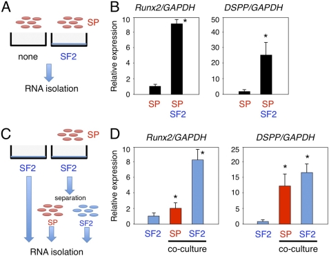
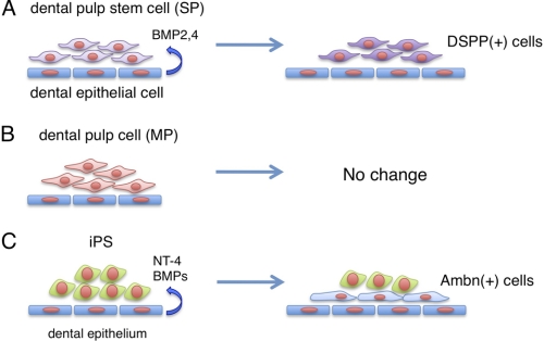
Epithelial-mesenchymal interactions regulate the growth and morphogenesis of ectodermal organs such as teeth. Dental pulp stem cells (DPSCs) are a part of dental mesenchyme, derived from the cranial neural crest, and differentiate into dentin forming odontoblasts. However, the interactions between DPSCs and epithelium have not been clearly elucidated. In this study, we established a mouse dental pulp stem cell line (SP) comprised of enriched side population cells that displayed a multipotent capacity to differentiate into odontogenic, osteogenic, adipogenic, and neurogenic cells. We also analyzed the interactions between SP cells and cells from the rat dental epithelial SF2 line. When cultured with SF2 cells, SP cells differentiated into odontoblasts that expressed dentin sialophosphoprotein. This differentiation was regulated by BMP2 and BMP4, and inhibited by the BMP antagonist Noggin. We also found that mouse iPS cells cultured with mitomycin C-treated SF2-24 cells displayed an epithelial cell-like morphology. Those cells expressed the epithelial cell markers p63 and cytokeratin-14, and the ameloblast markers ameloblastin and enamelin, whereas they did not express the endodermal cell marker Gata6 or mesodermal cell marker brachyury. This is the first report of differentiation of iPS cells into ameloblasts via interactions with dental epithelium. Co-culturing with dental epithelial cells appears to induce stem cell differentiation that favors an odontogenic cell fate, which may be a useful approach for tooth bioengineering strategies.


Figures

FIGURE 1.
FIGURE 1.
Isolation of SP cells from mDP cell line. A, flow cytometry analysis of SP cells. mDP cells made up ∼0.9% of the total cell population with a relatively lower level of Hoechst 33342 fluorescence (SP cells), while 13.8% of the population was maintained as MP cells. Using repeated cell sorting, the SP cell population was enriched by 11.6% at the first sorting, 30.3% at the second sorting, and 82.3% at the third sorting. B, expression of the stem cell markers Sca-1 and Oct3/4 in dental pulp, SP, and MP cells.
FIGURE 2.
FIGURE 2.
Odontoblast and osteoblast differentiation in SP cells. A, differentiation of SP cells to odontoblasts. Expression of the odontoblast marker DSPP and the undifferentiated mesenchymal marker Bcrp1 in dental pulp (black bar) and SP cells (gray bar) cultured with or without BMP2 or BMP4. B, differentiation of SP cells to osteoblasts in osteoblast induction medium (Osteogenic cond.). ALP and von Kossa staining of dental pulp, SP, and MP cells. C, expressions of osteoblast markers in dental pulp, SP, and MP cells cultured in regular (−) or osteoblast induction medium (+). *, p < 0.05.
FIGURE 3.
FIGURE 3.
In vitro epithelial-mesenchymal interaction system using dental epithelial cells (SF2) and dental mesenchymal stem cells (SP) to promote odontogenic cell differentiation. A and C, schematic diagram of the co-culture system. B, comparisons of Runx2 and DSPP gene expressions between the SP monolayer culture and SP and SF2 cell co-culture system. C, total RNA samples were separately prepared from SP and SF2 cells, using the anti-HA antibody. D, expressions of Runx2 and DSPP in co-cultured SF2 (blue) and SP (red) cells. The expression level of GAPDH was used an internal control. *, p < 0.05.
FIGURE 4.
FIGURE 4.
Co-culture conditions for screening of odontogenic cell differentiation using in vitro cell-cell interaction system. A, SP cells were cultured on SF2 cells in monolayers, then fixed with 4% paraformaldehyde (PFA) or treated with ammonia (denudation). B, DSPP expression in SP cells co-cultured under different conditions. C, four sets of co-culture conditions using cell chambers were analyzed. D, DSPP expression in SF2 cells (blue) and SP cells (red) cultured in lower dishes, with co-culture partner cells in the upper chambers. The expression level of GAPDH was used an internal control. *, p < 0.05.
FIGURE 5.
FIGURE 5.
In vitro epithelial-mesenchymal interaction system shows that crosstalk BMP signaling is essential for odontogenic cell differentiation. A, total RNA was isolated from SP cells co-cultured with SF2 cells in the presence or absence of Noggin recombinant protein. B, DSPP expression in SP cells co-cultured with SF2 cells after blocking BMP signaling. C, four sets of culture conditions using cell chambers were analyzed. D, BMP2 and BMP4 expressions in SF2 (blue) and SP (red) cells, with co-culture partner cells in the upper chambers. *, p < 0.05.
FIGURE 6.
FIGURE 6.
Epithelial cell shapes of iPS cells after co-culturing with SF2-24 cells. A, phase micrographs of monolayer SF2-24 cells and iPS cells cultured with SF2-24 feeder cells for 4 days, followed by DAPI staining. B and C, low and high magnification phase micrographs of iPS cells on MMC-treated SF2-24 feeder cells after 6 (6Day) and 10 days (10Day). Enlarged image shows a part of the iPS cells with epithelial cell shapes. C, epithelial cell cluster formed by iPS cell-derived epithelial cells (area within yellow dashed line). Bar, 50 mm.
FIGURE 7.
FIGURE 7.
Effects of culture conditions on ameloblast induction of iPS cells. A, iPS cells were co-cultured with SF2-24 cells, MMC-treated (MMC) MEFs, MMC-treated SF2-24 cells or PFA-treated SF2-24 cells. B, Ambn expression in mouse iPS (upper panel) and rat-derived SF2-24 (bottom panel) cells in different co-culture conditions for 10 days. C, time course analysis of gene expressions of stem cell (blue), endo/mesoderm (black), and ameloblast (red) markers in iPS cells co-cultured with SF2-24 cells for 7 (7Day) and 10 days (10Day).
FIGURE 8.
FIGURE 8.
Expression of Ambn, an ameloblast specific protein, in iPS cells co-cultured with SF2-24 cells. A, phase micrographs of iPS cell colonies cultured with mitomycin C-treated SF2-24 cells. Hoechst staining (blue), Ambn staining (red), and merged images. B, high magnifications of phase and merged images in A. Bottom panel, relative expression levels of Ambn protein in SF2-24 and iPS cells cultured in ameloblast induction system. *, p < 0.05; Bar, 100 mm.
FIGURE 9.
FIGURE 9.
Promotion of ameloblast induction of iPS cells using conditioned SF2-24 cells. A, iPS cells were cultured on mitomycin C-treated MEFs in iPS cell culture medium supplemented with (CM) or without (M) conditioned medium from SF2-24 cells. B, expression of mouse Ambn gene in iPS cells cultured in iPS cell culture medium supplemented with (CM) or without (M) conditioned medium from SF2-24 cells. C, creation of Ambn deletions. All recombinant Ambn proteins have V5 and His tags at the C terminus. D, expression of mouse Ambn gene in iPS cells cultured in iPS cell culture medium supplemented with (CM) or without (M) condition medium from SF2-24 cells, recombinant Ambn-expressing SF2-7 cells or recombinant Ambn proteins. *, p < 0.05 (compared with non-transfected SF2-7 cells). E, expression of mouse Ambn and CK14 genes in iPS cells cultured in SF2-24 conditioned medium supplemented with K252a, PD98059, anti-NT-4, or Noggin. *, p < 0.05 (compared with CM only).
FIGURE 10.
FIGURE 10.
Proposed models of odontogenic induction from dental mesenchymal stem cells and iPS cells by co-culturing with dental epithelial cells. A, dental epithelial cells induce DSPP-expressing odontoblasts from SP cells. B, no odontogenic induction was observed in differentiated (MP) cells co-cultured with dental epithelial cells. C, dental epithelial cells induce Ambn-expressing ameloblasts from iPS cells.

References

    1. Thesleff I. (2003) Epithelial-mesenchymal signaling regulating tooth morphogenesis. J. Cell Sci. 116, 1647–1648 - PubMed
    1. Fukumoto S., Kiba T., Hall B., Iehara N., Nakamura T., Longenecker G., Krebsbach P. H., Nanci A., Kulkarni A. B., Yamada Y. (2004) Ameloblastin is a cell adhesion molecule required for maintaining the differentiation state of ameloblasts. J. Cell Biol. 167, 973–983 - PMC - PubMed
    1. Yuasa K., Fukumoto S., Kamasaki Y., Yamada A., Fukumoto E., Kanaoka K., Saito K., Harada H., Arikawa-Hirasawa E., Miyagoe-Suzuki Y., Takeda S., Okamoto K., Kato Y., Fujiwara T. (2004) Laminin α2 is essential for odontoblast differentiation regulating dentin sialoprotein expression. J. Biol. Chem. 279, 10286–10292 - PubMed
    1. Nakamura T., de Vega S., Fukumoto S., Jimenez L., Unda F., Yamada Y. (2008) Transcription factor epiprofin is essential for tooth morphogenesis by regulating epithelial cell fate and tooth number. J. Biol. Chem. 283, 4825–4833 - PubMed
    1. Fisher L. W., Fedarko N. S. (2003) Six genes expressed in bones and teeth encode the current members of the SIBLING family of proteins. Connect Tissue Res. 44, Suppl. 1, 33–40 - PubMed

Publication types

MeSH terms