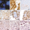
Neoplastic cells are a rare component in human glioblastoma microvasculature
- PMID: 22298889
- PMCID: PMC3292896
- DOI: 10.18632/oncotarget.427
Neoplastic cells are a rare component in human glioblastoma microvasculature
Abstract
Microvascular proliferation is a key biological and diagnostic hallmark of human glioblastoma, one of the most aggressive forms of human cancer. It has recently been suggested that stem-like glioblastoma cells have the capacity to differentiate into functional endothelial cells, and that a significant proportion of the vascular lining in tumors has a neoplastic origin. In principle, this finding could significantly impact the efficacy and development of antiangiogenic therapies targeting the vasculature. While the potential of stem-like cancer cells to form endothelium in culture seems clear, in our clinical experience using a variety of molecular markers, neoplastic cells do not contribute significantly to the endothelial-lined vasculature of primary human glioblastoma. We sought to confirm this impression by analyzing vessels in glioblastoma previously examined using chromogenic in situ hybridization (CISH) for EGFR and immunohistochemistry for mutant IDH1. Vessels containing cells expressing these definitive neoplastic markers were identified in a small fraction of tumors, but only 10% of vessel profiles examined contained such cells and when identified these cells comprised less than 10% of the vascular cellularity in the cross section. Interestingly, these rare intravascular cells showing EGFR amplification by CISH or mutant IDH1 protein by immunohistochemistry were located in the middle or outer portions of vessel walls, but not amongst the morphologic boundaries of the endothelial lining. To more directly address the capacity of glioblastoma cells to contribute to the vascular endothelium, we performed double labeling (Immunofluorescence/FISH) for the endothelial marker CD34 and EGFR gene locus. This analysis did not identify EGFR amplified CD34+ endothelial cells within vascular linings, and further supports our observation that incorporation of glioblastoma cells into the tumor vessels is, at best, extremely rare of questionable clinical or therapeutic significance.
Figures


Similar articles
-
Tumor vessel biology in pediatric intracranial ependymoma.J Neurosurg Pediatr. 2010 Apr;5(4):335-41. doi: 10.3171/2009.11.PEDS09260. J Neurosurg Pediatr. 2010. PMID: 20367336
-
Evaluation of Microvascular Density in Glioblastomas in Relation to p53 and Ki67 Immunoexpression.Int J Mol Sci. 2024 Jun 20;25(12):6810. doi: 10.3390/ijms25126810. Int J Mol Sci. 2024. PMID: 38928515 Free PMC article.
-
CD133+ glioblastoma stem-like cells induce vascular mimicry in vivo.Curr Neurovasc Res. 2011 Aug 1;8(3):210-9. doi: 10.2174/156720211796558023. Curr Neurovasc Res. 2011. PMID: 21675958
-
Vascular heterogeneity and targeting: the role of YKL-40 in glioblastoma vascularization.Oncotarget. 2015 Dec 1;6(38):40507-18. doi: 10.18632/oncotarget.5943. Oncotarget. 2015. PMID: 26439689 Free PMC article. Review.
-
The glioblastoma vasculature as a target for cancer therapy.Biochem Soc Trans. 2014 Dec;42(6):1647-52. doi: 10.1042/BST20140278. Biochem Soc Trans. 2014. PMID: 25399584 Review.
Cited by
-
Correlation between the prognostic value and the expression of the stem cell marker CD133 and isocitrate dehydrogenase1 in glioblastomas.J Neurooncol. 2013 Dec;115(3):333-41. doi: 10.1007/s11060-013-1234-z. Epub 2013 Oct 16. J Neurooncol. 2013. PMID: 24129546
-
The Expression of Leptin and Its Receptor During Tumorigenesis of Diffuse Gliomas such as Astrocytoma and Oligodendroglioma- Grade II, III and IV (NOS).Asian Pac J Cancer Prev. 2019 Feb 26;20(2):479-485. doi: 10.31557/APJCP.2019.20.2.479. Asian Pac J Cancer Prev. 2019. PMID: 30803210 Free PMC article.
-
Glioblastoma multiforme therapy and mechanisms of resistance.Pharmaceuticals (Basel). 2013 Nov 25;6(12):1475-506. doi: 10.3390/ph6121475. Pharmaceuticals (Basel). 2013. PMID: 24287492 Free PMC article.
-
Tumor Niches: Perspectives for Targeted Therapies in Glioblastoma.Antioxid Redox Signal. 2023 Nov;39(13-15):904-922. doi: 10.1089/ars.2022.0187. Epub 2023 Jun 19. Antioxid Redox Signal. 2023. PMID: 37166370 Free PMC article. Review.
-
Molecular crosstalk between tumour and brain parenchyma instructs histopathological features in glioblastoma.Oncotarget. 2016 May 31;7(22):31955-71. doi: 10.18632/oncotarget.7454. Oncotarget. 2016. PMID: 27049916 Free PMC article.
References
-
- Ohgaki H., Kleihues P. Population-based studies on incidence, survival rates, and genetic alterations in astrocytic and oligodendroglial gliomas. J Neuropathol Exp Neurol. 2005;64:479–89. - PubMed
-
- CBTRUS Primary Brain AND Central Nervous System Tumors Diagnosed in the United States in 2004-2007. 2011. Available from: http://www.cbtrus.org/2007-2008/2007-20081.html.
-
- Stupp R., Mason W.P., van den Bent M.J., Weller M., Fisher B., Taphoorn M.J., Belanger K., Brandes A.A., Marosi C., Bogdahn U., Curschmann J., Janzer R.C., Ludwin S.K., Gorlia T., Allgeier A., Lacombe D., et al. Radiotherapy plus concomitant and adjuvant temozolomide for glioblastoma. N Engl J Med. 2005;352:987–96. - PubMed
-
- Ohgaki H., Dessen P., Jourde B., Horstmann S., Nishikawa T., Di Patre P.L., Burkhard C., Schuler D., Probst-Hensch N.M., Maiorka P.C., Baeza N., Pisani P., Yonekawa Y., Yasargil M.G., Lutolf U.M., Kleihues P. Genetic pathways to glioblastoma: a population-based study. Cancer Res. 2004;64:6892–9. - PubMed
-
- Verhaak R.G., Hoadley K.A., Purdom E., Wang V., Qi Y., Wilkerson M.D., Miller C.R., Ding L., Golub T., Mesirov J.P., Alexe G., Lawrence M., O'Kelly M., Tamayo P., Weir B.A., Gabriel S., et al. Integrated genomic analysis identifies clinically relevant subtypes of glioblastoma characterized by abnormalities in PDGFRA, IDH1, EGFR, and NF1. Cancer Cell. 2010;17:98–110. - PMC - PubMed
Publication types
MeSH terms
Substances
Grants and funding
LinkOut - more resources
Full Text Sources
Medical
Research Materials
Miscellaneous

