. 2011 Dec;2(4):492-516.
doi: 10.1007/s12975-011-0125-x.

Blood-brain barrier pathophysiology in traumatic brain injury

Affiliations

Blood-brain barrier pathophysiology in traumatic brain injury

Adam Chodobski et al. Transl Stroke Res. 2011 Dec.

Abstract

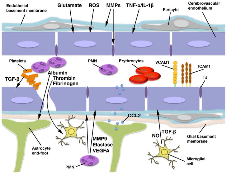
The blood-brain barrier (BBB) is formed by tightly connected cerebrovascular endothelial cells, but its normal function also depends on paracrine interactions between the brain endothelium and closely located glia. There is a growing consensus that brain injury, whether it is ischemic, hemorrhagic, or traumatic, leads to dysfunction of the BBB. Changes in BBB function observed after injury are thought to contribute to the loss of neural tissue and to affect the response to neuroprotective drugs. New discoveries suggest that considering the entire gliovascular unit, rather than the BBB alone, will expand our understanding of the cellular and molecular responses to traumatic brain injury (TBI). This review will address the BBB breakdown in TBI, the role of blood-borne factors in affecting the function of the gliovascular unit, changes in BBB permeability and post-traumatic edema formation, and the major pathophysiological factors associated with TBI that may contribute to post-traumatic dysfunction of the BBB. The key role of neuroinflammation and the possible effect of injury on transport mechanisms at the BBB will also be described. Finally, the potential role of the BBB as a target for therapeutic intervention through restoration of normal BBB function after injury and/or by harnessing the cerebrovascular endothelium to produce neurotrophic growth factors will be discussed.


Conflict of interest statement

Financial and competing interests disclosure

The authors have no financial and/or competing interests to disclose.

Figures

Figure 1
Figure 1
Schematic representation of the blood-brain barrier (BBB)/gliovascular unit in the injured brain. The BBB is formed by the brain endothelial cells connected by tight junctions (TJs). However, astrocytes, whose end-feet make an intimate contact with the cerebrovascular endothelium, are critical for normal function of the BBB. Microglial processes are also closely associated with the brain endothelium in 4–13% of cerebral microvessels, and glial and endothelial cells functionally interact with each other in a paracrine manner. Brain parenchymal cells are normally shielded from periphery by the BBB. However, the disruption of vascular integrity occurring after neurotrauma allows blood-borne factors, such as albumin and fibrinogen, as well as thrombin, which is cleaved from prothrombin by Factor Xa, to enter the brain. Fibrinogen, after its conversion to fibrin, acts on microglia, causing the rearrangement of microglial cytoskeleton and increased phagocytosis. Thrombin predominantly acts on microglia, stimulating their proliferation and inducing the production of nitric oxide (NO). It also increases the microglial synthesis of proinflammatory mediators, such as tumor necrosis factor-α (TNF-α) and interleukin (IL)-6 and -12. In addition, thrombin may target the cerebrovascular endothelium and increase the permeability of the BBB. Similar to thrombin, albumin promotes the proliferation of microglial cells and increases the production of NO and proinflammatory mediators, such as IL-1β. Albumin also acts on astrocytes by binding to transforming growth factor-β (TGF-β) receptor II, which may play a role in post-traumatic cortical epileptogenesis. Among the putative factors contributing to post-traumatic dysfunction of the BBB are glutamate, reactive oxygen species (ROS), matrix metalloproteinases (MMPs), proinflammatory cytokines TNF-α and IL-1β, and vascular endothelial growth factor A (VEGFA). After injury, glutamate is released from various parenchymal cells and from invading neutrophils (polymorphonuclear leukocytes; PMN). It increases the permeability of the BBB and has been shown to promote apoptosis of brain endothelial cells, although this latter action of glutamate has been questioned. ROS not only increase the permeability of brain endothelium, but also play an important role in promoting post-traumatic invasion of inflammatory cells by, for example, upregulating the endothelial expression of cell adhesion molecules, such as intercellular adhesion molecule-1 (ICAM1). MMPs are produced by multiple types of parenchymal cells and can also be released from invading leukocytes. MMPs disrupt the integrity of the BBB by attacking basal lamina proteins and degrading tight junctions. TNF-α and IL-1β increase the permeability of the BBB, but, more importantly, they also play a key role in progression of post-traumatic neuroinflammation. These cytokines increase the endothelial expression of cell adhesion molecules, such as E-selectin, ICAM1, and vascular cell adhesion molecule-1 (VCAM1). They also induce the endothelial and astrocytic production of CXC and CC chemokines, including CXCL1, -2, and CCL2, which attract circulating inflammatory cells and facilitate their migration across the BBB. Astrocyte-derived CCL2 can be transported across the cerebrovascular endothelium and then presented on its luminal surface. This chemokine can also increase the permeability of the BBB. VEGFA increases the permeability of brain endothelium by changing the distribution and downregulating the expression of tight junction proteins. After injury, VEGFA is predominantly produced by astrocytes, but is also carried by invading neutrophils. The major source of TGF-β is the aggregating platelets, but TGF-β is also produced by microglia and, to a lesser extent, by astrocytes. TGF-β has been shown to increase the permeability of the cerebrovascular endothelium; however, other investigators postulated that this growth factor has an opposite effect on the BBB, which is to enhance and maintain the barrier properties of brain endothelium. Invading neutrophils exert an adverse effect on BBB function. These inflammatory cells not only produce proinflammatory cytokines, such as TNF-α, and generate large amounts of ROS, but they also release various proteolytic enzymes, including MMP9 and neutrophil elastase.
Figure 2
Figure 2
Production of CXC and CC chemokines by the cerebrovascular endothelium and astrocytes in a rat model of traumatic brain injury (TBI). A The immunoreactive product for neutrophil chemoattractant CXCL1 (arrows) associated with a microvessel in the traumatized brain parenchyma at 6 hours post-TBI. Double immunostaining with anti-CXCL1 antibody and an antibody to RECA-1, a marker for vascular endothelium, is shown. Similar results were obtained for CXCL2 and CCL2, a major chemoattractant for monocytes. This distinct pattern of immunopositive staining for CXC and CC chemokines was not associated with pericytes, perivascular macrophages, or smooth muscle cells, suggesting that these chemokines are produced by the brain endothelium. B Double immunostaining with anti-CXCL1 antibody and an antibody to TGN38, a Golgi marker, demonstrates that the CXCL1-immunoreactive product in cerebral microvessels (outlined) is predominantly associated with the Golgi complex (arrows). The localization of CXC and CC chemokines to the Golgi complex is consistent with increased synthesis of these secreted proteins occurring after injury. C Post-traumatic expression of CCL2 in astrocytes. Double immunostaining with anti-CCL2 antibody and an antibody to glial fibrillary acidic protein (GFAP), an astrocyte marker, is shown. Neither CXCL1 nor CXCL2 were expressed in astrocytes after injury. D CCL2 expressed in a cortical astrocyte at 6 hours post-TBI co-localizes with the Golgi complex. E Astrocytic expression of CCL2 at 24 hours after TBI. Note that at this time after injury, the CCL2-immunopositive product is predominantly localized extracellularly along astrocyte processes (arrows). This pattern of CCL2-immunopositive staining likely reflects the binding of this chemokine to heparan sulfate proteoglycans expressed on the surface of astrocytic cells and/or within the extracellular matrix. Scale bars: panels AC, 10 μm; panels D, E 5 μm. Reprinted with permission from [178].
Figure 3
Figure 3
Migration of neutrophils across pial microvessels in a rat model of traumatic brain injury (TBI). Upon invasion of traumatized brain parenchyma, neutrophils release vascular endothelial growth factor A (VEGFA). The VEGFA-immunoreactive product appears as haloes surrounding neutrophils, which likely represent the growth factor sequestered within the extracellular matrix due to the VEGFA binding to heparan sulfate proteoglycans. A The cerebral cortex adjacent to the post-traumatic lesion at 8 hours post-TBI. Double immunostaining with anti-VEGFA antibody and an antibody to CD11b, a subunit of integrin receptor expressed on neutrophils, monocytes/macrophages, and microglia, is shown. Note that neutrophils not only invade the traumatized cortex after crossing the blood-brain barrier (BBB) in brain parenchymal microvessels, but also accumulate in the subarachnoid space (SAS) after crossing the BBB in pial microvessels. B A higher magnification view of selected area from panel A. This confocal microscopy image shows neutrophils accumulating in the SAS, from where these inflammatory cells appear to invade the brain parenchyma. An arrow points at a neutrophil that is in the initial stage of crossing the pial/glial lining of the cerebral cortex and starts releasing VEGFA. Note that other neutrophils accumulating in the SAS that are located further from the cortical surface clearly carry preformed VEGFA, but do not release it. C A coronal brain section cut at the level of the hippocampus and stained with anti-VEGFA and anti-CD11b antibodies. The rat was sacrificed at 24 hours after TBI. Similar to their accumulation in the SAS, neutrophils enter the cistern of velum interpositum (CVI), a slit-shaped space above the third ventricle with highly vascularized pia mater, from where these leukocytes also appear to invade the injured brain parenchyma. Scale bars: panels A, C, 100 μm; panel B, 10 μm. Reprinted with permission from [77].

References

    1. Saatman KE, Duhaime AC, Bullock R, Maas AI, Valadka A, Manley GT, et al. Classification of traumatic brain injury for targeted therapies. J Neurotrauma. 2008;25(7):719–38. - PMC - PubMed
    1. Abbott NJ, Rönnbäck L, Hansson E. Astrocyte-endothelial interactions at the blood-brain barrier. Nat Rev Neurosci. 2006;7(1):41–53. - PubMed
    1. Wolburg H, Noell S, Mack A, Wolburg-Buchholz K, Fallier-Becker P. Brain endothelial cells and the glio-vascular complex. Cell Tissue Res. 2009;335(1):75–96. - PubMed
    1. Lassmann H, Zimprich F, Vass K, Hickey WF. Microglial cells are a component of the perivascular glia limitans. J Neurosci Res. 1991;28(2):236–43. - PubMed
    1. Neuwelt E, Abbott NJ, Abrey L, Banks WA, Blakley B, Davis T, Engelhardt B, Grammas P, Nedergaard M, Nutt J, et al. Strategies to advance translational research into brain barriers. Lancet Neurol. 2008;7(1):84–96. - PubMed

LinkOut - more resources