. 2012 May;12(5):1102-12.
doi: 10.1111/j.1600-6143.2011.03959.x. Epub 2012 Feb 2.

Mechanisms of alloimmunization and subsequent bone marrow transplantation rejection induced by platelet transfusion in a murine model

Affiliations

Mechanisms of alloimmunization and subsequent bone marrow transplantation rejection induced by platelet transfusion in a murine model

S R Patel et al. Am J Transplant. 2012 May.

Abstract

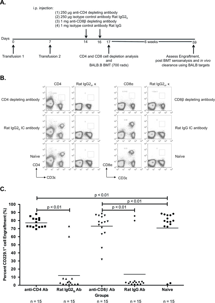
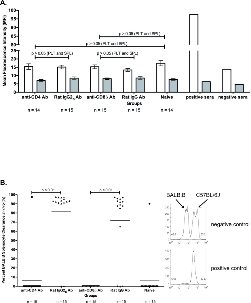
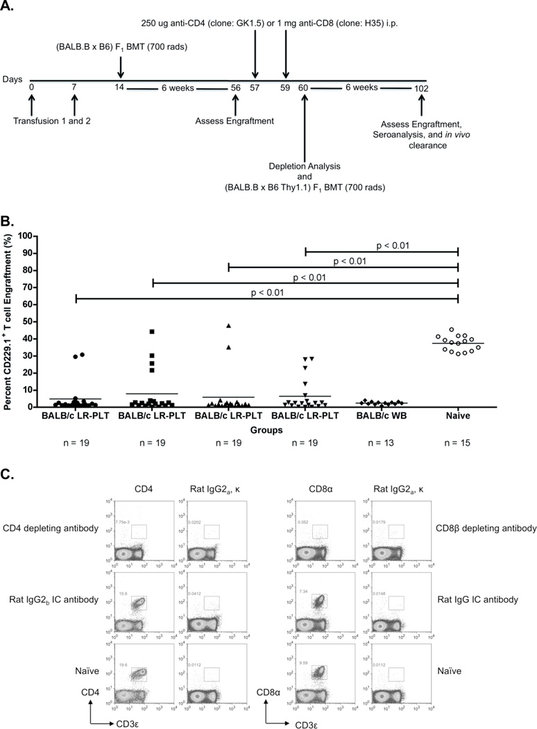
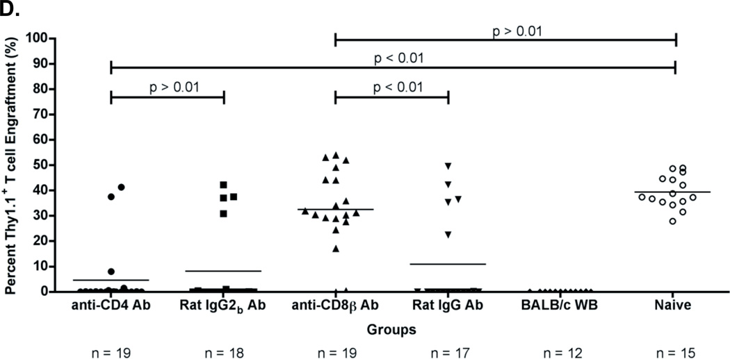
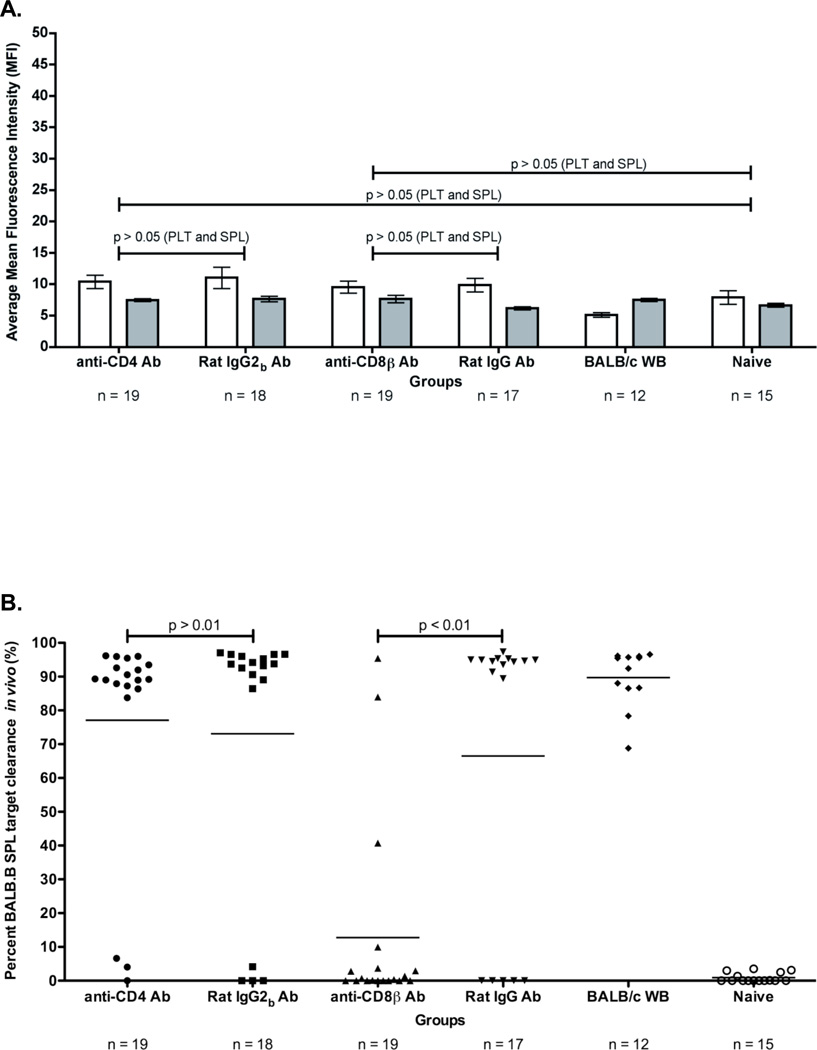
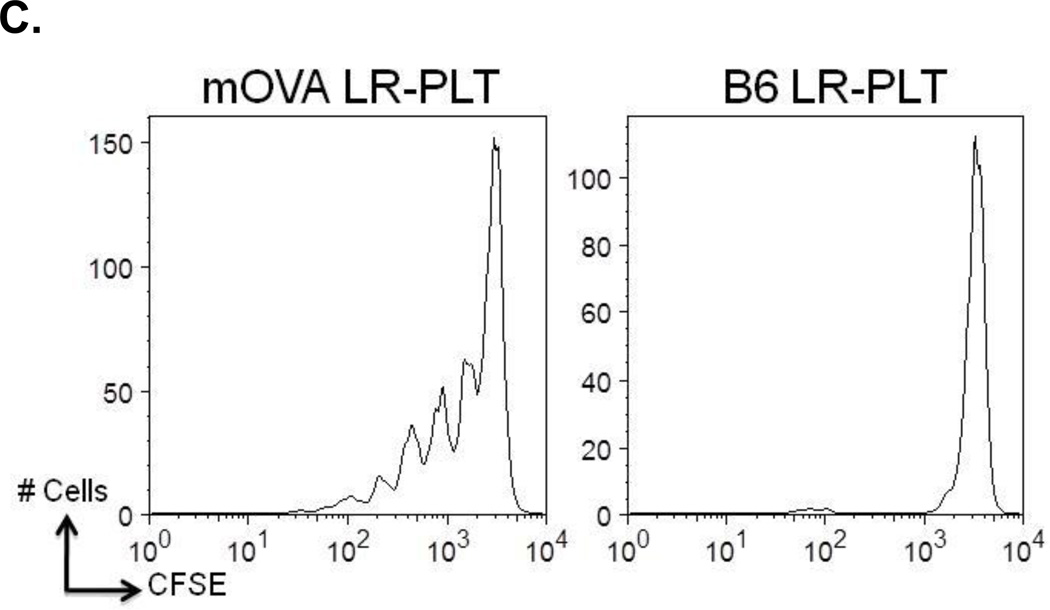
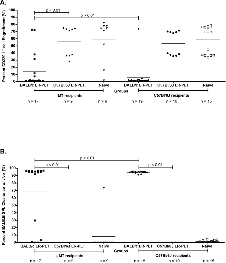
For many nonmalignant hematological disorders, HLA-matched bone marrow transplantation (BMT) is curative. However, due to lack of neoplasia, the toxicity of stringent conditioning regimens is difficult to justify, and reduced intensity conditioning is used. Unfortunately, current reduced intensity regimens have high rates of BMT rejection. We have recently reported in a murine model that mHAs on transfused platelet products induce subsequent BMT rejection. Most nonmalignant hematological disorders require transfusion support prior to BMT and the rate of BMT rejection in humans correlates with the number of transfusions given. Herein, we perform a mechanistic analysis of platelet transfusion-induced BMT rejection and report that unlike exposure to alloantigens during transplantation, platelet transfusion primes alloimmunity but does not stimulate full effector function. Subsequent BMT is itself an additional and distinct immunizing event, which does not induce rejection without antecedent priming from transfusion. Both CD4(+) and CD8(+) T cells are required for priming during platelet transfusion, but only CD8(+) T cells are required for BMT rejection. In neither case are antibodies required for rejection to occur.


Conflict of interest statement

Disclosures: The authors of this manuscript have no conflicts of interest to disclose as described by the American Journal of Transplantation

Figures

Figure 1
Figure 1. Depletion of CD4+ or CD8+ cells prevents rejection of a BALB.B BMT in BALB LR-PLT transfused recipients
(A) Experimental model testing requirement of CD4+ and CD8+ depletion in platelet transfusion induced BMT rejection. While BALB/c donors were MHC- and mHA-mismatched, BALB.B BM donors were MHC-matched:mHA-mismatched to the recipients. Designated recipients received two platelet transfusions a week apart. After the second transfusion, indicated recipients were treated i.p. with anti-CD4 (clone: GK1.5) or anti-CD8β (clone: H35) depleting antibodies, or isotype control antibodies Rat IgG2b or Rat IgG, respectively. Depletion of CD4+ or CD8+ T cells was monitored in the peripheral blood, spleen, peripheral lymph nodes, and BM. Twenty-four hours after the second treatment, designated recipients received a BALB.B BMT under reduced intensity conditions. Seroanalysis and in vivo survival of BALB.B targets was also performed after BMT (see figure 2). (B) Depletion analysis of CD4+ and CD8+ T cells in BMT recipients. Peripheral blood leukocytes from BMT recipients were stained for CD4+ or CD8+ T cells using anti-CD4 (clone: RM4-5) and anti-CD8α (clone: 53-6.7) antibodies. (C) BALB.B BMT engraftment results. Percent CD229.1+ cells in the peripheral blood represent engraftment; the mean of each group is represented as a horizontal line. Statistics were generated using column statistics and a one-way ANOVA with Dunnett’s post-test. Illustrated is the combined data from three independent experiments.
Figure 2
Figure 2. BALB specific alloimmunization after depletion and BMT
(A) BALB specific alloantibody production. anti-BALB antibodies in sera of transfused and transplanted recipients were assessed using BALB.B splenocyte (white) and platelet (grey) targets in an indirect immunofluorescence staining. (B) In vivo survival of BALB.B targets. Immunity against BALB expressing targets was assessed by in vivo survival of BALB.B splenocyte targets labeled with CFDA. Error bars in (A) represent the mean + SEM. The mean of each group in (B) is represented as a horizontal line. Representative histograms are shown to illustrate typical outcomes of the in vivo clearance assay. Statistics were generated using column statistics and a one-way ANOVA with Dunnett’s post-test. The data shown for both panels A and B is the combined data from three separate experiments.
Figure 3
Figure 3. Rejection of a BALB.B BMT is mediated by a CD8+ cellular response that requires the presence of CD4+ cells prior to BMT
(A) Experimental model testing the requirement of CD4+ and CD8+ T cells as the rejection vectors. Using the depletion model described in Figure 1A, BALB/c platelet donors were MHC- and mHA-mismatched, whereas (BALB.B x C57BL/6J) and (BALB.B x B6 Thy1.1) F1 BM donors were MHC-matched but mHA-haplomismatched. Recipients were transfused twice, a week apart. One week after the second transfusion, recipients received a (BALB.B x C57BL/6J) F1 BMT. Six weeks later, indicated recipients were treated i.p. with anti-CD4 or anti-CD8β depleting antibodies, or isotype control antibodies Rat IgG2b or Rat IgG, respectively. Depletion of CD4+ or CD8+ T cells was monitored in the peripheral blood. Recipients were then given a (BALB.B x B6 Thy1.1) F1 BMT. Seroanalysis and in vivo survival of BALB.B targets was performed after BMT (see figure 4). (B) (BALB.B x C57BL/6J) F1 BMT engraftment results. Engraftment was assessed in the peripheral blood of transplant recipients. BMT engraftment was measured as a percentage of CD229.1+ T cells two standard deviations above the rejecting BALB/c whole blood transfused recipients. All four BALB/c LR-PLT transfused recipients in (B) were treated the same. The BALB/c LR-PLT transfused recipients were split into the four treatment groups to compare engraftment during the first and second transplantation in panels (B) and (D), respectively. The symbol for each group in panel (B) corresponds with that in panel (D). (C) Analysis of CD4+ and CD8+ T cell depletion in re-transplant recipients. Peripheral blood leukocytes from recipients receiving the second BMT were stained for CD4+ or CD8+ T cells using anti-CD4 (clone: RM4-5) and anti-CD8α (clone: 53-6.7) antibodies. (D) Engraftment results for (BALB.B x B6 Thy1.1) F1 BMT recipients. Engraftment was defined as having a percentage of Thy1.1+ T cells two standard deviations above the mean of the rejecting recipients treated with the corresponding isotype control antibody. The mean of each group in (B) and (D) is represented as a horizontal line. Statistics were generated using column statistics and a one-way ANOVA with Dunnett’s post-test. Illustrated is the combined data from three independent experiments.
Figure 4
Figure 4. Alloimmunization against BALB mHA(s) after depletion and re-transplantation
(A) BALB specific alloantibody production. Anti-BALB antibodies in the sera of transfused and transplanted recipients were assessed using BALB.B splenocyte (white) and platelet (grey) targets in an indirect immunofluorescence staining. (B) In vivo survival of BALB.B targets. Immunity against BALB expressing targets was assessed by in vivo survival of BALB.B splenocyte targets labeled with CFDA. Error bars in (A) represent the mean + SEM. The mean of each group in (B) is represented as a horizontal line. Statistics were generated using column statistics and a one-way ANOVA with Dunnett’s post-test. Illustrated are data from three combined independent experiments. (C) CD8+ T cell crosspriming to a mHA on transfused LR-PLTs was tested utilizing the OT-I T cell transgenic, which is specific for the SIINFEKL (OVA257–264) peptide presented by MHC Class I, H-2Kb. OT-I splenocytes were labeled with CFDA (Invitrogen, Eugene, OR). 15 × 106 CFSE labeled OT-I splenocytes were adoptively transferred via tail vein injection and mice were transfused with mOVA or B6 LR-PLTs. Cell division was determined 3 days later by gating on OT-I T cells and evaluating CFSE.
Figure 5
Figure 5. B cells are not required for BALB LR-PLT transfusions to induce rejection of a BALB.B BMT
(A) Engraftment results. Engraftment is measured as a percentage of CD229.1+ cells in the peripheral blood of transplant recipients. (B) Alloimmunization to BALB mHAs. BALB specific immunity was measured by in vivo survival of BALB.B splenocyte targets. The mean of each group in (A) and (B) is represented as a horizontal line. Statistics were generated using one-way ANOVA with Dunnett’s post-test. While the illustrated results in (A) and (B) for BALB/c LR-PLT transfused recipients are the combined data from three independent experiments, the data depicted for C57BL/6J LR-PLT transfused recipients are the combined results from two different experiments.

Similar articles

Cited by

References

    1. Bhatia M, Walters MC. Hematopoietic cell transplantation for thalassemia and sickle cell disease: past, present and future. Bone Marrow Transplant. 2008;41(2):109–117. - PubMed
    1. Horan JT, Liesveld JL, Fenton P, Blumberg N, Walters MC. Hematopoietic stem cell transplantation for multiply transfused patients with sickle cell disease and thalassemia after low-dose total body irradiation, fludarabine, and rabbit anti-thymocyte globulin. Bone Marrow Transplant. 2005;35(2):171–177. - PubMed
    1. Marsh JC, Ball SE, Darbyshire P, Gordon-Smith EC, Keidan AJ, Martin A, et al. Guidelines for the diagnosis and management of acquired aplastic anaemia. Br J Haematol. 2003;123(5):782–801. - PubMed
    1. Or R, Aker M, Shapira MY, Resnick I, Bitan M, Samuel S, et al. Allogeneic stem cell transplantation for the treatment of diseases associated with a deficiency in bone marrow products. Springer Semin Immunopathol. 2004;26(1–2):133–142. - PubMed
    1. Wagner JE, Eapen M, MacMillan ML, Harris RE, Pasquini R, Boulad F, et al. Unrelated donor bone marrow transplantation for the treatment of Fanconi anemia. Blood. 2007;109(5):2256–2262. - PMC - PubMed

Publication types

MeSH terms