

Widespread microstructural white matter involvement in amyotrophic lateral sclerosis: a whole-brain DTI study
- PMID: 22300932
- PMCID: PMC8013257
- DOI: 10.3174/ajnr.A2918
Widespread microstructural white matter involvement in amyotrophic lateral sclerosis: a whole-brain DTI study
Abstract
Background and purpose: The extensive application of advanced MR imaging techniques to the study of ALS has undoubtedly improved our knowledge of disease pathophysiology, even if the actual spread of the neurodegenerative process throughout the central nervous system is not fully understood. The present study aimed to detect WM patterns of microstructural abnormalities to better investigate the pathologic process in ALS, within but also beyond CSTs, in a whole-brain analysis.
Materials and methods: DTI was performed in 19 patients with ALS and 20 matched healthy controls, by using whole-brain TBSS and VOI analyses.
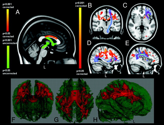
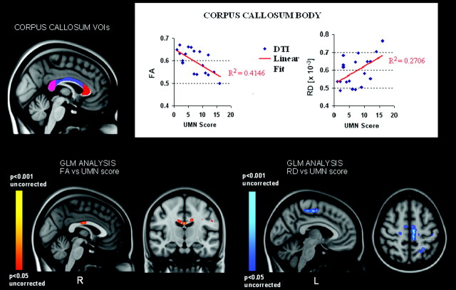
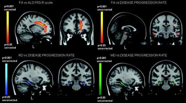
Results: We observed a significant decrease of FA in the body of CC of the ALS group (P < .05). At the VOI level, both FA decrease and RD increase in the body of CC significantly correlated with the UMN score (P = .003 and P = .02). Additionally, significant voxelwise positive correlations between FA and the ALSFRS-R were detected in the WM tracts underneath the left premotor cortex (P < .05).
Conclusions: The correlations between reduction of FA and increase of RD in the body of CC with the UMN score indicate that the WM degeneration in the CC is strictly related to the ALS pyramidal impairment, while the correlation between FA and ALSFRS-R in the associative tracts underneath the left premotor cortex might reflect the progressive spread of the disease from the motor toward the extramotor areas.
Figures
Similar articles
-
Advantages of QBI in TBSS analyses.Magn Reson Imaging. 2014 Feb;32(2):184-9. doi: 10.1016/j.mri.2013.09.002. Epub 2013 Oct 11. Magn Reson Imaging. 2014. PMID: 24211186
-
Diffusion tensor MRI of the corpus callosum in amyotrophic lateral sclerosis.J Magn Reson Imaging. 2014 Mar;39(3):641-7. doi: 10.1002/jmri.24218. Epub 2013 Jul 10. J Magn Reson Imaging. 2014. PMID: 23843179 Free PMC article.
-
A distinct MR imaging phenotype in amyotrophic lateral sclerosis: correlation between T1 magnetization transfer contrast hyperintensity along the corticospinal tract and diffusion tensor imaging analysis.AJNR Am J Neuroradiol. 2012 Apr;33(4):733-9. doi: 10.3174/ajnr.A2855. Epub 2011 Dec 22. AJNR Am J Neuroradiol. 2012. PMID: 22194369 Free PMC article.
-
Radial diffusivity as an imaging biomarker for early diagnosis of non-demented amyotrophic lateral sclerosis.Eur Radiol. 2018 Dec;28(12):4940-4948. doi: 10.1007/s00330-018-5506-z. Epub 2018 Jun 8. Eur Radiol. 2018. PMID: 29948064
-
Altered white matter microarchitecture in amyotrophic lateral sclerosis: A voxel-based meta-analysis of diffusion tensor imaging.Neuroimage Clin. 2018 Apr 4;19:122-129. doi: 10.1016/j.nicl.2018.04.005. eCollection 2018. Neuroimage Clin. 2018. PMID: 30035009 Free PMC article.
Cited by
-
Usefulness of diffusion tensor imaging findings as biomarkers for amyotrophic lateral sclerosis.Sci Rep. 2020 Mar 23;10(1):5199. doi: 10.1038/s41598-020-62049-0. Sci Rep. 2020. PMID: 32251314 Free PMC article.
-
Microstructural effects of Ramadan fasting on the brain: a diffusion tensor imaging study.Diagn Interv Radiol. 2015 May-Jun;21(3):256-61. doi: 10.5152/dir.2014.14361. Diagn Interv Radiol. 2015. PMID: 25835077 Free PMC article.
-
Widespread structural and functional connectivity changes in amyotrophic lateral sclerosis: insights from advanced neuroimaging research.Neural Plast. 2012;2012:473538. doi: 10.1155/2012/473538. Epub 2012 Jun 10. Neural Plast. 2012. PMID: 22720174 Free PMC article. Review.
-
MRI DTI and PDFF as Biomarkers for Lower Motor Neuron Degeneration in ALS.Front Neurosci. 2021 Aug 26;15:682126. doi: 10.3389/fnins.2021.682126. eCollection 2021. Front Neurosci. 2021. PMID: 34512239 Free PMC article.
-
Frontal and Cerebellar Atrophy Supports FTSD-ALS Clinical Continuum.Front Aging Neurosci. 2020 Nov 26;12:593526. doi: 10.3389/fnagi.2020.593526. eCollection 2020. Front Aging Neurosci. 2020. PMID: 33324193 Free PMC article.
References
-
- Davidoff RA. The pyramidal tract. Neurology 1990;40:332–39 - PubMed
-
- Ellis CM, Simmons A, Jones DK, et al. . Diffusion tensor MRI assesses corticospinal tract damage in ALS. Neurology 1999;53:1051–58 - PubMed
-
- Basser PJ, Pierpaoli C. Microstructural and physiological features of tissues elucidated by quantitative-diffusion tensor MRI. J Magn Reson 1996;111:209–19 - PubMed
-
- Graham JM, Papadakis N, Evans J, et al. . Diffusion tensor imaging for the assessment of upper motor neuron integrity in ALS. Neurology 2004;63:2111–19 - PubMed
MeSH terms
LinkOut - more resources
Full Text Sources
Medical
Miscellaneous