. 2012 Jun;33(6):1134-8.
doi: 10.3174/ajnr.A2914. Epub 2012 Feb 2.

Zuckerkandl tubercle of the thyroid: a common imaging finding that may mimic pathology

Affiliations

Zuckerkandl tubercle of the thyroid: a common imaging finding that may mimic pathology

T C Lee et al. AJNR Am J Neuroradiol. 2012 Jun.

Abstract

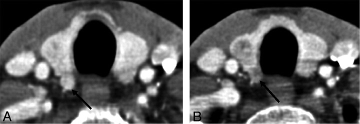
Background and purpose: The posterior thyroid tubercle, also known as ZT, is an important surgical landmark due to its close proximity to the recurrent laryngeal nerve. A recent case of ZT with a nodular configuration caused clinical concern but was shown on biopsy to be normal thyroid tissue. The purpose of this study was to review a series of CT neck studies to identify how often ZT-specifically, a nodular subtype-was present.

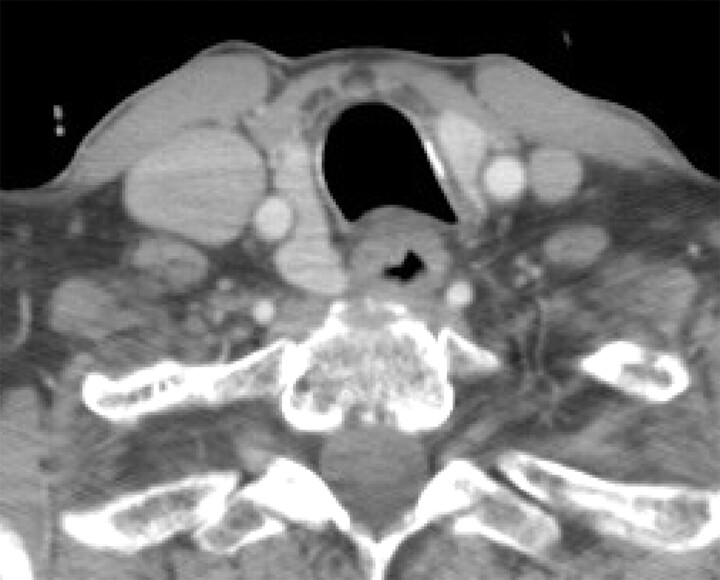
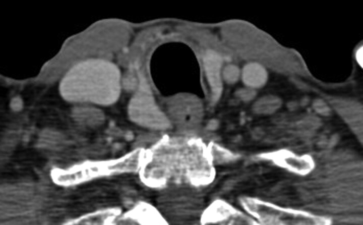
Materials and methods: A total of 96 neck CTs from sequential patients were retrospectively identified from January 2010 to July 2010. ZT was defined on imaging as a thyroid lobe extending posterior to the tracheoesophageal groove. A nodular subtype was defined as having a narrowed neck at the level of the tracheoesophageal groove.

Results: There were 31 women and 45 men (mean age of 56.1 years, range 22-100 years) who met inclusion criteria. Sixty-seven patients had thyroid glands that extended posterior to the tracheoesophageal groove at CT imaging; this finding was bilateral in 43 patients, more commonly on the right (60 versus 49). Thirty-two of these patients (42.1%; 16 male, 16 female) had a nodular subtype.

Conclusions: ZT is an important surgical landmark of the thyroid that has important radiologic variations. It has a nodular shape in over a third of CT neck studies in our series. Recognition of this feature of the thyroid gland at CT imaging can obviate the need for biopsy and avoid potential recurrent laryngeal nerve injury.


Figures

Fig 1.
Fig 1.
Drawings of 2 different thyroid glands as seen from a posterior point of view, with their relationships to the thyroid cartilage and a cut portion of the esophagus. The arrows indicate ZT and black dots indicate parathyroid glands.
Fig 2.
Fig 2.
Axial schematic representation of the thyroid (T = trachea, E = esophagus, black dot represents the right recurrent laryngeal nerve). A, Thyroid without apparent ZT. B, Thyroid with apparent right ZT. C, Thyroid with nodular right ZT (CCA = right common carotid artery within a lateral indentation of the thyroid contour).
Fig 3.
Fig 3.
Axial CT scan images demonstrate 3 different appearances of the ZT (arrows).
Fig 4.
Fig 4.
Patient A, with known ovarian carcinoma recurrence in the abdomen, presented with a few weeks' history of voice hoarseness and was noted to have a nodular area posterior to the right thyroid lobe. There was clinical concern for a metastasis or exophytic thyroid nodule.
Fig 5.
Fig 5.
CT-guided biopsy of Patient A in 2010 with questionable thyroid lesion (A) was performed because a recent sonography-guided fine-needle aspiration was read as indeterminate by pathology and there was persistent clinical concern despite stability from a CT in 2006. The patient was placed on the CT table with neck extended, and a metallic grid marker was placed on the section, showing the epicenter of the questionable lesion. B, A metallic grid marker is in place for biopsy planning. There is shoulder streak artifact through the questionable lesion, which lowers the attenuation. C, CT-guided percutaneous core biopsy needle was placed in the epicenter. Pathology results were consistent with normal thyroid tissue, with no malignant cells.
Fig 6.
Fig 6.
Patient A's pulmonary embolism chest CT from 2006 shows similar appearance of the questionable thyroid lesion.
Fig 7.
Fig 7.
Patient B has similar appearance of the right thyroid lobe on a CT from December 2009, with a posterior extension on the right.
Fig 8.
Fig 8.
Patient B's thyroid appeared similar as far back as 2005, allowing for slight difference in the obliquity of the axial section.
Fig 9.
Fig 9.
A, Thyroid gland with a nodular ZT at the posterior aspect of the right lobe (arrow). B, Follow-up CT study demonstrating development of a discrete hypoattenuated nodule in the right lobe and a hypoattenuated nodule in the previously identified ZT (arrow).
Fig 10.
Fig 10.
A and B, CT images on a patient status post total thyroidectomy for thyroid cancer show a small enhancing lesion near the right tracheoesophageal groove (arrows), which likely represents a tubercle of Zuckerhandl. C and E, Axial and coronal CT demonstrates enlargement of a paratracheal lesion after an interval of 6 months (arrows in C and E); a nearby lymph node also showed enlargement (arrow in D) and proved to be recurrent disease.

References

    1. Mirilas P, Skandalakis JE. Zuckerkandl's tubercle: Hannibal ad Portas. Am Coll Surg 2003;196;5:796–801 - PubMed
    1. Yalcin B, Tatar I, Ozan H. The Zuckerkandl tubercle and the recurrent laryngeal nerve. Am J Surg 2008;196:311–12 - PubMed
    1. Costanzo M, Caruso LA, Veroux M, et al. . The lobe of Zuckerkandl: an important sign of recurrent laryngeal nerve. Ann Ital Chir 2005;76:337–40, discussion 340–41 - PubMed
    1. Musajo FG, Mangiante G, Ischia A, et al. . Zuckerkandl tubercle of the thyroid gland (anatomo-surgical study: preliminary considerations). Chir Ital 1989;41:129–36 - PubMed
    1. Buck RT, Siddiqui AR. Thyroid abnormality secondary to tortuous carotid artery. Eur J Nucl Med 1986;12:51–52 - PubMed