. 2012 Apr;86(7):3528-40.
doi: 10.1128/JVI.06713-11. Epub 2012 Feb 1.

Herpes simplex virus 1 tegument protein US11 downmodulates the RLR signaling pathway via direct interaction with RIG-I and MDA-5

Affiliations

Herpes simplex virus 1 tegument protein US11 downmodulates the RLR signaling pathway via direct interaction with RIG-I and MDA-5

Junji Xing et al. J Virol. 2012 Apr.

Abstract

The interferon (IFN)-mediated antiviral response is a major defense of the host immune system. In order to complete their life cycle, viruses must modulate host IFN-mediated immune responses. Herpes simplex virus 1 (HSV-1) is a large DNA virus containing more than 80 genes, many of which encode proteins that are involved in virus-host interactions and show immune modulatory capabilities. In this study, we demonstrate that the US11 protein, an RNA binding tegument protein of HSV-1, is a novel antagonist of the beta IFN (IFN-β) pathway. US11 significantly inhibited Sendai virus (SeV)-induced IFN-β production, and its double-stranded RNA (dsRNA) binding domain was indispensable for this inhibition activity. Additionally, wild-type HSV-1 coinfection showed stronger inhibition than US11 mutant HSV-1 in SeV-induced IFN-β production. Coimmunoprecipitation analysis demonstrated that the US11 protein in HSV-1-infected cells interacts with endogenous RIG-I and MDA-5 through its C-terminal RNA-binding domain, which was RNA independent. Expression of US11 in both transfected and HSV-1-infected cells interferes with the interaction between MAVS and RIG-I or MDA-5. Finally, US11 dampens SeV-mediated IRF3 activation. Taken together, the combined data indicate that HSV-1 US11 binds to RIG-I and MDA-5 and inhibits their downstream signaling pathway, preventing the production of IFN-β, which may contribute to the pathogenesis of HSV-1 infection.


Figures
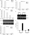
Fig 1
Fig 1
US11 inhibits SeV-mediated activation of the IFN-β and NF-κB promoter activities. (A, C, and D) HEK 293T cells were transfected with 500 ng of IFN-β promoter reporter plasmid p125-luc (A), NF-κB-Luc (C), or (pRDIII-I)4-Luc (D), together with Renilla luciferase plasmid pRL-TK (50 ng) and pCMV-HA empty vector or plasmids encoding the indicated viral proteins (1,000 ng). At 24 h after transfection, cells were left untreated or infected with 100 HAU ml−1 SeV as indicated, and luciferase activity was measured 16 h postinfection. (B) As in panel A, except an increased amount of US11-HA expression plasmid, as indicated, was used. The expression of US11 was analyzed by Western blotting using anti-HA and anti-β-actin (as a control) monoclonal antibodies. Data are expressed as relative luciferase activities with standard deviations for three independent experiments performed in duplicate. (E) HEK293T cells were transfected with pCMV-HA empty vector or US11-HA expression plasmid. At 24 h posttransfection, cells were mock infected or infected with 100 HAU ml−1 SeV for 16 h before RT-PCR was performed using GAPDH and IFN-β primers. (F) Medium from infected cells in panel E was isolated and analyzed by ELISA for IFN-β secretion as described in Materials and Methods. The data represent means + standard deviations for three replicates.
Fig 2
Fig 2
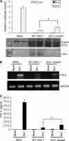
Effects of WT or US11 mutant HSV-1 infection on SeV-induced IFN-β production. (A) HEK 293T cells in a 24-cell plate were cotransfected with the IFN-β reporter plasmid p125-luc and Renilla luciferase plasmid pRL-TK and were mock infected or infected with 100 HAU/ml SeV as indicated at 24 h after transfection. At 8 h postinfection, the HEK 293T cells were then mock infected or infected with either WT or US11 mutant HSV-1 at an MOI of 1 for another 16 h until the luciferase activity was measured. Data are expressed as relative luciferase activities with standard deviations for three independent experiments performed in duplicate. Additionally, the expression of both the US11 and VP22 proteins after WT or US11 mutant HSV-1 infection was verified by Western blotting using rabbit anti-US11 or anti-VP22 pAbs. β-Actin as a loading control was also detected. Statistical analysis was performed using Student's t test. *, P < 0.031. (B) HEK 293T cells were seeded onto a 35-mm cell culture plate after 24 h and were infected with SeV or HSV-1 as described for panel A. Then, total RNA was extracted at indicated time points for semiquantitative RT-PCR analysis as described for Fig. 1E. (C) HEK 293T cells in a 24-cell plate were mock infected or infected with 100 HAU/ml SeV. At 8 h postinfection, the cells were then mock infected or infected with either WT or US11 mutant HSV-1 at an MOI of 1 for another 16 h. Medium from the same infected cells was isolated and analyzed by ELISA for IFN-β secretion as described in Materials and Methods. “Mock” cells were treated with medium without SeV and HSV-1 infection. The data represent means plus standard deviations for three replicates. Statistical analysis was performed using Student's t test. *, P < 0.025.
Fig 3
Fig 3
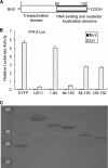
The C-termimal dsRNA binding domain of US11 is responsible for inhibiting SeV-mediated IFN-β promoter activity. (A) Schematic representation of US11 functional domains and deletions. (B) Luciferase assays in HEK 293T cells were performed as for Fig. 1A to measure the activation of the IFN-β promoter following SeV infection in the presence of full-length and deletion mutants of US11, including US11-EYFP, US11(1--83)-EYFP, US11(84-152)-EYFP, US11(84-125)-EYFP, and US11(126-152)-EYFP. Data are expressed as relative luciferase activities with standard deviations for three independent experiments performed in duplicate. (C) The expression of these US11 deletion mutants was verified by Western blotting using rabbit anti-YFP polyclonal antibody.
Fig 4
Fig 4
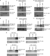
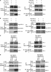
US11 interacts with both RIG-I and MDA-5 in transfected and HSV-1-infected cells. (A, B, C, and D) US11 associates with both RIG-I and MDA-5 in transfected cells. HEK293T cells (∼5 × 10⁶) were cotransfected with 10 μg of plasmid US11-HA and with 10 μg of plasmid pEF-Flag-RIG-I, encoding full-length RIG-I (A and C), or with plasmid pEF-Flag-MDA-5, encoding full-length MDA-5 (B and D), respectively. At 36 h after transfection, cells were lysed and clarified supernatants were left untreated (A and B) or treated with RNase A at 150 μg/ml (C and D). The samples were then subjected to immunoprecipitation assays using anti-HA MAb (IP: HA) or nonspecific mouse monoclonal antibody (IgG2b). Cell lysates and immunoprecipitated proteins were separated in denaturing 12% polyacrylamide gels and transferred to nitrocellulose membranes. The transferred proteins were probed with anti-HA and anti-Flag MAbs. (E and F) US11 interacts with overexpressed RIG-I and MDA-5 in HSV-1-infected cells. HEK293T cells were transfected with pEF-Flag-RIG-I (E) or pEF-Flag-MDA-5 (F), respectively. At 20 h after transfection, cells were infected with HSV-1 strain F at an MOI of 10 for 16 h. The cells were then lysed, and the extracts were subjected to immunoprecipitation using anti-Flag MAb (IP: Flag) or nonspecific mouse monoclonal antibody (IgG2b). Precipitates were analyzed by Western blotting using anti-Flag MAb or rabbit anti-US11 pAb. (G and H) US11 interacts with endogenous RIG-I and MDA-5 in HSV-1-infected cells. HEK293T cells were infected with WT or US11 mutant HSV-1 at an MOI of 10 for 16 h. The cells were then lysed, and the extracts were subjected to immunoprecipitation using anti-RIG-I pAb (IP: RIG-I), anti-MDA-5 pAb (IP: MDA-5), or control IgG. Precipitates were analyzed by Western blotting.
Fig 5
Fig 5
The carboxyl terminus of US11 interacts with the carboxyl termini of RIG-I and MDA-5. (A and B) HEK293T cells were cotransfected with plasmid pEF-Flag-RIG-IC (A) containing the carboxyl-terminal domain (aa 218 to 925) or pEF-Flag-RIG-IN (B) containing the amino-terminal domain (aa 1 to 229) and with plasmid US11-HA. (C and D) HEK293T cells were cotransfected with plasmid pEF-Flag-MDA-5H (C) containing the carboxyl-terminal helicase domain (aa 287 to 1025) or pEF-Flag-MDA-5C (D) containing the amino-terminal CARD domain (aa 1 to 287) and with plasmid US11-HA. Immunoprecipitation and Western blot analysis were performed as described for Fig. 5A. (E and F) HEK293T cells were cotransfected with plasmid pEF-Flag-RIG-IC (E) or pEF-Flag-MDA-5H (F) and with plasmid US11(84-152)-EYFP or pEYFP-N1, respectively. At 24 h after transfection, immunoprecipitation with anti-Flag MAb or nonspecific mouse monoclonal antibody (IgG2b) and Western blot analysis were performed as described for Fig. 5A.
Fig 6
Fig 6
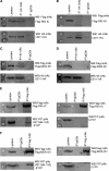
US11 blocks the formation of complex between RIG-I and MAVS or between MDA-5 and MAVS. (A and B) HEK293T cells (∼5 × 10⁶) were cotransfected with 10 μg of plasmid pEF-Flag-RIG-I (A) or pEF-Flag-MDA-5 (B) or 10 μg of plasmid pMyc-MAVS and 10 μg of plasmid US11-HA or transfected with empty vector. At 36 h posttransfection, cells were lysed and the clarified supernatants were subjected to immunoprecipitation assays using anti-Myc MAb (IP: Myc) or nonspecific mouse monoclonal antibody (IgG1). US11, MAVS, or RIG-I or MDA-5 was detected by Western blotting using anti-HA, anti-Myc, or anti-Flag MAbs, respectively. (C and D) HEK293T cells were cotransfected with plasmids pMyc-MAVS and pEF-Flag-RIG-I (C) or pEF-Flag-MDA-5 (D), respectively. Twenty hours after transfection, cells were infected with either WT or US11 mutant HSV-1 at an MOI of 10 for another 16 h. The cells were subsequently lysed and subjected to immunoprecipitation assays as described for panel A. Additionally, rabbit anti-US11 pAb was used for detection of US11 expression after either WT or US11 mutant HSV-1 infection.
Fig 7
Fig 7
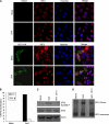
US11 impedes the activation of IRF3 downstream of the RLR signaling pathway. (A) US11 blocks IRF3 nuclear translocation induced by SeV infection. HeLa cells were transfected with the US11-HA expression plasmid or pCMV-HA vector. Twenty-four hours posttransfection, cells were then mock infected or infected with 100 HAU ml−1 SeV for 8 h. Cells were stained with mouse anti-HA MAb and rabbit anti-IRF3 antibody. FITC-conjugated goat anti-mouse (green) and TRITC-conjugated goat anti-rabbit (red) were used as the secondary antibody. Cell nuclei (blue) were stained with Hoechst 33258. The images were obtained by fluorescence microscopy using a ×40 objective. (B) Cells expressing US11-HA or vector from panel A were scored for nuclear translocation of IRF3. At least 100 cells were counted for each sample. Data shown are from one representative experiment of at least three. (C and D) US11 inhibits SeV-induced IRF3 phosphorylation and dimerization. HEK293T cells were transfected with the US11-HA expression plasmid. Twenty-four hours posttransfection, cells were then mock infected or infected with 100 HAU ml−1 SeV for 8 h. Protein extracts were subjected to SDS-PAGE (C) or native PAGE (D) for subsequent analysis with anti-phospho-IRF3 (Ser396) (C) or anti-IRF3 (D) antibody. IRF3 and actin as a loading control were also detected.

Similar articles

Cited by

References

    1. Alexopoulou L, Holt AC, Medzhitov R, Flavell RA. 2001. Recognition of double-stranded RNA and activation of NF-kappaB by Toll-like receptor 3. Nature 413:732–738 - PubMed
    1. Andrejeva J, et al. 2004. The V proteins of paramyxoviruses bind the IFN-inducible RNA helicase, mda-5, and inhibit its activation of the IFN-beta promoter. Proc. Natl. Acad. Sci. U. S. A. 101:17264–17269 - PMC - PubMed
    1. Barral PM, et al. 2007. MDA-5 is cleaved in poliovirus-infected cells. J. Virol. 81:3677–3684 - PMC - PubMed
    1. Barral PM, Sarkar D, Fisher PB, Racaniello VR. 2009. RIG-I is cleaved during picornavirus infection. Virology 391:171–176 - PMC - PubMed
    1. Basler CF, et al. 2003. The Ebola virus VP35 protein inhibits activation of interferon regulatory factor 3. J. Virol. 77:7945–7956 - PMC - PubMed

Publication types

MeSH terms