. 2012 Apr;86(8):4708-14.
doi: 10.1128/JVI.05887-11. Epub 2012 Feb 1.

Human immunodeficiency virus type 1 capsid mutation N74D alters cyclophilin A dependence and impairs macrophage infection

Affiliations

Human immunodeficiency virus type 1 capsid mutation N74D alters cyclophilin A dependence and impairs macrophage infection

Zandrea Ambrose et al. J Virol. 2012 Apr.

Abstract

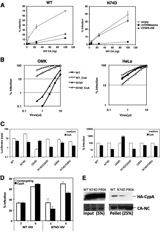
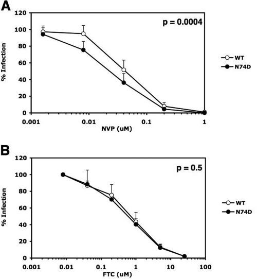
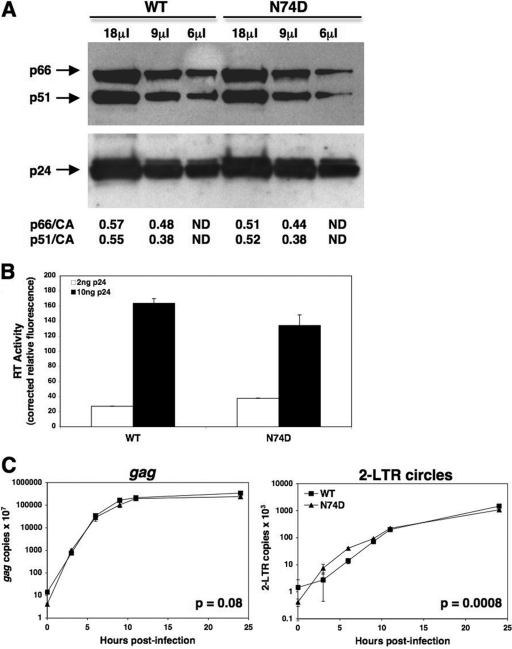
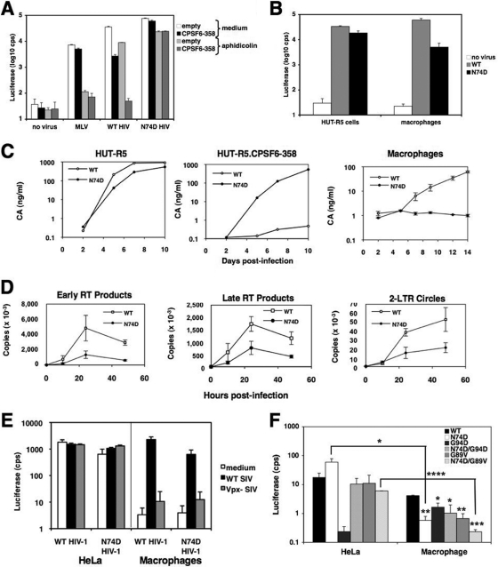
The antiviral factor CPSF6-358 interferes with the nuclear entry of human immunodeficiency virus type 1 (HIV-1). HIV-1 acquires resistance to CPSF6-358 through the N74D mutation of the capsid (CA), which alters its nuclear entry pathway. Here we show that compared to wild-type (WT) HIV-1, N74D HIV-1 is more sensitive to cyclosporine, has increased sensitivity to nevirapine, and is impaired in macrophage infection prior to reverse transcription. These phenotypes suggest a difference in the N74D reverse transcription complex that manifests early after infection and prior to interaction with the nuclear pore. Overall, our data indicate that N74D HIV-1 replication in transformed cells requires cyclophilin A but is dependent on other interactions in macrophages.


Figures

Fig 1
Fig 1
Sensitivity of N74D HIV-1 to TRIMs and CsA. (A) HeLa cells expressing empty vector, CPSF6-358, or rhTRIM5α were infected in duplicate with various amounts of WT HIV-1 or the N74D mutant (as measured by p24 enzyme-linked immunosorbent assay [ELISA]) encoding the HSA gene in place of nef and pseudotyped with vesicular stomatitis virus G (VSV-G) and assayed for HSA expression 48 h later. (B) Owl monkey kidney (OMK) or HeLa cells incubated with or without 2.5 μM CsA were infected with different volumes of WT or N74D HIV-1 encoding red fluorescent protein (RFP) and pseudotyped with VSV-G and assayed by a fluorescence-activated cell sorter (FACS) for RFP-positive cells after 48 h. (C) HeLa cells were infected with WT HIV-1 or N74D, G94D, N74D/G94D, G89V, and N74D/G89V mutants encoding the luciferase gene and pseudotyped with VSV-G (HIV-luc/VSV-G) in the presence or absence of 2.5 μM CsA and assayed for luciferase expression 48 h later. Infectivity data shown are absolute luciferase values (left) and normalized values for infectivity in the absence of CsA (right) cps, counts per second. (D) Endogenous CypA expression in HeLa cells was knocked down by small interfering RNA (siRNA) prior to infection with the WT or N74D HIV-1 encoding RFP and pseudotyped with VSV-G. Control cells were treated with nontargeting siRNA. Cells were infected with increasing microliters of virus, and infection was measured 48 h later by flow cytometry. A Western blot of cell lysates harvested on the day of infection was performed to verify protein knockdown. (E) WT, N74D, and P90A CA-NC complexes were incubated with 293T cell extracts expressing CypA tagged with hemagglutinin (HA). Reaction mixtures were isolated after centrifugation through 50% sucrose and probed by a Western blot with anti-HA antibodies (top panels). Coomassie blue staining of total protein is shown in the bottom panels. The results are representative of two independent experiments. Error bars in panels A and C represent the standard deviations of duplicate wells of an experiment.
Fig 2
Fig 2
N74D HIV-1 is more susceptible than WT HIV-1 to the NNRTI NVP. Infections of TZM-bl cells were performed in duplicate with equal multiplicities of infection (MOIs), as determined in Ghost cells of WT and N74D HIV-luc/VSV-G in the presence or absence of NVP (A) or FTC (B). Results are the percentages of infection at each drug concentration relative to infection in the absence of the drug. Error bars represent the standard deviations from the averages of 4 and 2 independent experiments for panels A and B, respectively. Statistical P values were determined using nonlinear regression analysis to compare the curves.
Fig 3
Fig 3
WT and N74D HIV-luc/VSV-G package similar amounts of RT per particle and carry out similar levels of reverse transcription in vivo. (A) A Western blot was performed on WT and N74D HIV-luc/VSV-G lysates using anti-RT antibodies (a gift from N. Sluis-Cremer) to visualize p66 and p51 (upper). The blot was stripped and reprobed with anti-CA antibodies (a gift from D. Ott) to visualize p24 (lower). The p66/CA and p51/CA ratios were quantified using ImageJ software. ND, not determined. (B) RT activity was measured from WT and N74D HIV-luc/VSV-G lysates, using the EnzCheck reverse transcriptase assay (Invitrogen), and read on a fluorescent plate reader. Equal p24 levels of each virus, as measured by ELISA, were pelleted, resuspended in culture medium containing 1% Triton-X, and used in the assay. Purified WT RT protein (a gift from N. Sluis-Cremer) was used as a standard control. (C) Accumulation of gag DNA (late RT products) and 2-LTR circles was measured in TZM-bl cells infected with equal MOIs of WT and N74D HIV-luc/VSV-G. Infections were synchronized by adding virus at 4°C for 30 min and then shifting the cells to 37°C for up to 24 h. Cells were trypsinized at different times, and total DNA was isolated. qPCR was performed as previously described (15). Error bars represent standard deviations of duplicate wells of an experiment. Statistical P values were determined using nonlinear regression analysis to compare the curves.
Fig 4
Fig 4
Infection of macrophages by the N74D mutant is inhibited prior to or at reverse transcription in a Vpx-independent manner. (A) HeLa and HeLa-CPSF6-358 cells were incubated with culture medium or with murine leukemia virus (MLV)-luc/VSV-G, WT HIV-luc/VSV-G, or N74D HIV-luc/VSV-G in the presence or absence of 2 μg/ml aphidicolin. Luciferase expression was measured 48 h later. (B) HUT-R5 cells and primary human macrophages were infected in duplicate with WT or N74D HIV-luc/VSV-G and analyzed for luciferase activity after 48 h. Representative of 4 independent experiments, using 3 donors for macrophages. (C) HUT-R5 cells, HUT-R5-CPSF6-358 cells, and macrophages were infected with WT HIV-1NL4-3/BaL (a gift from N. Landau) or N74D HIV-1NL4-3/BaL. CA was measured in the culture supernatants by p24 ELISA. Macrophages were infected in duplicate. Representative of 2 independent experiments, using 2 donors for macrophages. (D) Macrophages were infected in duplicate with WT or N74D HIV-luc/VSV-G and lysed at different time points. Early (RU5) and late (gag) RT products, as well as 2-LTR circles, were measured by qPCR. Viral DNA copies are expressed per μg of total DNA obtained from the cells. Representative of 3 independent experiments, using 2 donors. (E) HeLa cells and macrophages were incubated with culture medium or equal MOIs of WT or Vpx-negative SIV-enhanced green fluorescent protein (EGFP)/VSV-G, as determined in HeLa cells, for 4 h. Then the cells were infected with equal MOIs of WT or N74D HIV-luc/VSV-G, as determined on Ghost cells. Luciferase activity was measured 48 h postinfection. Error bars represent standard deviations of duplicates. Representative of 3 independent experiments. (F) HeLa cells or macrophages were infected with the same amounts of WT HIV-luc/VSV-G or the N74D, G94D, N74D/G94D, G89V, or N74D/G89V mutant and analyzed for luciferase activity after 48 h. Representative of 4 independent experiments. Statistical significance values of panel F were calculated by using Student's two-sided t test. Asterisks directly above bars indicate differences between WT and mutant infections in a given cell type, whereas asterisks above lines indicate differences in infectivity for the same virus between cell types. ****, P < 0.0001; ***, P < 0.001; **, P < 0.01; *, P < 0.05. Error bars in all panels represent standard deviations of duplicate wells of an experiment.

Similar articles

Cited by

References

    1. Aberham C, Weber S, Phares W. 1996. Spontaneous mutations in the human immunodeficiency virus type 1 gag gene that affect viral replication in the presence of cyclosporins. J. Virol. 70:3536–3544 - PMC - PubMed
    1. Ambrose Z, Julias JG, Boyer PL, Kewalramani VN, Hughes SH. 2006. The level of reverse transcriptase (RT) in human immunodeficiency virus type 1 particles affects susceptibility to nonnucleoside RT inhibitors but not to lamivudine. J. Virol. 80:2578–2581 - PMC - PubMed
    1. Boone LR, Innes CL, Glover PL, Linney E. 1989. Development and characterization of an Fv-1-sensitive retrovirus-packaging system: single-hit titration kinetics observed in restrictive cells. J. Virol. 63:2592–2597 - PMC - PubMed
    1. Bowerman B, Brown PO, Bishop JM, Varmus HE. 1989. A nucleoprotein complex mediates the integration of retroviral DNA. Genes Dev. 3:469–478 - PubMed
    1. Brass AL, et al. 2008. Identification of host proteins required for HIV infection through a functional genomic screen. Science 319:921–926 - PubMed

Publication types

MeSH terms

LinkOut - more resources