. 2012 Mar 1;188(5):2254-65.
doi: 10.4049/jimmunol.1103049. Epub 2012 Feb 1.

Structural and functional characterization of a novel nonglycosidic type I NKT agonist with immunomodulatory properties

Affiliations

Structural and functional characterization of a novel nonglycosidic type I NKT agonist with immunomodulatory properties

Jerome Kerzerho et al. J Immunol. .

Erratum in

  • J Immunol. 2012 Oct 15;189(8):4194. Alari-Pahisa, Elisenda [corrected to Alari-Pahissa, Elisenda]

Abstract

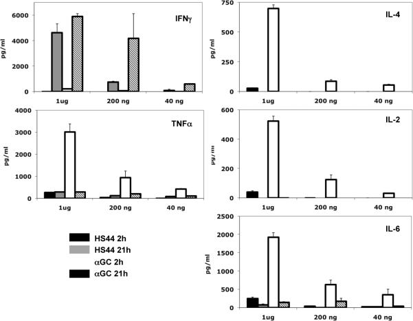
Activation of type I NKT (iNKT) cells by CD1d-presented agonists is a potent immunotherapeutic tool. α-Galactosylceramide (α-GalCer) is the prototypic agonist, but its excessive potency with simultaneous production of both pro- and anti-inflammatory cytokines hampers its potential therapeutic use. In search for novel agonists, we have analyzed the structure and function of HS44, a synthetic aminocyclitolic ceramide analog designed to avoid unrestrained iNKT cell activation. HS44 is a weaker agonist compared with α-GalCer in vitro, although in vivo it induces robust IFN-γ production, and highly reduced but still functional Th2 response. The characteristic cytokine storm produced upon α-GalCer activation was not induced. Consequently, HS44 induced a very efficient iNKT cell-dependent antitumoral response in B16 animal model. In addition, intranasal administration showed the capacity to induce lung inflammation and airway hyperreactivity, a cardinal asthma feature. Thus, HS44 is able to elicit functional Th1 or Th2 responses. Structural studies show that HS44 binds to CD1d with the same conformation as α-GalCer. The TCR binds to HS44 similarly as α-GalCer, but forms less contacts, thus explaining its weaker TCR affinity and, consequently, its weaker recognition by iNKT cells. The ability of this compound to activate an efficient, but not massive, tailored functional immune response makes it an attractive reagent for immune manipulation.


Figures

FIGURE 1
FIGURE 1. Expansion of iNKT cells upon treatment with HS44
Spleen cells from B6 mice were cultured in the presence or absence of 1μg/ml of HS44 or OCH or 100ng/ml of α-GalCer for 4 days. Flow cytometry analysis of splenocytes labeled with CD1d-PBS57-PE tetramer and anti-TCR-FITC. iNKT percentages (double positive cells) were calculated among electronically gated lymphocyte population. A representative of 2 different experiments is shown.
FIGURE 2
FIGURE 2. Expression of cytokines by HS44 treated iNKT cells
A) Spleen cells from B6 mice were cultured in the presence or absence of increasing concentrations α-GalCer, HS44 or OCH. At day 4, supernatants were collected and IFN-γ and IL-4 were measured by ELISA. Data are the mean ± s.e.m. of 4 well cultures for α-GalCer and OCH and 8 well cultures for HS44, from 3 different mice. A representative of 2 separate experiments is shown. B) iNKT cells were negatively enriched from the spleens of BALB/c mice. Enriched iNKT cells were cultured in the presence or absence of increasing concentration of α-GalCer, HS44 or OCH. 24 hours and 48 hours later, the expression of IL-4, IFN-γ, IL-2, IL-10, IL-5 and IL-17A cytokines were assessed by real-time PCR. Data are the mean ± s.e.m., representative of three separate experiments.
FIGURE 3
FIGURE 3. Serum cytokine production after HS44 administration
Indicated amounts of HS44 or α-GalCer were i.p. administered to B6 mice. 2h and 21h later, blood samples were collected and cytokine levels were determined by CBA assay. Data indicate the mean of three different mice. A representative of three different experiments is shown.
FIGURE 4
FIGURE 4. HS44 antitumoral activity controls establishment of melanoma metastases
C57BL/6 mice were intravenously treated with indicated amounts of either HS44 or α-GalCer or with vehicle 3 days before i.v. challenge with 5×10⁵ B16 melanoma cells. 2 weeks later, the lungs were extracted for metastases quantification. A representative experiment out of two, with five mice per condition is shown. Horizontal continous lines show statistical differences between relevant treatments, with * p<0,05, ** p< 0,01. Discontinous lines indicate relevant treatments without statistically significant differences. Photographic images from a representative mice and treatment are shown
FIGURE 5
FIGURE 5. Intranasal administration of HS44 induces airway inflammation and airway hyperreactivity (AHR)
(A, B) A group of BALB / c mice (n=4) were treated i.n. with 1 μg of HS44 or α-GalCer (Positive control) or PBS (Negative control) on day 0. 24 hours later, the mice were assessed for the development of airway hyperreactivity by measuring PenH (A) or lung resistance (RL) and dynamic compliance (Cdyn) (B). Data are the mean ± s.e.m., representative of three separate experiments with * P < 0.05 and ** P < 0.005. (C) Bronchoalveolar lavage (BAL) fluid from the mice in panel A was analyzed 24h after AHR measurement. Results show the total number of cells in BAL fluid. Total, total cell number; Mac, monocyte / macrophage; Eos, eosinophils; Lym, lymphocytes; PMN, neutrophils (* P < 0.05, ** P< 0.01). (D) Lung histology. Lung tissue of mice from panel A were stained with hematoxylin and eosin (H&E) (upper panel) or with periodic acid Schiff (PAS) (lower panel) and analyzed for the presence of inflammation and mucus respectively.
FIGURE 6
FIGURE 6. TCR binding kinetics and HS44 ternary complex crystal structure and, stereo view of HS44 electron density and HS44 binding to mCD1d
(A) A representative sensorgram of the TCR binding to mCD1d-HS44 complex is shown (from three independent experiments). Each colored curve depicts a different concentration of injected TCR. (B) Crystal structure of the mCD1d-HS44-TCR ternary complex is shown. HS44 shown as yellow sticks, between mCD1d (gray) and TCR (α chain in cyan; TCR β chain in orange). Chemical structures of HS44 and α-GalCer for comparison are depicted with the galactose and aminocyclitol head groups highlighted in red. (C) Representation of the final 2Fo-Fc map drawn around glycolipid HS44 from the ternary mCD1d-HS44-TCR complex. The 2FoFc electron density map is contoured for HS44 at 1 σ and drawn as a blue mesh in a side view. Hydrophobic mCD1d residues interacting with the lipid backbone and charged residues contacting the polar moieties of the glycolipid aminocyclitol are depicted. (D) HS44 and α-GalCer are shown superimposed in the mCD1d binding groove. The aminocyclitol head of HS44 (yellow) and galactose head of α-GalCer (green) are exposed similarly for TCR recognition. Binding to CD1d is very similar for both ligands. H-bonds between mCD1d and HS44 are highlighted in blue dash lines and involve the CD1d residues D80, D153 and T156. Left panel represents a top view onto the CD1d binding groove with the semi-transparent molecular surface of CD1d shown to visualize the ligand binding inside the groove. α-GalCer is shown in green; HS44 is shown in yellow; mCD1d heavy chain and β2m are shown in gray. Oxygen depicted in red and nitrogen in blue.
FIGURE 7
FIGURE 7. Comparison of TCR binding to α-GalCer and HS44
Interaction of the TCR α-chain (cyan) with α-GalCer from PDB ID 3H6 (green) and HS44 (yellow). There are 4 H-bonds between α-GalCer and TCR compared to only 3 direct H-bonds (dashed blue lines) between HS44 and TCR, while HS44 forms also one water-mediated H bond with the TCR (N30). Lower panels depict the conserved contacts between TCR α and ß chains and CD1 residues, mainly through CDR3α and CDR2ß, as well as one salt bridge involving E96 of CDR3ß and K148 of CD1d.

Similar articles

Cited by

References

    1. Bendelac A, Savage PB, Teyton L. The biology of NKT cells. Annu Rev Immunol. 2007;25:297. - PubMed
    1. Zeng Z, Castano AR, Segelke BW, Stura EA, Peterson PA, Wilson IA. Crystal structure of mouse CD1: An MHC-like fold with a large hydrophobic binding groove. Science. 1997;277:339. - PubMed
    1. Zajonc DM, Kronenberg M. Carbohydrate specificity of the recognition of diverse glycolipids by natural killer T cells. Immunol Rev. 2009;230:188. - PubMed
    1. Zhou D, Mattner J, Cantu C, 3rd, Schrantz N, Yin N, Gao Y, Sagiv Y, Hudspeth K, Wu YP, Yamashita T, Teneberg S, Wang D, Proia RL, Levery SB, Savage PB, Teyton L, Bendelac A. Lysosomal glycosphingolipid recognition by NKT cells. Science. 2004;306:1786. - PubMed
    1. Gapin L. iNKT cell autoreactivity: what is `self' and how is it recognized? Nat Rev Immunol. 2010;10:272. - PMC - PubMed

Publication types

MeSH terms

Associated data