. 2012 Mar 1;188(5):2371-9.
doi: 10.4049/jimmunol.1100136. Epub 2012 Feb 1.

The MC159 protein from the molluscum contagiosum poxvirus inhibits NF-κB activation by interacting with the IκB kinase complex

Affiliations

The MC159 protein from the molluscum contagiosum poxvirus inhibits NF-κB activation by interacting with the IκB kinase complex

Crystal M H Randall et al. J Immunol. .

Abstract

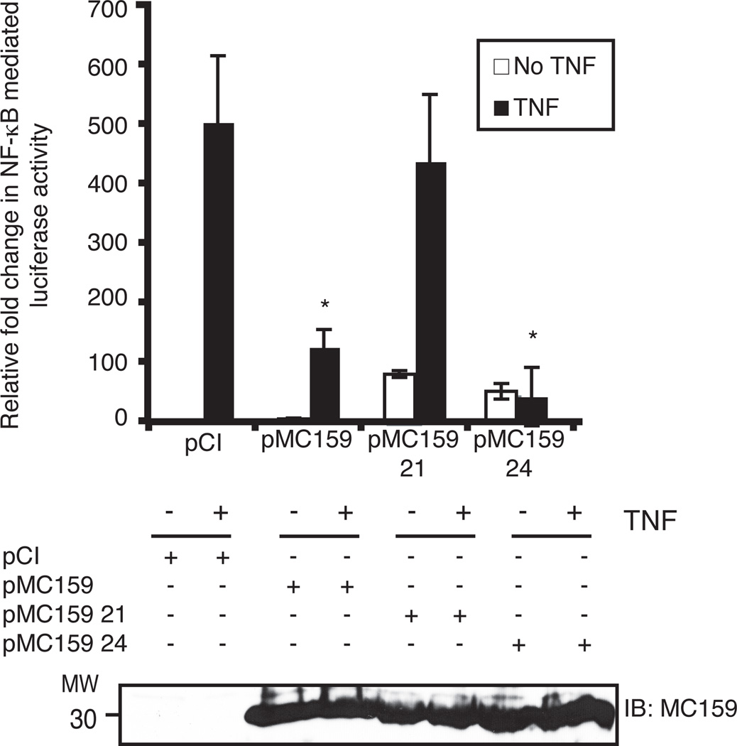
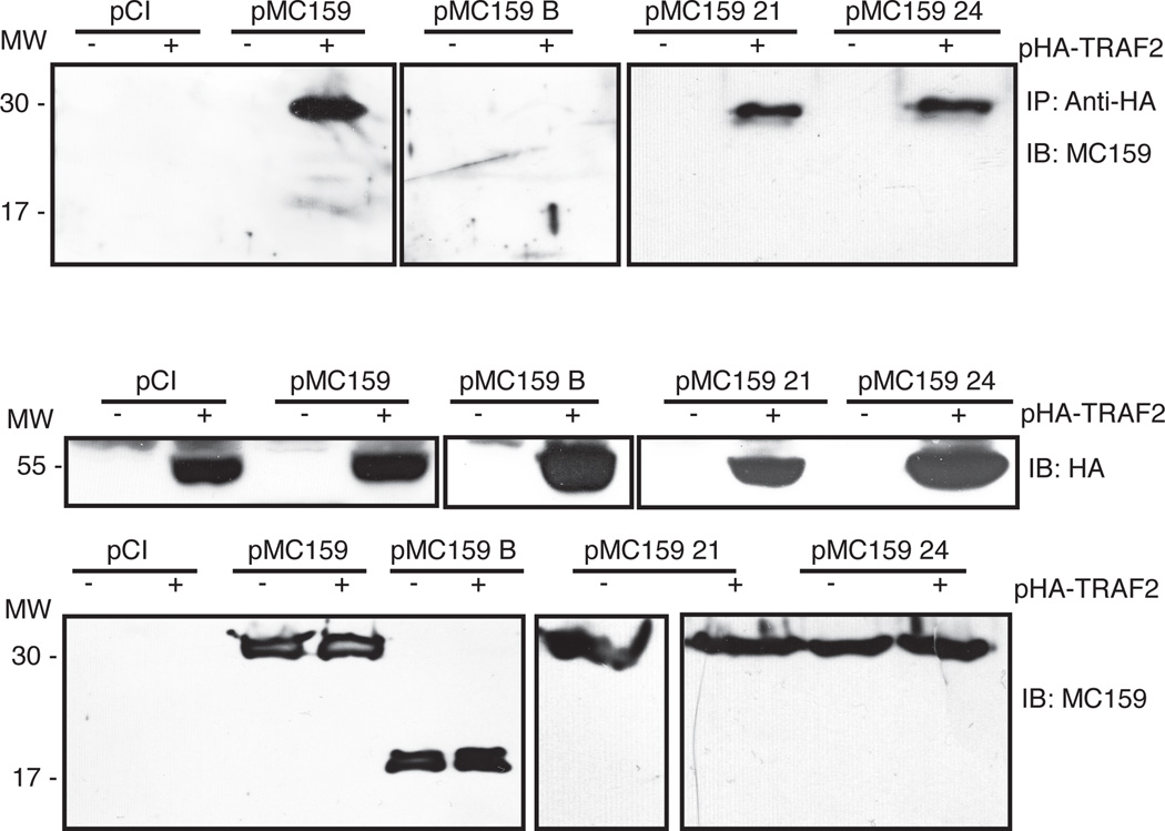
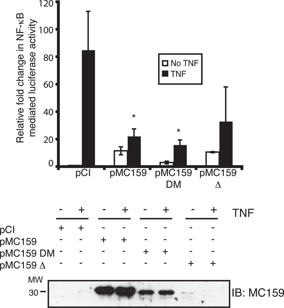
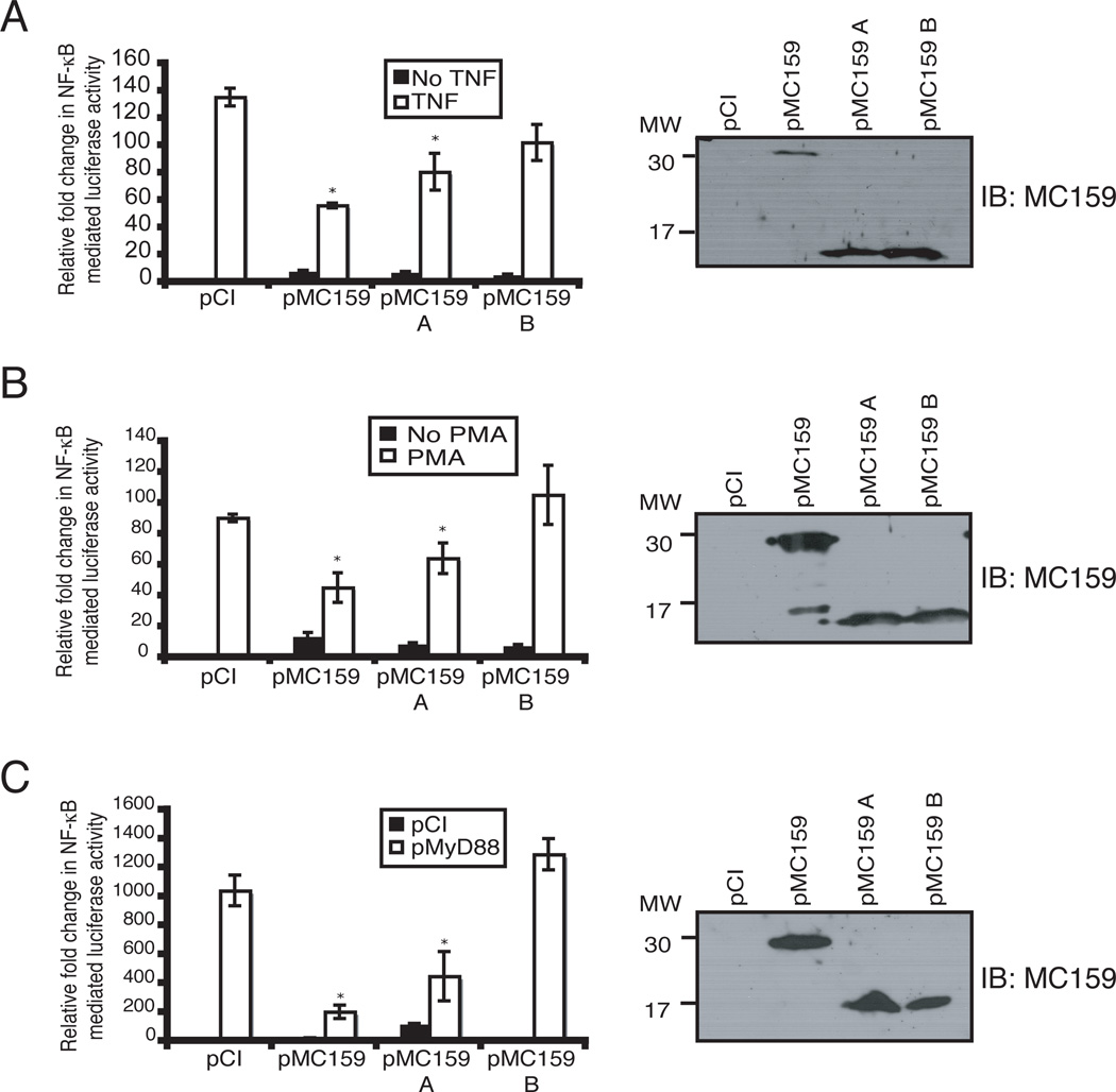
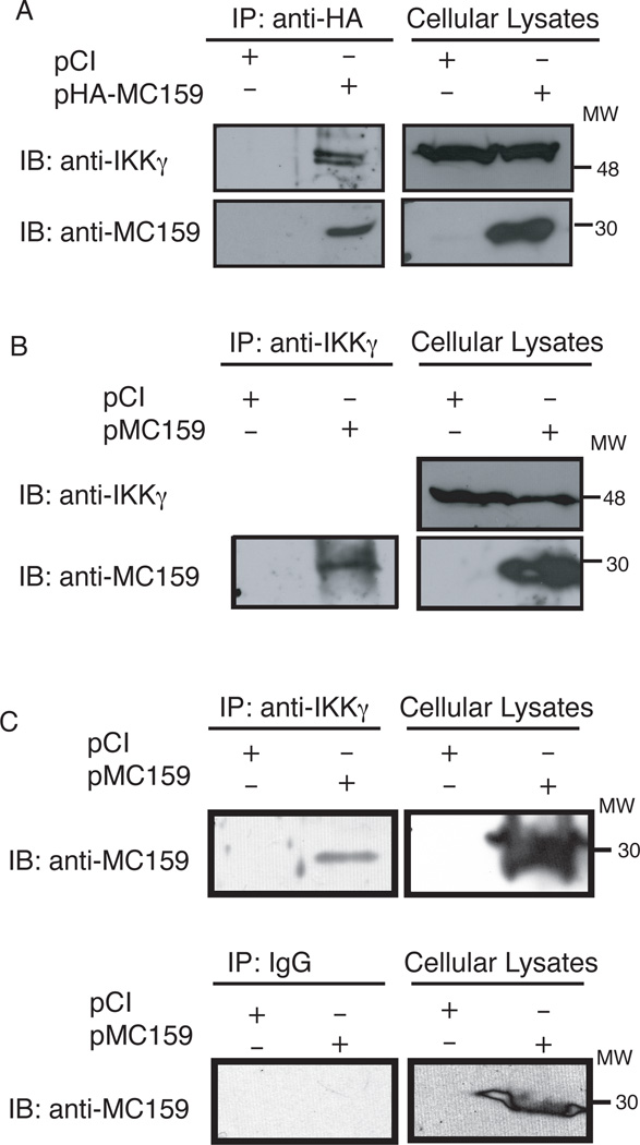
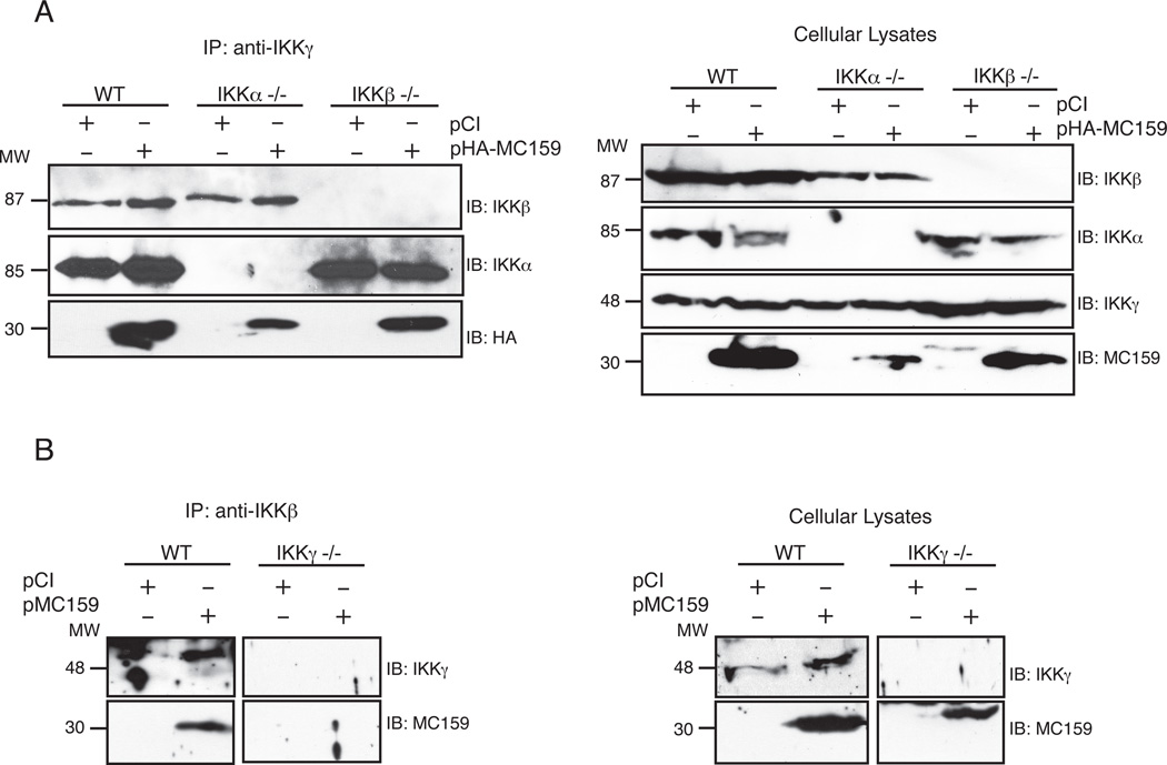
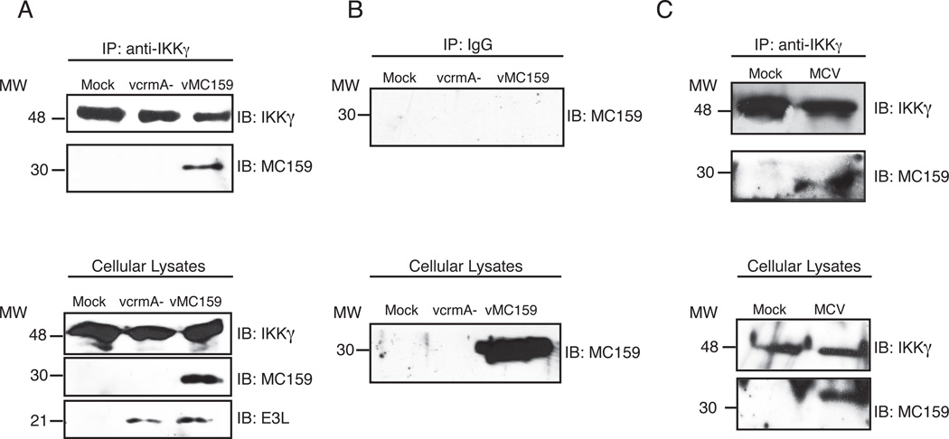
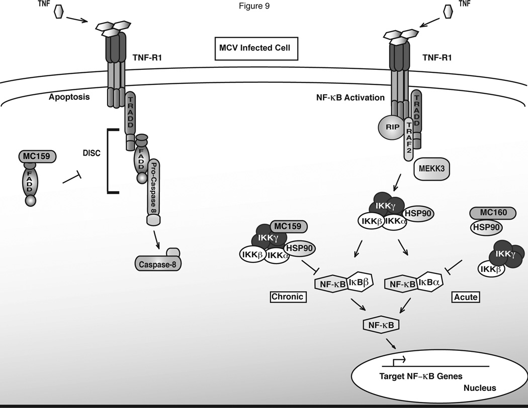
Molluscum contagiosum virus (MCV) causes persistent neoplasms in healthy and immunocompromised people. Its ability to persist likely is due to its arsenal of viral immunoevasion proteins. For example, the MCV MC159 protein inhibits TNF-R1-induced NF-κB activation and apoptosis. The MC159 protein is a viral FLIP and, as such, possesses two tandem death effector domains (DEDs). We show in this article that, in human embryonic kidney 293 T cells, the expression of wild-type MC159 or a mutant MC159 protein containing the first DED (MC159 A) inhibited TNF-induced NF-κB, or NF-κB activated by PMA or MyD88 overexpression, whereas a mutant protein lacking the first DED (MC159 B) did not. We hypothesized that the MC159 protein targeted the IκB kinase (IKK) complex to inhibit these diverse signaling events. Indeed, the MC159 protein, but not MC159 B, coimmunoprecipitated with IKKγ. MC159 coimmunoprecipitated with IKKγ when using mouse embryonic fibroblasts that lack either IKKα or IKKβ, suggesting that the MC159 protein interacted directly with IKKγ. MC159-IKKγ coimmunoprecipitations were detected during infection of cells with either MCV isolated from human lesions or with a recombinant MC159-expressing vaccinia virus. MC159 also interacts with TRAF2, a signaling molecule involved in NF-κB activation. However, mutational analysis of MC159 failed to reveal a correlation between MC159-TRAF2 interactions and MC159's inhibitory function. We propose that MC159-IKK interactions, but not MC159-TRAF2 interactions, are responsible for inhibiting NF-κB activation.


Figures

Figure 1
Figure 1. The MC159 RXDL motif confers resistance to TNF-induced NF-κB activation
Subconfluent 293T cellular monolayers were transfected with pNF-κBluc, pRL-null, and either pCI, pMC159, pMC159 21 or pMC159 24. At 24 h later, cells were incubated with regular medium or medium containing TNF (10 ng/ml). After an additional 12 h incubation, cells were lysed and luciferase activities were measured. Statistically significant data for MC159 inhibition of NF-κB-mediated luciferase activity (p<0.05) are indicated with an asterisk. Lysates also were analyzed by immunoblotting. Proteins were subjected to SDS-12% PAGE, transferred to PVDF membranes and incubated with an anti-MC159 antiserum.
Figure 2
Figure 2. TRAF2-MC159 interactions are insufficient to inhibit NF-κB
One well of subconfluent 293T cellular monolayers in a 6-well plate were transfected with pHA-TRAF2 and either pCI, pMC159, pMC159 B, pMC159 21 or pMC159 24. At 24 h post-transfection, cells were lysed in 500 µl DED lysis buffer. Immunoprecipitations (IP) were performed by incubating 450 µl of clarified cellular lysates with anti-HA antiserum and protein G-Sepharose beads. The remaining 50 µl of clarified lysates were used for detection of TRAF2 and MC159 proteins by immunoblotting. Immunoprecipitates (top) or 30 µg of corresponding clarified cellular lysates (middle and bottom panels) were subjected to SDS-12% PAGE. The separated proteins were transferred to PVDF membranes, and immunoblotted (IB) with either anti-HA or anti-MC159 antibodies.
Figure 3
Figure 3. The MC159 protein inhibits NF-κB activation independent of TRAF3
Subconfluent 293T cellular monolayers were transfected with pNF-κBluc, pRL-null, and either pCI, pMC159, pMC159 DM or pMC159 Δ. At 24 h later, transfected cells were incubated with regular medium or medium containing TNF (10 ng/ml) for 12 h, after which time cells were lysed and luciferase activities were measured. Statistically significant data sets for MC159 inhibition of NF-κB-mediated luciferase activity (p<0.05) are indicated with an asterisk. Lysates also were analyzed for MC159 proteins by immunoblotting. Following SDS-12% PAGE, proteins were transferred to PVDF membranes and incubated with an anti-MC159 antibody.
Figure 4
Figure 4. The MC159 DEDA inhibits TNF-, PMA- or MyD88- induced NF-κB activation
Subconfluent 293T cellular monolayers cells were transfected with pNF-κBluc, pRL-null, and either pCI, pMC159, pMC159 A or pMC159 B. Transfected cells were incubated with regular medium or medium containing (A) TNF (10 ng/ml) for 8 h or (B) or PMA (50 ng/ml) for 8 h. (C) Subconfluent 293T cell monolayers were transfected with pNF-κBluc, pRL-null, pMyD88 and either pCI, pMC159, pMC159 A or pMC159 B for 24 h. For all experiments, cells were lysed using passive lysis buffer and luciferase activities were measured. Statistically significant data sets for MC159 inhibition of NF-κB mediated luciferase activity (p<0.05) are indicated with an asterisk. Lysates were analyzed by immunoblotting. Following SDS-12% PAGE, proteins were transferred to PVDF membranes and incubated with an anti-MC159 antibody.
Figure 5
Figure 5. The MC159 protein co-immunoprecipitates with IKKγ
One well of subconfluent 293T cellular monolayers in a 6-well plate were transfected with pCI or pHA-MC159. At 24 h post-transfection, cells were lysed in 500 µl DED lysis buffer. Immunoprecipitations (IP) were performed by incubating 450 µl of clarified cellular lysates with protein G-Sepharose beads and either (A) anti-HA antibodies, (B) anti-IKKγ antibodies or (C) anti-IKKγ antibodies or IgG antibodies. The remaining 50 µl of clarified cellular lysates were used for detection of IKKγ and MC159 proteins by immunoblotting. Immunoprecipitates (top) or 30 µg of corresponding clarified cellular lysates (middle and bottom panels) were subjected to SDS-12% PAGE. Immunoprecipitated samples (left panels) or clarified cellular lysates (right panels) were analyzed by immunoblotting. Following SDS-12% PAGE, proteins were transferred to PVDF membranes, and incubated with either mouse anti-IKKγ or anti-MC159 antibodies.
Figure 6
Figure 6. IKKγ co-immunoprecipitates IKKα and IKKβ in the presence of MC159 protein
A 10-cm2 dish of subconfluent MEF cellular monolayers were transfected with pCI, pMC159 or pMC159 B. At 24 h post-transfection cells were lysed in 150 µl DED lysis buffer. 50 µl of clarified lysates were collected to new tubes, and 30 µg of this sample were analyzed for MC159 and IKK proteins by immunoblotting (bottom panel). Immunoprecipitations (IP) were performed using 100 µl clarified lysates incubated with protein G-Sepharose beads and anti-IKKγ. Immunoprecipitated samples (top panel) or clarified cellular lysates (bottom panel) were analyzed by immunoblotting. Following SDS-12% PAGE, proteins were transferred to PVDF membranes, and incubated with either mouse anti-IKKγ, anti-IKKα, anti-IKKβ or anti-MC159 antibodies.
Figure 7
Figure 7. The MC159 protein associates with IKKγ independent of IKKα and IKKβ
(A) A10-cm2 dish of subconfluent monolayers of WT, IKKα −/−, or IKKβ −/− MEF cells were transfected with pCI or pHA-MC159. At 24 h post-transfection, cells were lysed in 150 µl DED lysis buffer. 50 µl of clarified lysates were collected to new tubes, and 30 µg of this sample were analyzed for MC159 and IKK proteins by immunoblotting (bottom panel). Immunoprecipitations (IP) were performed using 100 µl clarified lysates incubated with protein G-Sepharose beads and anti-IKKγ. (B) A10-cm2 dish of subconfluent monolayers of WT or IKKγ −/− MEF cells were transfected with pCI or pHA-MC159. At 24 h post-transfection, cells were lysed in 150 µl DED lysis buffer. 50 µl of clarified lysates were collected to new tubes, and 30 µg of this sample were analyzed for MC159 and IKKγ proteins by immunoblotting (bottom panel). Immunoprecipitations (IP) were performed using 100 µl clarified lysates incubated with protein G-Sepharose beads and anti-IKKβ antiserum. For A and B, immunoprecipitated samples (left panels) or clarified cellular lysates (right panels) were analyzed by immunoblotting. Following SDS-12% PAGE, proteins were transferred to PVDF membranes; incubated with either mouse anti-IKKγ, anti-IKKα, anti-IKKβ, anti-HA or anti-MC159 antibodies.
Figure 8
Figure 8. The MC159 protein associates with IKKγ during poxvirus infection
(A and B) One well of a 6-well plate of 293T cellular monolayers were either mock infected or infected with vcrmA- or vMC159 (MOI = 10). (C) One well of a 6-well plate of MRC-5 cell monolayers were mock-infected or with 300 µl MCV lesion preparation. For all experiments, at 24 h post-infection cells were lysed in 500 µl DED lysis buffer. 50 µl of clarified lysates were used for immunoblotting to detect the presence of IKKγ, MC159 or E3 proteins. With the remaining 450 µl of clarified lysates, immunoprecipitations (IP) were performed on clarified lysates using protein G-Sepharose beads and (A, C) anti-IKKγ antiserum or (B) IgG. Immunoprecipitated samples (top panels) or clarified cellular lysates (bottom panels) were analyzed by immunoblot. Following SDS-12% PAGE, proteins were transferred to PVDF membranes, and incubated with rabbit anti-IKKγ, anti-MC159 or anti-E3 antibodies.
Figure 9
Figure 9. A model of MC159 and MC160 inhibition of TNF-R1 signaling during MCV infection
TNF binds to the TNF-R1 receptor to initiate either apoptosis or NF-κB activation. For apoptosis, TNF-TNF-R1 interactions initiate recruitment and activation of the death inducing signaling complex (DISC), composed of TRADD, FADD and procaspase-8. The MC159 protein binds to FADD and prevents DISC assembly to inhibit TNF-induced apoptosis during MCV infection. NF-κB activation is initiated by TNF binding TNF-R1, which recruits TRADD, TRAF2, RIP and MEKK, resulting in the IKK complex activation and the degradation of IκB proteins thus enabling NF-κB activation. We propose the following model for MC159- and MC160- mediated inhibition of TNF signaling during MCV infection: During the initial stage of an MCV infection, the MC160 protein inhibits acute NF-κB activation by binding to Hsp90 to cause IKKα degradation, subsequently preventing the degradation of IκBα (53). Later during MCV infection, the MC159 protein inhibits chronic NF-κB activation by binding to the IKKγ subunit of the IKK complex, thereby inhibiting IκBβ degradation.

Similar articles

Cited by

References

    1. Bugert JJ, Lohmuller C, Darai G. Characterization of early gene transcripts of molluscum contagiosum virus. Virology. 1999;257:119–129. - PubMed
    1. Senkevich TG, Bugert JJ, Sisler JR, Koonin EV, Darai G, Moss B. Genome sequence of a human tumorigenic poxvirus: prediction of specific host response-evasion genes. Science. 1996;273:813–816. - PubMed
    1. Senkevich TG, Koonin EV, Bugert JJ, Darai G, Moss B. The genome of molluscum contagiosum virus: analysis and comparison with other poxviruses. Virology. 1997;233:19–42. - PubMed
    1. Tyring SK. Molluscum contagiosum: the importance of early diagnosis and treatment. Am J Obstet Gynecol. 2003;189:S12–S16. - PubMed
    1. Reynolds MG, Holman RC, Yorita Christensen KL, Cheek JE, Damon IK. The Incidence of Molluscum contagiosum among American Indians and Alaska Natives. PLoS One. 2009;4:e5255. - PMC - PubMed

Publication types