. 2012 Mar 1;188(5):2127-35.
doi: 10.4049/jimmunol.1102412. Epub 2012 Feb 1.

Vitamin D inhibits monocyte/macrophage proinflammatory cytokine production by targeting MAPK phosphatase-1

Affiliations

Vitamin D inhibits monocyte/macrophage proinflammatory cytokine production by targeting MAPK phosphatase-1

Yong Zhang et al. J Immunol. .

Abstract

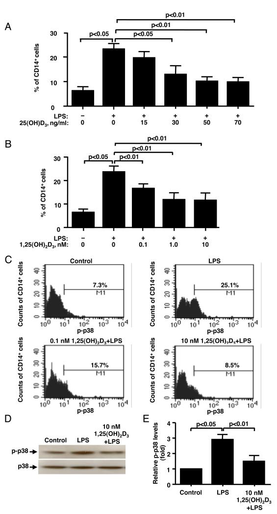
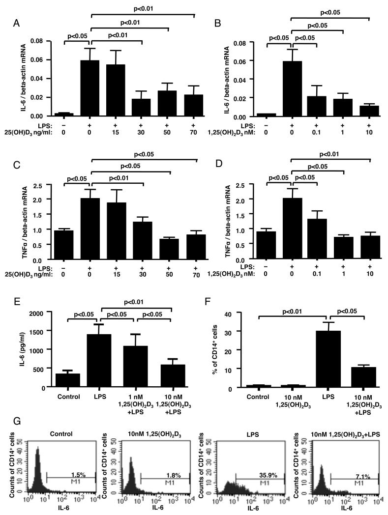
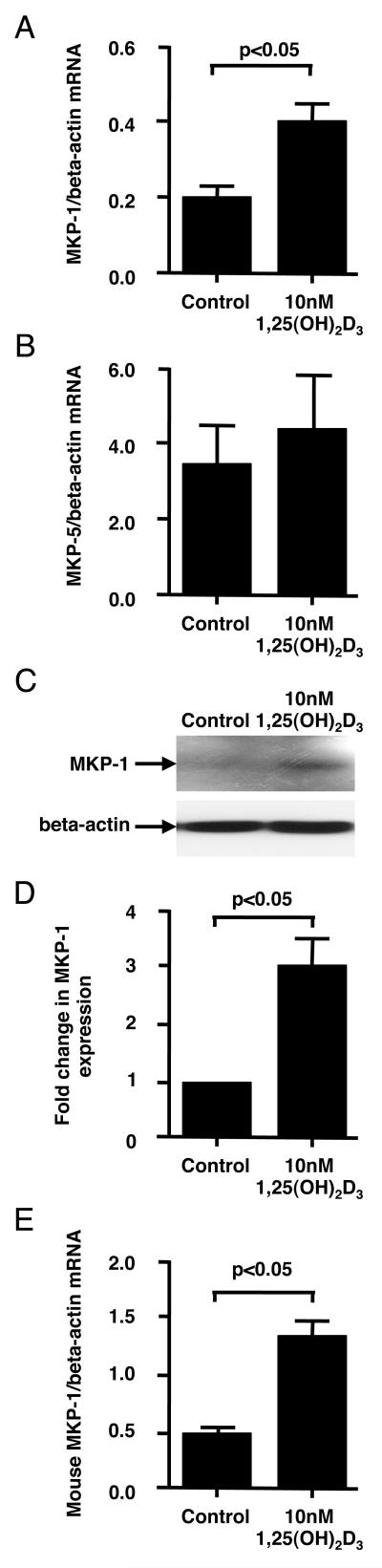
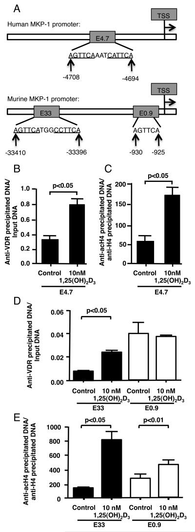
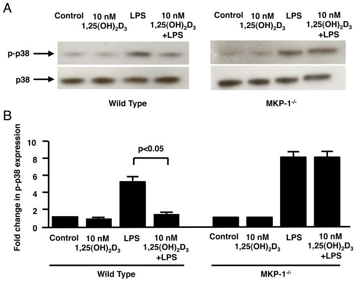
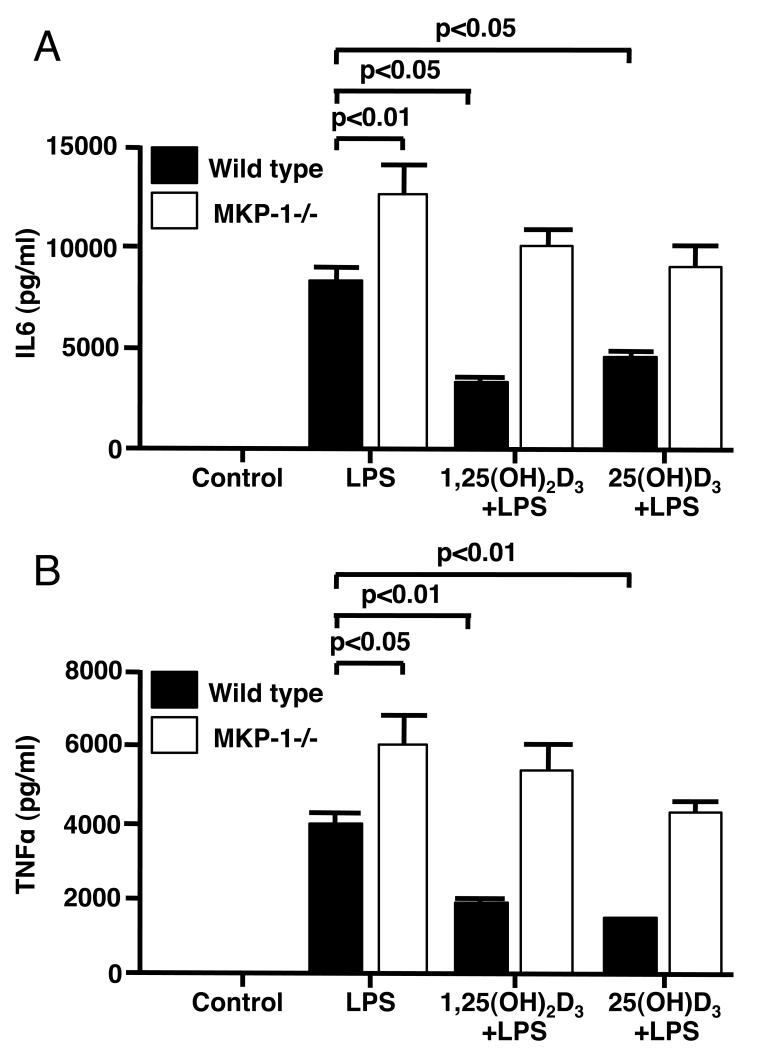
It is estimated that 1 billion people around the world are vitamin D deficient. Vitamin D deficiency has been linked to various inflammatory diseases. However, the mechanism by which vitamin D reduces inflammation remains poorly understood. In this study, we investigated the inhibitory effects of physiologic levels of vitamin D on LPS-stimulated inflammatory response in human blood monocytes and explored potential mechanisms of vitamin D action. We observed that two forms of the vitamin D, 1,25(OH)(2)D(3), and 25(OH)D(3), dose dependently inhibited LPS-induced p38 phosphorylation at physiologic concentrations, IL-6 and TNF-α production by human monocytes. Upon vitamin D treatment, the expression of MAPK phosphatase-1 (MKP-1) was significantly upregulated in human monocytes and murine bone marrow-derived macrophages (BMM). Increased binding of the vitamin D receptor and increased histone H4 acetylation at the identified vitamin D response element of the murine and human MKP-1 promoters were demonstrated. Moreover, in BMM from MKP1(-/-) mice, the inhibition of LPS-induced p38 phosphorylation by vitamin D was completely abolished. Vitamin D inhibition of LPS-induced IL-6 and TNF-α production by BMM from MKP-1(-/-) mice was significantly reduced as compared with wild-type mice. In conclusion, this study identified the upregulation of MKP-1 by vitamin D as a novel pathway by which vitamin D inhibits LPS-induced p38 activation and cytokine production in monocytes/macrophages.


Figures

Figure 1
Figure 1. Vitamin D inhibits LPS-induced p38 phosphorylation in human monocytes
PBMC were cultured in hormone-free medium containing 25(OH)D3 (A) or 1,25(OH)2D3, (B) for 24 h, followed by stimulation with 10 ng/ml LPS for 10 min. (A, B) As shown by flow cytometry, vitamin D pretreatment inhibits LPS-induced p38 phosphorylation in CD14+ cells (n=4). (C) Representative flow cytometry data on the effects of LPS and vitamin D/LPS on p38 activation in human monocytes is shown. (D) Pretreatment with vitamin D significantly inhibits p-p38 expression by the cells as shown by Western blot. Whole cell extracts from LPS or vitamin D/LPS treated adherent PBMC fraction were prepared, and blotted against phosphorylated p38 and total p38. (E) Fold changes in the densitometry of phosphorylated p38 normalized to total p38 MAPK expression are provided. Values represent mean ± SEM (n=4 experiments).
Figure 2
Figure 2. Vitamin D inhibits LPS-induced cytokine production in human monocytes
PBMC were cultured in hormone-free medium containing 25(OH)D3, (A, C) or 1,25(OH)2D3, (B, D, E) for 24 h, followed by 24 h of treatment with 10 ng/ml of LPS. IL-6 (A, B) and TNF-a (C, D) mRNA levels in the total PBMC were detected by real-time PCR after 24 h of stimulation with LPS (n=4). IL-6 protein levels (E) in the culture supernatants following LPS stimulation were detected by ELISA (n=4). (F) IL-6 expression in CD14+ cells was detected in human monocytes by flow cytometry after 24 h of pretreatment with 10 nM 1,25(OH)2D3 followed by 6 h of stimulation with LPS. The percentage of CD14+ cells expressing IL-6 was calculated. Values represent mean±SEM (n=4 experiments). (G) Representative flow cytometry data on the effects of LPS and vitamin D/LPS on IL-6 production by human monocytes is shown.
Figure 3
Figure 3. Vitamin D induces MKP-1 by human and mouse monocytes/macrophages
Human PBMC were cultured in hormone-free medium containing 10 nM 1,25(OH)2D3 or vehicle control for 24 h. Adherent PBMC fraction was collected. (A, B) Human MKP-1 and MKP-5 mRNA levels were tested by real-time PCR and normalized to beta-actin mRNA levels (n=6 experiments). (C) Human MKP-1 protein levels were tested by Western blot. (D) Fold changes in the densitometry of human MKP-1 to beta-actin expression from Western blot are provided (n=4 experiments). (E) Murine BMM cells were cultured in DMEM for 18 h, and then treated with 10 nM 1,25(OH)2D3 or vehicle control for 6 h. mRNA levels of murine MKP-1 were tested and normalized to beta-actin mRNA (n=3 experiments). All values represent mean±SEM.
Figure 4
Figure 4. Vitamin D regulates MKP-1 expression
(A) Schematic representation of the potential VDRE in human and murine MKP-1 promoter. TSS – transcriptional start site. (B, C). The recruitment of VDR to E4.7 of human MKP-1 promoter and histone H4 acetylation at this site as determined by ChIP assay after 24 h of treatment of human adherent PBMC with vitamin D. (D, E) The recruitment of VDR to E33 and E0.9 VDRE sites of the murine MKP-1 promoter and histone H4 acetylation at these sites as determined by ChIP assay after 6 h of treatment of murine BMM with vitamin D. The quantity of anti-VDR antibody precipitated DNA was normalized to Input DNA, anti-acetylated histone H4 antibody precipitated DNA was normalized to anti-histone H4 antibody precipitated DNA. Values represent mean ± SEM (n=3 experiments).
Figure 5
Figure 5. Vitamin D inhibits LPS-induced p38 phosphorylation in bone marrow macrophages from wild type but not MKP-1−/− mice
(A) BMMs from wild-type and MKP1−/− mice were cultured in DMEM containing 10 nM 1,25(OH)2D3 or vehicle control for 24 h, followed by 10 ng/ml LPS stimulation for 10 min. Whole cell extracts were prepared, and blotted against phosphorylated p38 and total p38. (B) Fold changes in the densitometry of phosphorylated p38 normalized to total p38 are provided. Values represent mean±SEM (n=3 experiments).
Figure 6
Figure 6. Inhibition of LPS-induced production of IL-6 and TNF-α in bone marrow macrophages from wild type mice by vitamin D was significantly compromised in MKP-1−/− mice
BMMs from wild-type and MKP1−/− mice were cultured in DMEM containing 10 nM 1,25(OH)2D3 or 75nM (30ng/ml) 25(OH)D3 for 24 h, followed by 10 ng/ml LPS stimulation for 24 h. IL-6 (A) and TNF-α (B) protein levels in the culture supernatants were detected by ELISA. Values represent mean±SEM (n=3 experiments).

References

    1. Holick MF. Vitamin D deficiency. N Engl J Med. 2007;357:266–281. - PubMed
    1. Adams JS, Hewison M. Unexpected actions of vitamin D: new perspectives on the regulation of innate and adaptive immunity. Nat Clin Pract Endocrinol Metab. 2008;4:80–90. - PMC - PubMed
    1. Miller J, Gallo RL. Vitamin D and innate immunity. Dermatol Ther. 2010;23:13–22. - PubMed
    1. Plum LA, DeLuca HF. Vitamin D, disease and therapeutic opportunities. Nat Rev Drug Discov. 2010;9:941–955. - PubMed
    1. Jones G. Pharmacokinetics of vitamin D toxicity. Am J Clin Nutr. 2008;88:582S–586S. - PubMed

Publication types

MeSH terms