. 2012;49(2):101-10.
doi: 10.1159/000332329. Epub 2012 Feb 1.

Pannexin protein expression in the rat middle cerebral artery

Affiliations

Pannexin protein expression in the rat middle cerebral artery

Alan R Burns et al. J Vasc Res. 2012.

Abstract

Background: Connexin proteins are well known to participate in cell-to-cell communication within the cerebral vasculature. Pannexins are a recently discovered family of proteins that could potentially be involved in cell-to-cell communication. Herein, we sought to determine whether pannexins are expressed in rat middle cerebral artery (MCA).

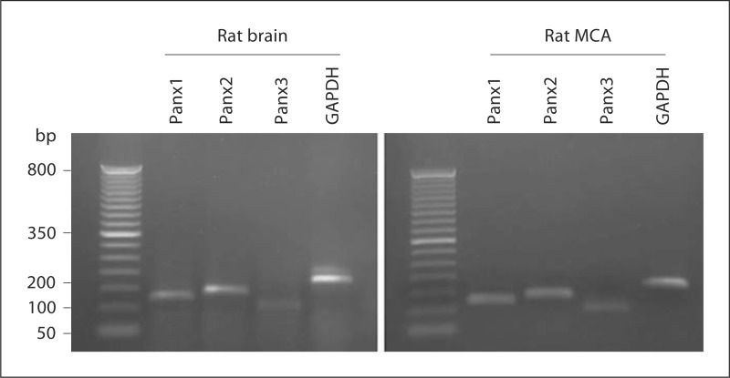
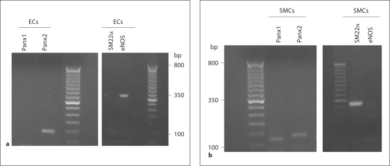
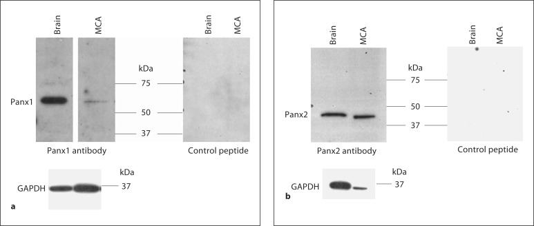
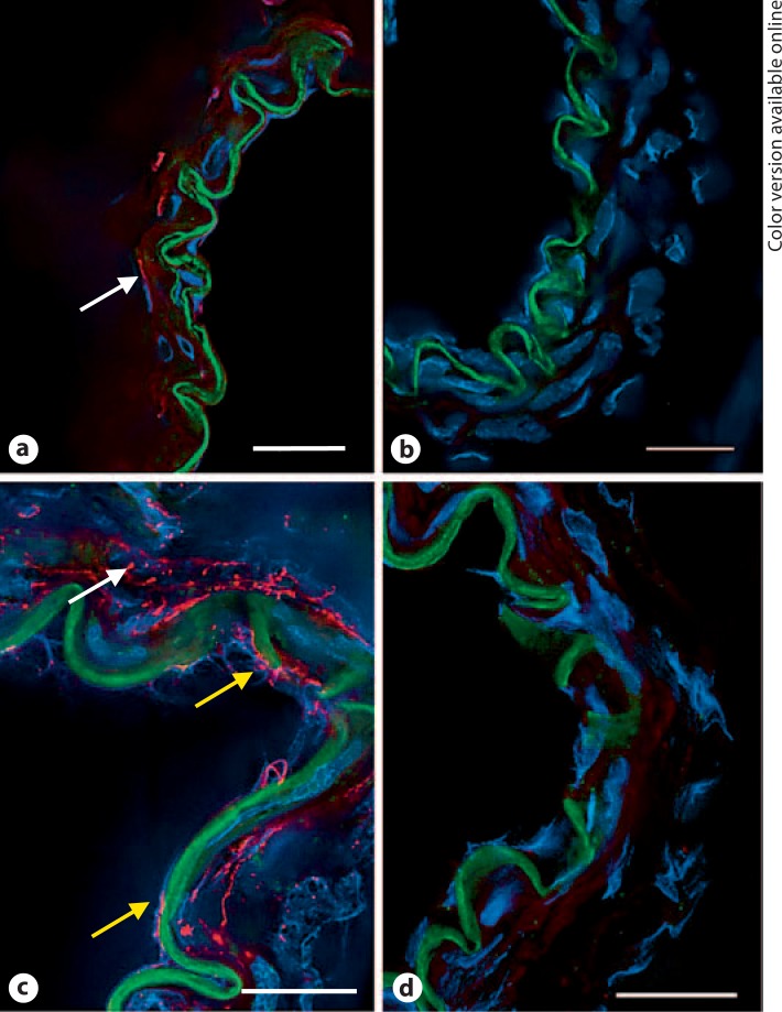
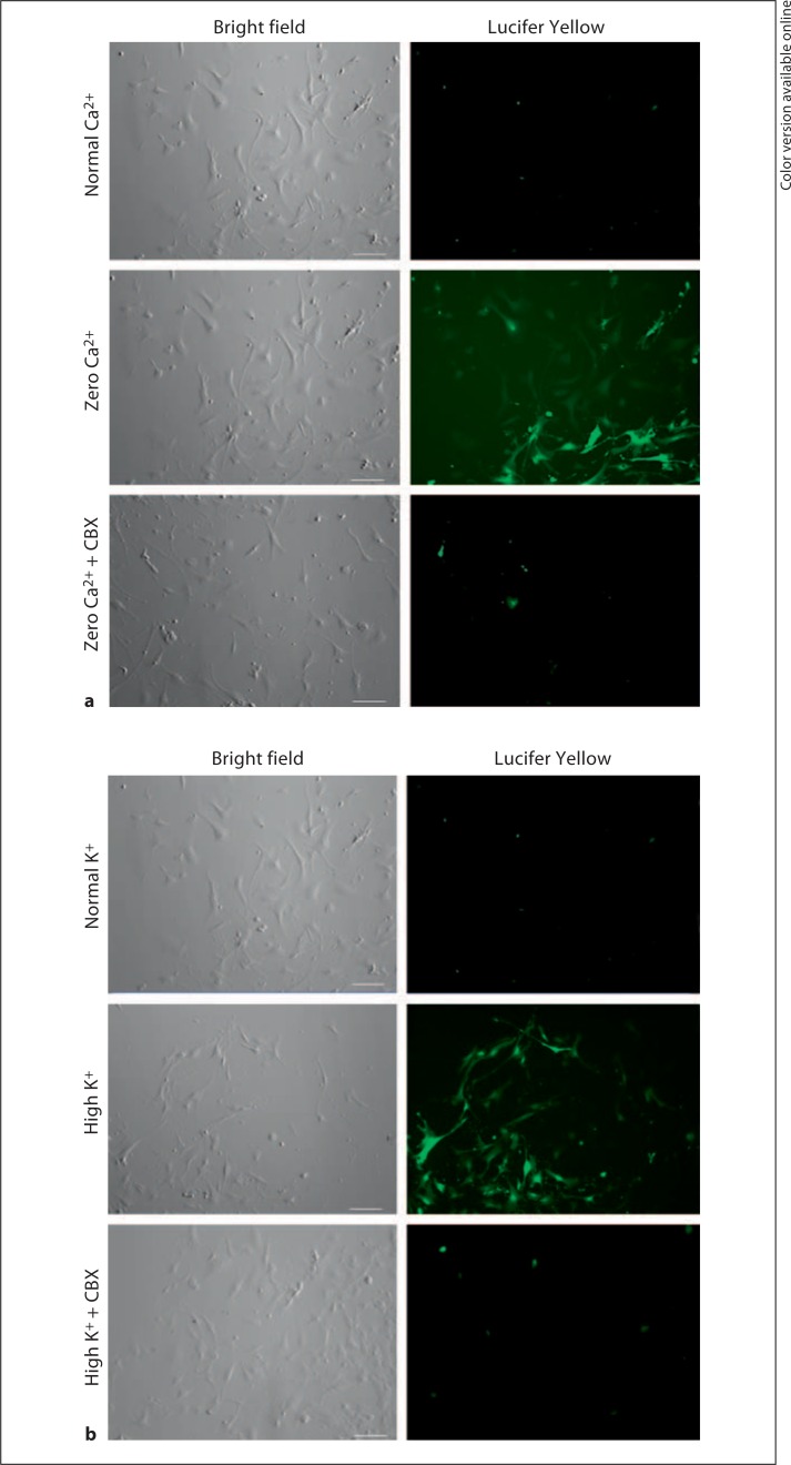
Methods: A combination of RT-PCR, immunoblotting and immunohistochemistry techniques was used to characterize the expression pattern of pannexins in rat MCA. A fluorescent dye uptake approach in cultured smooth muscle cells was used to determine whether these cells have functional hemichannels.

Results: We report for the first time that pannexins are expressed in the cerebral vasculature. We reveal that pannexin 1 is expressed in smooth muscle but not in endothelium and pannexin 2 is expressed in both endothelium and smooth muscle. Fluorescent dye entered cultured smooth muscle cells in the absence of extracellular calcium or when the cells were depolarized, which was prevented by the putative hemichannel blocker carbenoxolone.

Conclusions: The identification of pannexins in rat MCA indicates that pannexin expression is not restricted to neuronal cells. Dye uptake in cultured smooth muscle cells exhibited properties similar to those of connexin and pannexin hemichannels, which may represent another form of cell-to-cell communication within the vasculature.


Figures

Fig. 1
Fig. 1
RT-PCR products for Panx1 (125 bp), Panx2 (140 bp) and Panx3 (101 bp) were found in rat brain and rat MCA. GAPDH (180 bp) was used as an internal control. Lane 1: 50-bp ladder (size marker). No product was detected if reverse transcriptase was omitted (data not shown). Data are representative of 3 separate preparations.
Fig. 2
Fig. 2
RT-PCR product for Panx2 (140 bp) was found in freshly dissociated endothelial cells (ECs; a, left panel) and RT-PCR products for Panx1 (125 bp) and Panx2 (140 bp) were found in freshly dissociated smooth muscle cells (SMCs; b, left panel) from rat MCA. The right panels show RT-PCR products indicating the absence of SM22α (325 bp) and the presence of endothelial nitric oxide synthase (eNOS; 356 bp) in dissociated endothelial cells (a, right panel) and vice versa in dissociated smooth muscle cells (b, right panel). No product was detected if reverse transcriptase was omitted (data not shown). Data are representative of 3 separate preparations.
Fig. 3
Fig. 3
a Tissue extracts from rat brain (10 μg) and MCA (20 μg) were run on 10% SDS-PAGE gels and probed with Panx1 antibody in the absence and presence of the control peptide. b Tissue extracts from rat brain (10 μg) and MCA (2.5 μg) were run on 10% SDS-PAGE gels and probed with Panx2 antibody in the absence and presence of the control peptide. Each band disappeared when the primary antibody was incubated with the control peptide. Each membrane was stripped and reprobed for GAPDH. The molecular mass is indicated. Data are representative of 4 separate preparations.
Fig. 4
Fig. 4
Pannexin immunofluorescence in frozen cross-sections of rat MCA. a Panx1 fluorescence (red) showed weak staining in smooth muscle (arrow) and was undetected in endothelial cells. b Red fluorescence was absent in sections in which the control rabbit IgG was substituted for the primary antibody. c Panx2 fluorescence (red) was detected in both endothelial cells (bottom two arrows) and smooth muscle cells (top arrow). d Red fluorescence was absent in sections incubated with a control rabbit IgG. Green fluorescence is the autofluorescence of the internal elastic lamina that separates the endothelium from smooth muscle. Blue fluorescence identifies nuclei labeled with DAPI. Data are representative of 3 separate preparations. Scale bar = 20 μm. (Color only in online version).
Fig. 5
Fig. 5
Evidence of functional hemichannels in rat aorta smooth muscle cells. a In the normal calcium-containing medium, no significant Lucifer Yellow uptake was observed (n = 10). However, in the medium containing zero calcium, hemichannel-mediated dye uptake was observed that was blocked in the presence of 100 μmol/l carbenoxolone (CBX; n = 8). b In normal extracellular potassium, no significant Lucifer Yellow uptake was observed (n = 7). However, when cells were depolarized with high potassium, hemichannel-mediated dye uptake was observed that was blocked in the presence of 100 μmol/l carbenoxolone (n = 8). Scale bar = 100 μm.

References

    1. Faraci FM, Heistad DD. Regulation of large cerebral arteries and cerebral microvascular pressure. Circ Res. 1990;66:8–17. - PubMed
    1. Panchin Y, Kelmanson I, Matz M, Lukyanov K, Usman N, Lukyanov S. A ubiquitous family of putative gap junction molecules. Curr Biol. 2000;10:R473–R474. - PubMed
    1. MacVicar BA, Thompson RJ. Non-junction functions of pannexin-1 channels. Trends Neurosci. 2010;33:93–102. - PubMed
    1. Vanden Abeele F, Bidaux G, Gordienko D, Beck B, Panchin YV, Baranova AV, Ivanov DV, Skryma R, Prevarskaya N. Functional implications of calcium permeability of the channel formed by pannexin 1. J Cell Biol. 2006;174:535–546. - PMC - PubMed
    1. Bruzzone R, Hormuzdi SG, Barbe MT, Herb A, Monyer H. Pannexins, a family of gap junction proteins expressed in brain. Proc Natl Acad Sci USA. 2003;100:13644–13649. - PMC - PubMed

Publication types