. 2012 Feb;33(2):261-70.
doi: 10.1038/aps.2011.156.

A novel sulfonamide agent, MPSP-001, exhibits potent activity against human cancer cells in vitro through disruption of microtubule

Affiliations

A novel sulfonamide agent, MPSP-001, exhibits potent activity against human cancer cells in vitro through disruption of microtubule

Zu-long Liu et al. Acta Pharmacol Sin. 2012 Feb.

Abstract

Aim: To evaluate the anti-cancer effects of a new sulfonamide derivative, 2-(N-(3-chlorophenyl)-4-methoxyphenylsulfonamido)-N-hydroxypropanamide (MPSP-001).

Methods: Human cancer cell lines (HepG2, THP-1, K562, HGC-27, SKOV3, PANC-1, SW480, Kba, HeLa, A549, MDA-MB-453, and MCF-7) were examined. The cytotoxicity of MPSP-001 was evaluated using the WST-8 assay. Cell cycle distribution was examined with flow cytometry. Mitotic spindle formation was detected using immunofluorescence microscopy. Apoptosis-related proteins were examined with Western blot using specific phosphorylated protein antibodies. Competitive tubulin-binding assay was performed to test whether the compound competitively bound to the colchicine site. Molecular docking was performed to explore the possible binding conformation.

Results: MPSP-001 potently inhibited the growth of the 12 different types of human cancer cells with the IC(50) values ranging from 1.9 to 15.7 μmol/L. The compound exerted potent inhibition on the drug-resistant Kb/VCR and MCF-7/ADR cells, as on Kba and MCF-7 cells. In HeLa, HGC-27, A549, and other cells, the compound (5 μmol/L) caused cell cycle arrest at the G(2)/M phase, and subsequently induced cell apoptosis. In Hela cells, it prevented the mitotic spindle formation. Furthermore, the compound dose-dependently inhibited polymerization of tubulin in vitro, and directly bound to the colchicine-site of β-tubulin. Molecular docking predicted that the compound may form two hydrogen bonds to the binding pocket. The compound showed synergistic effects with colchicine and taxol in blocking mitosis of HeLa cells.

Conclusion: MPSP-001 shows a broad-spectrum of anti-tumor efficacy in vitro and represents a novel structure with anti-microtubule activity.


Figures

Figure 1
Figure 1
Chemical structure of MPSP-001.
Figure 2
Figure 2
Effects of MPSP-001 on cell cycle distribution and cell death. (A) The concentration effects of MPSP-001 on cell cycle progression of HeLa cells. HeLa cells were treated with different concentrations of MPSP-001 for 16 h. Then, the cells were fixed and stained with PI and analyzed by flow cytometry. Percentages of cells in different phases were shown. The data are representative of three independent experiments. (B) The time effects of MPSP-001 on cell cycle progressions of HeLa cells. HeLa cells were treated with 5 μmol/L MPSP-001 for different time. The cells were then fixed and stained with PI, and analyzed by flow cytometry. Percentages of cells in different phases were shown. The data are representative of three independent experiments. (C) MPSP-001 induced apoptosis with the cleavage of PARP. The time (left panel) and concentration (right panel) effects of MPSP-001 on cell apoptosis of HeLa cells. Protein samples were separated by SDS-PAGE for immunoblot analysis using antibody against PARP and GAPDH were stained as the internal cytosolic control.
Figure 3
Figure 3
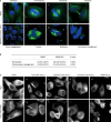
Effects of MPSP-001 on mitotic spindle formation and tubulin disruption. (A) Illustrations of the effects of MPSP-001 on the mitotic spindle in HeLa cells (e-h) using immuno-fluorescence microscopy technique. HeLa cells grown in log phase were treated with 5 μmol/L MPSP-001 for 16 h, followed by fixation and immunofluorescence staining for tubulin (white) and DNA (blue). Examples of a normal interphase (a), normal prometaphase (b), normal metaphase (c) and normal telophase (d) were shown. Examples of a damaged interphase (e) and various forms of abnormal mitoses include a tripolar prometaphase (f), a mutipolar metaphase (g) and an abnormal bipolar metaphase with lagging chromosomes or chromosomes not aligned to the metaphase plate (h) were shown (×600). (B) Statistical analysis of the effects of MPSP-001 on the mitotic spindles in HeLa cells. (C) Disruption of tubulin by MPSP-001, colchicine, and paclitaxel in HeLa cells and tubulin-GFP-HeLa cells (over-expression of the fusion protein of tubulin and GFP). HeLa cells and tubulin-GFP-HeLa cells grown in log phase were treated with 0.1% DMSO (a, f), 250 nmol/L Taxol (b, g), 100 nmol/L colchicine (c, h), 100 nmol/L vincristine (d, i) and 5 μmol/L MPSP-001 for 16 h, followed by fixation and immunofluorescence staining for tubulin (In Tubulin-GFP-HeLa cells, immunofluorescence staining was omitted) .Then the morphology of interphase tubulin (white) was observed (×600).
Figure 4
Figure 4
Effects of MPSP-001 on in vitro tubulin polymerization and competitive binding of colchicine site. (A) Effects of MPSP-001 (25 μmol/L, 100 μmol/L), Taxol (10 μmol/L), colchicines (10 μmol/L) and vincristine (10 μmol/L) on bovine brain tubulin polymerization were measured turbidimetrically. Changes in absorbance at 340 nm (A340) were measured and plotted as a function of time. (B) MPSP-001 binding to tubulin directly and inhibiting tubulin polymerization. Tubulin was co-incubated with indicated concentrations of VCR and MPSP-001 for 1 h, then 5 μmol/L colchicine was added. The fluorescence was measured by spectrofluorometer. All assays were repeated twice and representative data were shown. (C) Interactions between α,β-tubulin and compound MPSP-001 in the docking complex in 3D pattern. Tubulin was shown in cartoon style with the β and α subunit colored in green and cyan, respectively; compound MPSP-001 was shown in stick style; the residues within 4 Å around compound MPSP-001 were shown in line style. Magenta dashed lines denoted the potential hydrogen bonds. (D) 2D representation were drawn using LIGPLOT. Dashed lines represented hydrogen bonds and spiked residues form hydrophobic contacts with the compound.
Figure 5
Figure 5
Synergistic effects of MPSP-001 in combination with colchicine and Taxol on blocking mitosis. (A) Flow cytometry analysis of the mitosis block effects using different concentrations of MPSP-001, colchicines and Taxol. HeLa cells were incubated with different concentrations of MPSP-001, colchicines and taxol for 16 h. Then cells were fixed and stained with PI and analyzed by flow cytometry. The G2/M distribution values were graphed. Data are the means of triplicates±SD. (B) Flow cytometry analysis of the effects of taxol (25 nmol/L), colchicine (10 nmol/L), MPSP-001 (1 μmol/L) or the combination of Taxol+MPSP-001 and colchicines+MPSP-001 on cell cycle distribution. HeLa cells were incubated with drugs for 16 h. The cells were then fixed and stained with PI and analyzed by flow cytometry.

Similar articles

Cited by

References

    1. Urruticoechea A, Alemany R, Balart J, Villanueva A, Vinals F, Capella G. Recent advances in cancer therapy: an overview. Curr Pharm Des. 2010;16:310. - PubMed
    1. Dumontet C, Jordan MA. Microtubule-binding agents: a dynamic field of cancer therapeutics. Nat Rev Drug Discov. 2010;9:790–803. - PMC - PubMed
    1. Gan PP, McCarroll JA, Po'uha ST, Kamath K, Jordan MA, Kavallaris M. Microtubule dynamics, mitotic arrest, and apoptosis: drug-induced differential effects of betaIII-tubulin. Mol Cancer Ther. 2010;9:1339–48. - PubMed
    1. Screpanti E, Santaguida S, Nguyen T, Silvestri R, Gussio R, Musacchio A, et al. A screen for kinetochore-microtubule interaction inhibitors identifies novel antitubulin compounds. PLoS One. 2010;5:e 11603. - PMC - PubMed
    1. Kanthou C, Tozer GM. Microtubule depolymerizing vascular disrupting agents: novel therapeutic agents for oncology and other pathologies. Int J Exp Pathol. 2009;90:284–94. - PMC - PubMed

Publication types

Substances