

Normal morphology, age-related changes and abnormal findings of the cervical spine. Part II: Magnetic resonance imaging of over 1,200 asymptomatic subjects
- PMID: 22302162
- PMCID: PMC3535246
- DOI: 10.1007/s00586-012-2176-4
Normal morphology, age-related changes and abnormal findings of the cervical spine. Part II: Magnetic resonance imaging of over 1,200 asymptomatic subjects
Abstract
Purpose: The aim of this study is to establish standard MRI values for the cervical spinal canal, dural tube, and spinal cord, to evaluate age-related changes in healthy subjects, and to assess the prevalence of abnormal findings in asymptomatic subjects.
Methods: The sagittal diameter of the spinal canal and the sagittal diameter and cross-sectional area of the dural tube and spinal cord were measured on MRIs of 1,211 healthy volunteers. These included at least 100 men and 100 women in each decade of life between the third (20s) and eighth (70s). Abnormal findings such as spinal cord compression and signal changes in the spinal cord were recorded.
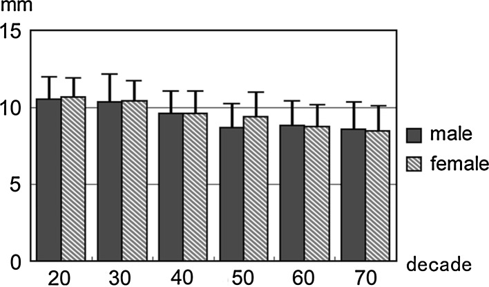
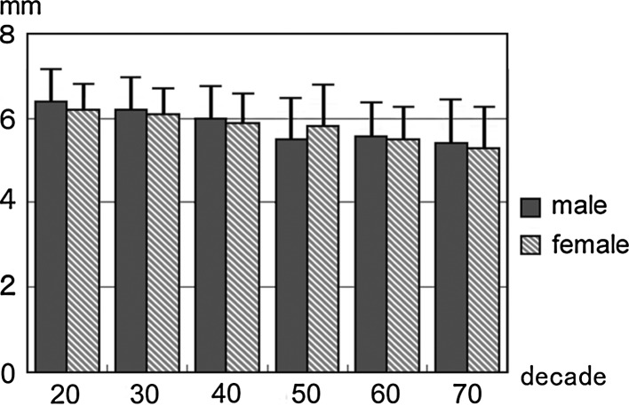
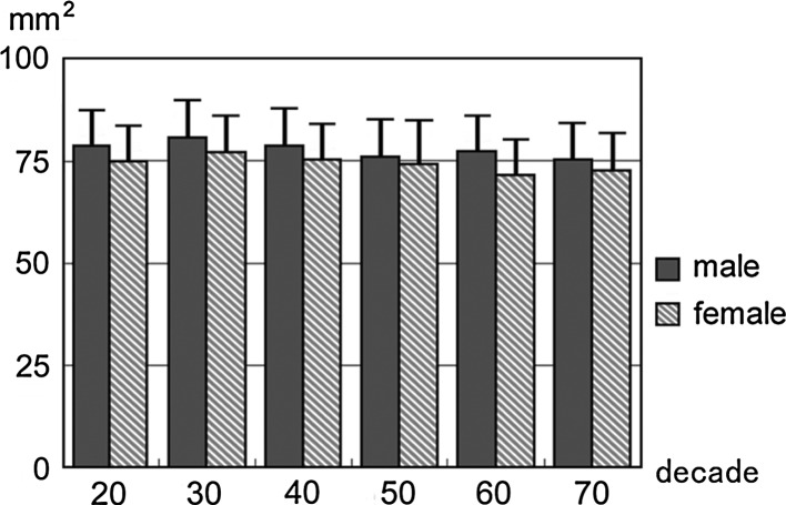
Results: The sagittal diameter of the spinal canal was 11.2 ± 1.4 mm [mean ± standard deviation (SD)]/11.1 ± 1.4 mm (male/female) at the mid-C5 vertebral level, and 9.5 ± 1.8/9.6 ± 1.6 mm at the C5/6 disc level. The cross-sectional area of the spinal cord was 78.1 ± 9.4/74.4 ± 9.4 mm² at the mid-C5 level and 70.6 ± 11.7/68.9 ± 11.3 mm² at the C5/6 disc level. Both the sagittal diameter and the axial area of the dural tube and spinal cord tended to decrease with increasing age. This tendency was more marked at the level of the intervertebral discs than at the level of the vertebral bodies, especially at the C5/6 intervertebral disc level. The spinal cord occupation rate in the dural tube at the C5 vertebral body level averaged 58.3 ± 7.0%. Spinal cord compression was observed in 64 cases (5.3%) and a T2 high-signal change was observed in 28 cases (2.3%).
Conclusions: Using MRI data of 1,211 asymptomatic subjects, the standard values for the cervical spinal canal, dural tube, and spinal cord for healthy members of each sex and each decade of life and the age-related changes in these parameters were established. The relatively high prevalence of abnormal MRI findings of the cervical spine in asymptomatic individuals emphasizes the dangers of predicating operative decisions on diagnostic tests without precisely correlating these findings with clinical signs and symptoms.
Figures


References
-
- Higo M, Sako T, Suzuki Y, et al. Roentgenological study of the antero-posterior diameter in cervical developmental canal stenosis. Rinsho Seikei Geka. 1984;19(4):361–366.
-
- Kimura I, Shingu H, Nasu Y, et al. Computed tomography of the spinal canal for the cervical spine and spinal cord injury. Rinsho Seikei Geka. 1983;18(5):541–551.
Publication types
MeSH terms
LinkOut - more resources
Full Text Sources
Medical
Research Materials
Miscellaneous
