. 2012 Apr 19;86(4):114.
doi: 10.1095/biolreprod.111.097253. Print 2012 Apr.

A zinc-dependent mechanism regulates meiotic progression in mammalian oocytes

Affiliations

A zinc-dependent mechanism regulates meiotic progression in mammalian oocytes

Miranda L Bernhardt et al. Biol Reprod. .

Abstract

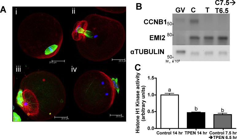
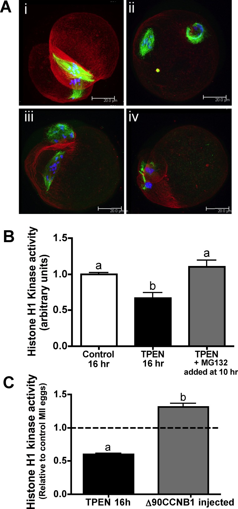
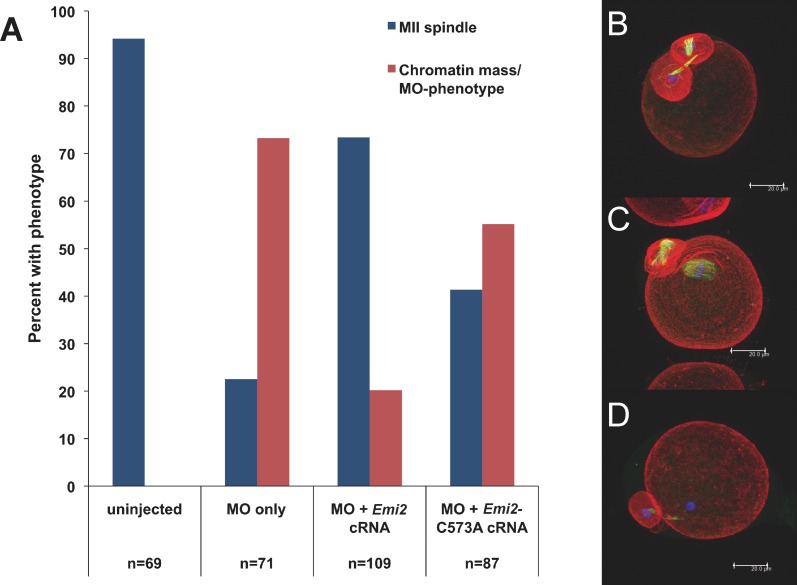
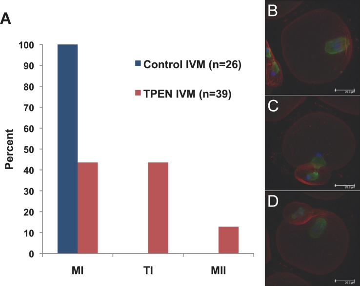
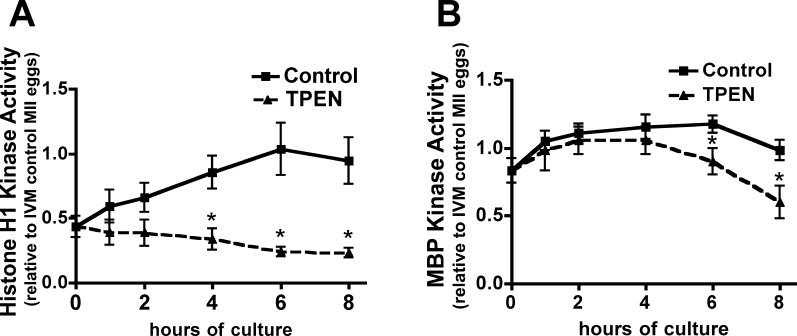
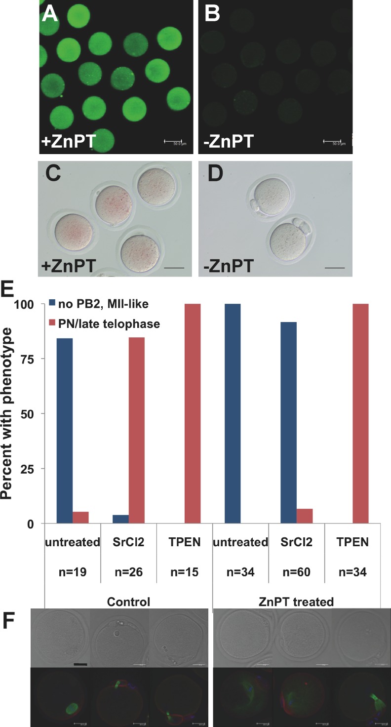
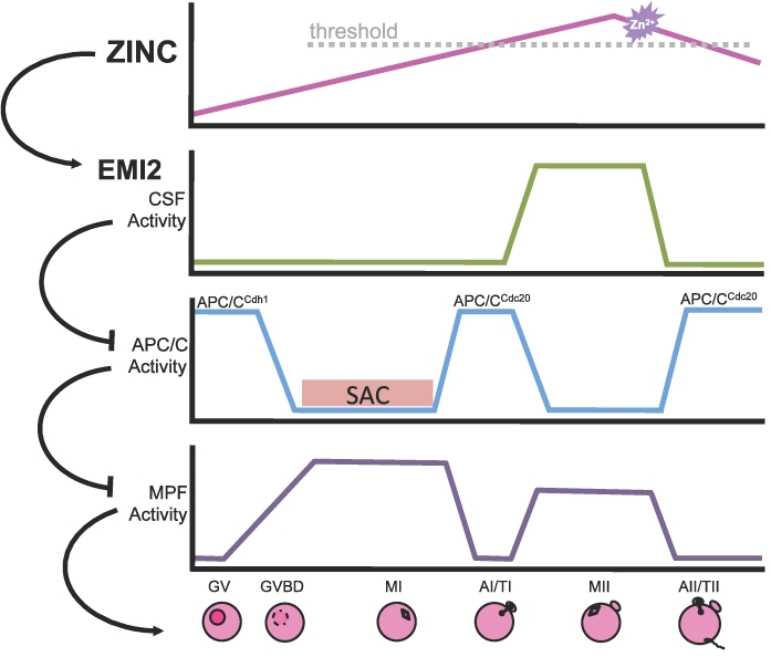
Precise coordination of meiotic progression is a critical determinant of an egg's capacity to be fertilized successfully, and zinc has emerged as a key regulatory element in this process. An early manifestation of a regulatory role for this transition metal is the significant increase in total intracellular zinc. This accumulation is essential for meiotic progression beyond telophase I and the establishment of meiotic arrest at metaphase II. The subsequent developmental event, fertilization, induces a rapid expulsion of labile zinc that is a hallmark event in meiotic resumption. In the present study, we show that the zinc fluxes work, in part, by altering the activity of the cytostatic factor (CSF), the cellular activity required for the establishment and maintenance of metaphase II arrest in the mature, unfertilized egg. We propose a model in which zinc exerts concentration-dependent regulation of meiosis through the CSF component EMI2, a zinc-binding protein. Together, the data support the conclusion that zinc itself, through its interaction with EMI2, is a central component of the CSF.


Figures

FIG. 1.
FIG. 1.
Zinc is required for successful MI-MII transition. A) COCs were cultured in control medium for 7.5 h and transferred to TPEN-containing medium at MI for an additional 6.5 h followed by removal of cumulus cells. Immunofluorescence analysis of the spindle shows that rather than forming normal MII spindles of controls (i), 30% of treated oocytes were arrested at TI (ii), whereas 51% had a single chromatin mass without complete retention of midbody microtubules (iii). Others had multiple chromatin masses (iv) or failed to complete cytokinesis. (Refer also to Supplemental Table S1.) Three-dimensional projections of confocal Z stacks with actin (red), tubulin (green), and DAPI (blue) are shown. Bar = 20 μm. B and C) Western blot analysis for CCNB1 and EMI2 and histone H1 kinase activity of oocytes from the same treatments as in A are shown. Western blots were repeated at least three times, showing similar results. Equal numbers of GV stage oocytes, which have low levels of CCNB1 and EMI2 protein, were also included for comparison. Graph presents densitometric analysis for at least seven individual oocytes per group. Error bars represent the SEM, and different letters indicate significant differences according to ANOVA with Bonferroni post-hoc test (P < 0.001).
FIG. 2.
FIG. 2.
Restoration of MPF activity following the MI-MII transition partially rescues zinc-insufficient oocytes. A) COCs were cultured in TPEN-containing medium for 10 h and transferred to medium containing both TPEN and MG132 for an additional 6 h followed by removal of cumulus cells. Spindle stains show that 69% of oocytes had some degree of MII spindle formation, with 19% showing aligned metaphase plates (i), 31% showing three or fewer misaligned chromosomes (ii), and 19% showing more than three misaligned chromosomes (iii). A majority of oocytes cultured in TPEN without MG132 for the entire culture period displayed the TI-arrested spindles associated with zinc insufficiency during IVM (iv). (Refer also to Supplemental Table S2.) Three-dimensional projections of confocal Z stacks with actin (red), tubulin (green), and DAPI (blue) are shown. Bar = 20 μm. B) Graph of histone H1 kinase activity showing densitometric analysis for at least six individual oocytes per group. Error bars represent the SEM, and different letters indicate significant differences according to ANOVA with Bonferroni post-hoc test (P < 0.01). C) Histone H1 kinase activity for oocytes cultured in TPEN containing medium for 10 to 12.5 h followed by injection with CCNB1(Δ90)-EGFP cRNA and transfer back to TPEN containing medium for 3.5 to 6 h is presented as in B.
FIG. 3.
FIG. 3.
Intact EMI2 ZBR is required for MI-MII transition. A) Cumulus-denuded GV oocytes were injected with Emi2 MO, held in IBMX for 5 h, and transferred to IVM medium. Following first polar body extrusion, groups of oocytes were injected with cRNA coding for T7-tagged wild-type Emi2 or for T7-tagged C573A mutated Emi2. BD) Following 15 h total IVM, oocytes were fixed; stained for actin (red), tubulin (green), and DNA (blue); imaged by confocal microscopy; and scored for spindle stage. In all, 94% of uninjected eggs had normal MII spindles, whereas 73% of MO-injected oocytes had chromatin masses and partial retention of midbody microtubules. (Refer also to Supplemental Fig. S2.) Wild-type Emi2 cRNA rescued MII spindle formation in 73% of oocytes, whereas ZBR-mutant Emi2 cRNA rescued MII formation in only 41%. Compiled data from three independent experiments are shown; the total number of oocytes scored for each treatment group is shown below the graph in A. Projections of confocal Z stacks are shown for MO injected (B), Emi2 cRNA rescue (C), and Emi2-C573A cRNA rescue (D). Bar = 20 μm.
FIG. 4.
FIG. 4.
TPEN inhibits the ability of EMI2 to maintain metaphase arrest in oocytes. A) Cumulus-denuded GV oocytes were injected with Emi2 cRNA, held in IBMX for 2–3 h, and transferred to IVM medium for 14 h using control medium or medium containing 10 μM TPEN. BD) Oocytes were fixed; stained for actin (red), tubulin (green), and DNA (blue); imaged by confocal microscopy; and scored for spindle stage. In all, 100% of Emi2 cRNA-injected oocytes cultured in control medium arrested at MI, whereas EMI2 expression caused MI arrest in only 44% of oocytes cultured in TPEN-containing medium. This experiment was repeated four times; data from one representative experiment are shown. Projections of confocal Z stacks are shown for Emi2 cRNA-injected oocytes cultured in TPEN containing medium arrested at MI (B), TI (C), and MII (D). Bar = 20 μm.
FIG. 5.
FIG. 5.
MPF and MAPK activity decline after TPEN treatment of MII eggs. In vivo-ovulated MII eggs were cultured in KSOM medium with or without 10 μM TPEN for 1–8 h. Graphs of histone H1 kinase activity (A) and MBP kinase activity (B) show densitometric analysis for four to seven individual eggs per data point. Error bars represent the SEM, and asterisks indicate significant differences according to Student t-test comparing TPEN-treated and control groups at individual time points (P < 0.05).
FIG. 6.
FIG. 6.
Increasing intracellular zinc in MII eggs prevents activation by SrCl2 but not by TPEN. In vivo-ovulated MII eggs were treated with 10 μM ZnPT for 5 min to increase intracellular zinc followed by activation with either 10 mM SrCl2 or 10 μM TPEN. A) To demonstrate the intracellular zinc increase, MII-stage eggs were loaded with FluoZin-3 AM for 60 min before ZnPT treatment followed by live imaging by confocal microscopy. B) FluoZin-3 AM-loaded eggs not treated with ZnPT and imaged with identical settings are shown. C and D) As an additional measure, eggs activated using SrCl2 were treated for 5 min with (C) or without (D) ZnPT 2 h after activation, stained with the colorimetric zinc stain dithizone, and imaged by bright-field microscopy. ZnPT dramatically increased staining in both cases. Bar = 50 μm. E and F) By 6 h post-SrCl2 or TPEN treatment, most control oocytes had formed second polar bodies and PN or PN-like structures, but only TPEN caused activation in ZnPT pretreated eggs. Compiled data from three independent experiments are shown; n signifies the total number of oocytes scored for each treatment group. Representative bright-field images and three-dimensional projections of confocal Z stacks with actin (red), tubulin (green), and DAPI (blue) are shown for each treatment group below the graph. Eggs treated with ZnPT displayed varying degrees of MII-spindle disorganization, ranging from elongated spindles with astral microtubules to scattered chromosomes with sparse tubulin staining. Bar = 20 μm.
FIG. 7.
FIG. 7.
A proposed model for the addition of zinc to the pathways controlling the dynamics of oocyte maturation in which zinc acts as a switch to regulate EMI2 during MII establishment, maintenance, and arrest. The top panel shows approximate relative zinc levels in GV oocytes, MII eggs, and early embryos as determined by x-ray fluorescence microscopy [14]. Subsequent panels represent dynamics of important cellular activities over the course of oocyte maturation and fertilization. In this model, when intracellular zinc is above a threshold level, the APC/C inhibitor EMI2 becomes activated, leading to CSF-mediated MII arrest. After activation, the zinc sparks (indicated by the purple starburst) cause a reduction in cellular zinc and could lead to reduced EMI2 activity upon fertilization, helping to drive the events of egg activation. AI/TI, anaphase I/telophase I; AII/TII, anaphase II/telophase II; GVBD, germinal vesicle breakdown; SAC, spindle assembly checkpoint.

References

    1. Outten CE, O'Halloran TV. Femtomolar sensitivity of metalloregulatory proteins controlling zinc homeostasis. Science 2001; 292: 2488 2492 - PubMed
    1. Berridge MJ, Bootman MD, Roderick HL. Calcium signalling: dynamics, homeostasis and remodelling. Nat Rev Mol Cell Biol 2003; 4: 517 529 - PubMed
    1. Grynkiewicz G, Poenie M, Tsien RY. A new generation of Ca2+ indicators with greatly improved fluorescence properties. J Biol Chem 1985; 260: 3440 3450 - PubMed
    1. Poenie M, Alderton J, Tsien RY, Steinhardt RA. Changes of free calcium levels with stages of the cell division cycle. Nature 1985; 315: 147 149 - PubMed
    1. Krezel A, Maret W. Zinc-buffering capacity of a eukaryotic cell at physiological pZn. J Biol Inorg Chem 2006; 11: 1049 1062 - PubMed

Publication types