Comparative Study
. 2012 Feb 1;32(5):1705-13.
doi: 10.1523/JNEUROSCI.5626-11.2012.

Contrast-enhanced magnetic resonance microangiography reveals remodeling of the cerebral microvasculature in transgenic ArcAβ mice

Affiliations
Comparative Study

Contrast-enhanced magnetic resonance microangiography reveals remodeling of the cerebral microvasculature in transgenic ArcAβ mice

Jan Klohs et al. J Neurosci. .

Abstract

Amyloid-β (Aβ) deposition in the cerebral vasculature is accompanied by remodeling which has a profound influence on vascular integrity and function. In the current study we have quantitatively assessed the age-dependent changes of the cortical vasculature in the arcAβ model of cerebral amyloidosis. To estimate the density of the cortical microvasculature in vivo, we used contrast-enhanced magnetic resonance microangiography (CE-μMRA). Three-dimensional gradient echo datasets with 60 μm isotropic resolution were acquired in 4- and 24-month-old arcAβ mice and compared with wild-type (wt) control mice of the same age before and after administration of superparamagnetic iron oxide nanoparticles. After segmentation of the cortical vasculature from difference images, an automated algorithm was applied for assessing the number and size distribution of intracortical vessels. With CE-μMRA, cerebral arteries and veins with a diameter of less than the nominal pixel resolution (60 μm) can be visualized. A significant age-dependent reduction in the number of functional intracortical microvessels (radii of 20-80 μm) has been observed in 24-month-old arcAβ mice compared with age-matched wt mice, whereas there was no difference between transgenic and wt mice of 4 months of age. Immunohistochemistry demonstrated strong fibrinogen and Aβ deposition in small- and medium-sized vessels, but not in large cerebral arteries, of 24-month-old arcAβ mice. The reduced density of transcortical vessels may thus be attributed to impaired perfusion and vascular occlusion caused by deposition of Aβ and fibrin. The study demonstrated that remodeling of the cerebrovasculature can be monitored noninvasively with CE-μMRA in mice.


Figures

Figure 1.
Figure 1.
High-resolution 3D TOF-MRA of the intracranial and extracranial vasculature of a 24-month-old wt control mouse (A) and an age-matched arcAβ mouse (B). The representative maximal intensity projections show the angiograms in sagittal, axial, and horizontal views. While the intracranial vasculature of arcAβ and wt mice shows no flow disturbances, flow voids are seen in extracranial vessels (white arrows). Sections of MIPs of the anterior cerebral artery of a 4-month-old (C) and a 24-month-old (D) wt control mouse. Discernable bifurcations are numbered in hierarchical order. More distal branches are visible in the angiogram of the 4-month-old mouse (C, white arrowhead). Scale bar, 1 mm.
Figure 2.
Figure 2.
Precontrast (A, D) and postcontrast (B, E) images were acquired before and after administration of a superparamagnetic iron oxide contrast agent. Grayscale windowing was kept identical for both images. Difference images (C, F) are obtained by subtraction of the postcontrast image from the precontrast image. Depicted are representative horizontal images of the cortex and olfactory bulb of a 24-month-old wt mouse (A–C) and an age-matched arcAβ mouse (D–F). After administration of the contrast agent, hypointensities representing intact blood vessels become visible which are not discernable on the precontrast image. The arrows point to focal hypointense areas that are present before administration of the contrast agent. Scale bar: 1 mm.
Figure 3.
Figure 3.
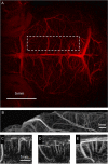
MIPs derived from a 3D stack of difference images viewed in horizontal (A), sagittal (B–D), and axial (E) orientation. The projections A and B were acquired over the whole acquisition volume, covering 2.2 mm of the upper part of the mouse brain. The horizontal view is dominated by projections of superficial veins like the transverse and superior sagittal sinus. Subcubes CE were taken from the axial MIP (location in the brain is indicated by the dashed line in A). The cerebral vasculature is highly organized, running perpendicular to the cortical surface.
Figure 4.
Figure 4.
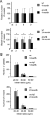
Semiautomated analysis of intracortical vessel density of wt and arcAβ mice at 4 and 24 months of age. A, The relative number of vessels was estimated for different connectivity thresholds corresponding to 3, 6, and 9 pixels, corresponding to minimal length of a vessel segment of 183, 366, and 546 μm, respectively. B, Number of vessels categorized according to their estimated vessel radius when the connectivity threshold was set to 3. The algorithm counts the fraction of vessels above an intensity threshold for different values of vessel connectivity in a VOI covering the cortex. A significant decrease in the number of vessels was observed in 24-month-old arcAβ compared with wt controls, while no significant differences were seen between arcAβ mice and wt controls at 4 months of age (mean ± SD; *p < 0.05, repeated-measures ANOVA and Tukey‘s test). There was no significant difference between the number of vessels in each category among the four groups tested.
Figure 5.
Figure 5.
Assessment of the endothelial marker vWF (red) with immunohistochemistry. Nuclear staining was done with DAPI (blue). Representative confocal images of 4-month-old wt (A), 24-month-old wt (B), and 24-month-old arcAβ mice (C). The white arrow points to vWF immunoreactivity in thrombus-like structure in a microvessel of a 24-month-old arcAβ mouse. There was no difference in vWF immunoreactivity between arcAβ and wt controls at 4 months of age and between arcAβ and wt controls at 24 months of age (D; mean ± SD; t test). Scale bar: 10 μm.
Figure 6.
Figure 6.
Assessment of Aβ and fibrinogen deposition in wt and arcAβ mice. Depicted are representative confocal images of 4-month-old arcAβ (A), 24-month-old wt (B), and 24-month-old arcAβ mouse brain sections (C–F) after thioflavin S staining (green) and antifibrinogen immunohistochemistry (red). The white arrows point to autofluorescence caused by lipofuscin aggregates, which are typical findings in neurons of aged animals. Fibrinogen accumulation was seen only in thioflavin S-positive vessels in 24-month-old arcAβ mice. Fibrinogen accumulation colocalized strongly with Aβ deposition (G; mean ± SD; *p < 0.05 Student's t test). Deposition of Aβ and fibrinogen were confined to small- and medium-sized arteries and were only scarcely visible in large vessels (D–F). The histogram depicts the cumulative frequency of the thioflavin S/fibrinogen immunoreactivity according to different categories of vessel diameter (H). Scale bars: AC, 10 μm; DF, 30 μm.

Similar articles

Cited by

References

    1. Baltes C, Radzwill N, Bosshard S, Marek D, Rudin M. Micro MRI of the mouse brain using a novel 400 MHz cryogenic quadrature RF probe. NMR Biomed. 2009;22:834–842. - PubMed
    1. Beckmann N, Schuler A, Mueggler T, Meyer EP, Wiederhold KH, Staufenbiel M, Krucker T. Age-dependent cerebrovascular abnormalities and blood flow disturbances in APP23 mice modeling Alzheimer's disease. J Neurosci. 2003;23:8453–8459. - PMC - PubMed
    1. Benveniste H, Einstein G, Kim KR, Hulette C, Johnson GA. Detection of neuritic plaques in Alzheimer's disease by magnetic resonance microscopy. Proc Natl Acad Sci U S A. 1999;96:14079–14084. - PMC - PubMed
    1. Bolan PJ, Yacoub E, Garwood M, Ugurbil K, Harel N. In vivo micro-MRI of intracortical neurovasculature. Neuroimage. 2006;32:62–69. - PubMed
    1. Bouras C, Kövari E, Herrmann FR, Rivara CB, Bailey TL, von Gunten A, Hof PR, Giannakopoulos P. Stereologic analysis of microvascular morphology in the elderly: Alzheimer disease pathology and cognitive status. J Neuropathol Exp Neurol. 2006;65:235–244. - PubMed

Publication types

MeSH terms

LinkOut - more resources