. 2012 Feb 15;26(4):338-43.
doi: 10.1101/gad.182121.111. Epub 2012 Feb 2.

Suppression of progenitor differentiation requires the long noncoding RNA ANCR

Affiliations

Suppression of progenitor differentiation requires the long noncoding RNA ANCR

Markus Kretz et al. Genes Dev. .

Abstract

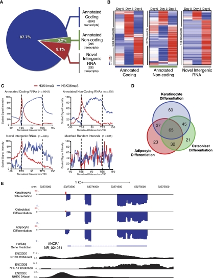
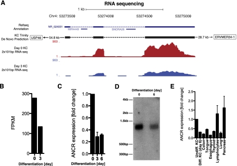
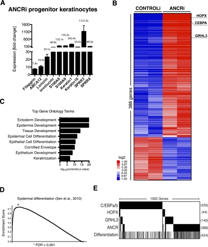
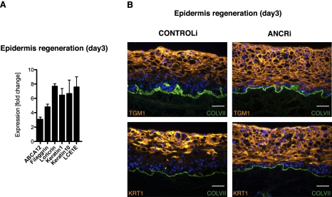
Long noncoding RNAs (lncRNAs) regulate diverse processes, yet a potential role for lncRNAs in maintaining the undifferentiated state in somatic tissue progenitor cells remains uncharacterized. We used transcriptome sequencing and tiling arrays to compare lncRNA expression in epidermal progenitor populations versus differentiating cells. We identified ANCR (anti-differentiation ncRNA) as an 855-base-pair lncRNA down-regulated during differentiation. Depleting ANCR in progenitor-containing populations, without any other stimuli, led to rapid differentiation gene induction. In epidermis, ANCR loss abolished the normal exclusion of differentiation from the progenitor-containing compartment. The ANCR lncRNA is thus required to enforce the undifferentiated cell state within epidermis.


Figures

Figure 1.
Figure 1.
Transcriptome characterization identifies ANCR as a lncRNA suppressed during terminal differentiation. (A) High-throughput paired-end RNA-Seq during three time points (days 0, 3, and 6) of human keratinocyte terminal differentiation was performed. Transcriptome assembly and analysis generated three classes of significantly expressed RNA: annotated coding transcripts, annotated noncoding transcripts, and unannotated novel intergenic RNAs not associated with any annotated gene. (B) Mean-centered, hierarchical clustering of differentially expressed transcripts within the three RNA classes during day 0, day 3, and day 6 of terminal differentiation. (C) Aggregate chromatin signatures of H3K4me3 and H3K36me3 over significantly expressed RNA from the three RNA classes and random genomic intervals that are length- and chromosome-matched to novel transcripts. (TSS) Transcription start site; (TSE) transcription end site. (D) Intergenic K4/K36 domain tiling arrays were performed in matched progenitor and differentiated populations of keratinocytes, adipocytes, and osteoblasts using the assembled terminal differentiation transcriptome and all annotated noncoding RNA as a reference. Significantly differentially expressed transcripts are overlapped. (E) Differential expression data from K4/K36 tiling arrays displayed as log2 fold change during differentiation relative to progenitor cells from the three cell types at the ANCR/NR_024031 genomic locus. Keratinocyte ChIP-seq signal data from H3K4me3 and H3K36me3 and DNaseI hypersensitivity sequencing over this locus are depicted below in black. (Blue) Probes down-regulated during differentiation; (red,) probes up-regulated during differentiation.
Figure 2.
Figure 2.
ANCR is a lncRNA repressed during keratinocyte differentiation. (A) Schematic of ANCR genomic locus on chromosome 4, including RefSeq gene prediction for NR_024031. The de novo assembled ANCR transcript region based on keratinocyte RNA-Seq reads using the Trinity algorithm as well as raw RNA-Seq reads plotted for day 0 and day 3 of differentiation. (B) Relative abundance of ANCR transcript in fragments per kilobase of exon model per million mapped fragments (FPKM) during keratinocyte differentiation. (C) ANCR transcript expression is reduced during keratinocyte differentiation as shown by quantitative RT–PCR (qRT–PCR); undifferentiated cells (day 0) and days 3 and 6 of calcium-induced differentiation. (D) Northern blot analysis demonstrating a single 855-kb ANCR RNA species reduced at day 6 of keratinocyte differentiation. (E) ANCR transcript abundance in day 6 differentiated keratinocytes (diff. KC), colon, tongue, esophagus, thyroid, lymphocytes, lung, and pancreas, normalized to ANCR levels in undifferentiated progenitor keratinocytes (undiff. KC).
Figure 3.
Figure 3.
ANCR regulates a global gene expression program associated with epidermal differentiation. (A) Increased mRNA expression of early and late differentiation genes in ANCR-depleted progenitor keratinocytes in cultured keratinocytes versus control siRNAs, as shown by qPCR analysis. (B) Heat map depicting transcript profiling of independent biologic replicate progenitor keratinocytes with siRNA-mediated ANCR depletion (ANCRi) versus control siRNA (CONTROLi). Expression changes for each probe are normalized to a mean-centered signal across all samples. (C) Gene ontology terms significantly enriched in the ANCR-depleted gene set. (D) Gene set enrichment analysis (GSEA) shows highly statistically significant overlap of the ANCRi gene set with a calcium-induced keratinocyte differentiation gene signature published previously (Sen et al. 2010). (E) Gene set overlap of ANCRi gene set with published gene signatures from loss of key transcription factors that are dysregulated by ANCR depletion. Black bars indicate significantly differentially expressed genes in the gene set. The 1000 genes total between these four gene sets are compared with each other as well as with the keratinocyte differentiation signature. The total number of genes in each gene signature falling within the 1000-gene set is listed to the right of each row.
Figure 4.
Figure 4.
ANCR depletion induces ectopic differentiation in the epidermal progenitor compartment. (A) Increased differentiation gene mRNA expression as a function of ANCR depletion, as shown by qPCR analysis from whole-tissue RNA extracts. (B) Ectopic expression of epidermal differentiation proteins in the epidermal basal and lower spinous layers in ANCR-depleted regenerating organotypic human epidermis (ANCRi) using ANCR-specific versus scrambled control shRNA constructs (CONTROLi). Differentiation proteins detected by immunofluorescence (orange), basement membrane collagen VII (green)], and nuclear Hoechst 33342 (blue). Bar, 50 μm.

References

    1. Bartolomei MS, Zemel S, Tilghman SM 1991. Parental imprinting of the mouse H19 gene. Nature 351: 153–155 - PubMed
    1. Blanpain C, Fuchs E 2006. Epidermal stem cells of the skin. Annu Rev Cell Dev Biol 22: 339–373 - PMC - PubMed
    1. Cabili MN, Trapnell C, Goff L, Koziol M, Tazon-Vega B, Regev A, Rinn JL 2011. Integrative annotation of human large intergenic noncoding RNAs reveals global properties and specific subclasses. Genes Dev 25: 1915–1927 - PMC - PubMed
    1. Cesana M, Cacchiarelli D, Legnini I, Santini T, Sthandier O, Chinappi M, Tramontano A, Bozzoni I 2011. A long noncoding RNA controls muscle differentiation by functioning as a competing endogenous RNA. Cell 147: 358–369 - PMC - PubMed
    1. Eisen MB, Spellman PT, Brown PO, Botstein D 1998. Cluster analysis and display of genome-wide expression patterns. Proc Natl Acad Sci 95: 14863–14868 - PMC - PubMed

Publication types

Associated data