

Bovine PrP expression levels in transgenic mice influence transmission characteristics of atypical bovine spongiform encephalopathy
- PMID: 22302882
- PMCID: PMC3541801
- DOI: 10.1099/vir.0.040030-0
Bovine PrP expression levels in transgenic mice influence transmission characteristics of atypical bovine spongiform encephalopathy
Abstract
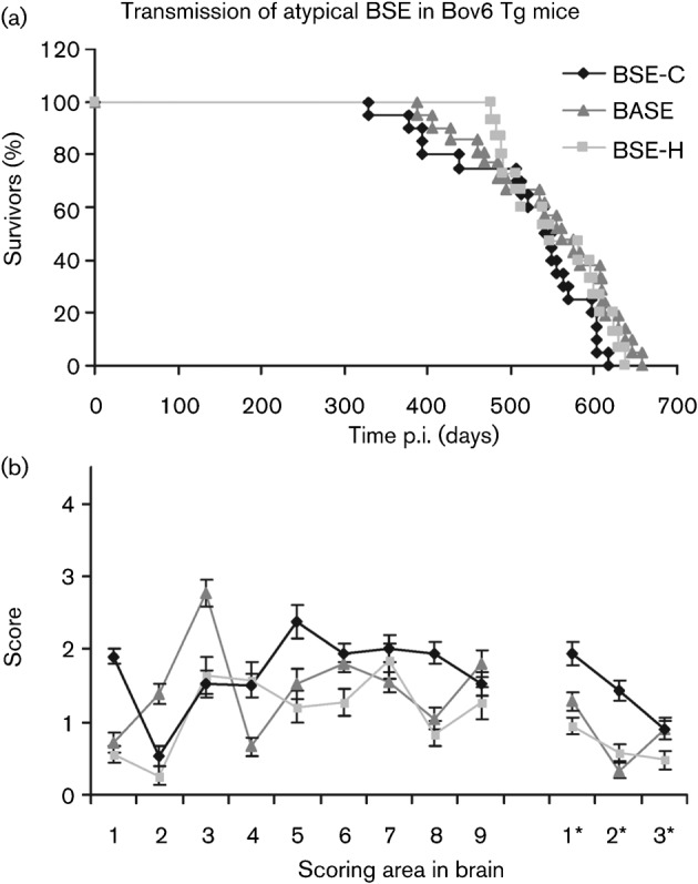
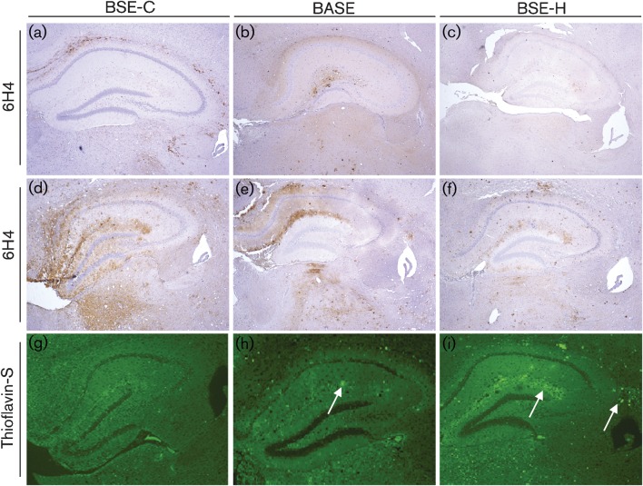
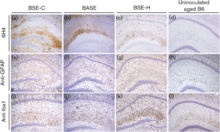
Until recently, transmissible spongiform encephalopathy (TSE) disease in cattle was thought to be caused by a single agent strain, bovine spongiform encephalopathy (BSE) (classical BSE or BSE-C). However, due to the initiation of a large-scale surveillance programme throughout Europe, two atypical BSE strains, bovine amyloidotic spongiform encephalopathy (BASE, also named BSE-L) and BSE-H have since been discovered. These atypical BSE isolates have been previously transmitted to a range of transgenic mouse models overexpressing PrP from different species at different levels, on a variety of genetic backgrounds. To control for genetic background and expression level in the analysis of these isolates, we performed here a comprehensive comparison of the neuropathological and molecular properties of all three BSE agents (BASE, BSE-C and BSE-H) upon transmission into the same gene-targeted transgenic mouse line expressing the bovine prion protein (Bov6) and a wild-type control of the same genetic background. Significantly, upon challenge with these BSE agents, we found that BASE did not produce shorter survival times in these mice compared with BSE-C, contrary to previous studies using overexpressing bovine transgenic mice. Amyloid plaques were only present in mice challenged with atypical BSE and neuropathological features, including intensity of PrP deposition in the brain and severity of vacuolar degeneration were less pronounced in BASE compared with BSE-C-challenged mice.
Figures
References
-
- Arsac J. N., Bétemps D., Morignat E., Féraudet C., Bencsik A., Aubert D., Grassi J., Baron T. (2009). Transmissibility of atypical scrapie in ovine transgenic mice: major effects of host prion protein expression and donor prion genotype. PLoS ONE 4, e7300 10.1371/journal.pone.0007300 - DOI - PMC - PubMed
-
- Balkema-Buschmann A., Ziegler U., McIntyre L., Keller M., Hoffmann C., Rogers R., Hills B., Groschup M. H. (2011a). Experimental challenge of cattle with German atypical bovine spongiform encephalopathy (BSE) isolates. J Toxicol Environ Health A 74, 103–109 10.1080/15287394.2011.529060 - DOI - PubMed
Publication types
MeSH terms
Substances
Grants and funding
- BBS/E/D/20251967/BB_/Biotechnology and Biological Sciences Research Council/United Kingdom
- Y1-AI-4893-02/AI/NIAID NIH HHS/United States
- BBS/E/D/20251968/BB_/Biotechnology and Biological Sciences Research Council/United Kingdom
- Y01 AI004893/AI/NIAID NIH HHS/United States
- 224-05-1307/PHS HHS/United States
LinkOut - more resources
Full Text Sources
Research Materials