. 2012 Feb 1;26(3):247-58.
doi: 10.1101/gad.180885.111.

Structural basis of agrin-LRP4-MuSK signaling

Affiliations

Structural basis of agrin-LRP4-MuSK signaling

Yinong Zong et al. Genes Dev. .

Abstract

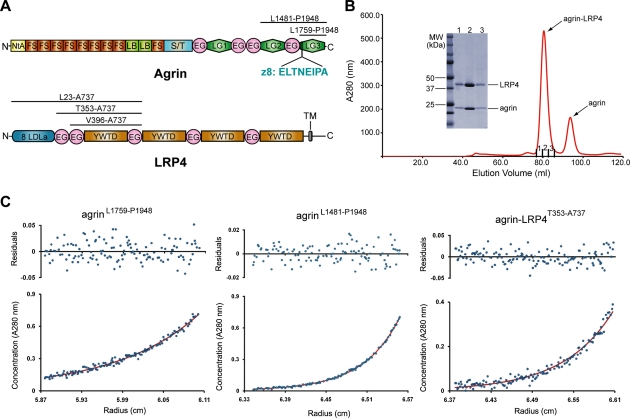
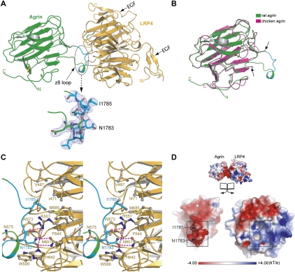
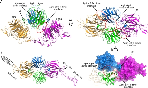
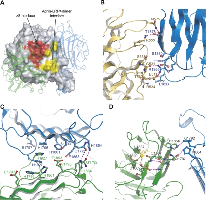
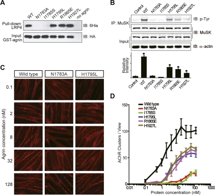
Synapses are the fundamental units of neural circuits that enable complex behaviors. The neuromuscular junction (NMJ), a synapse formed between a motoneuron and a muscle fiber, has contributed greatly to understanding of the general principles of synaptogenesis as well as of neuromuscular disorders. NMJ formation requires neural agrin, a motoneuron-derived protein, which interacts with LRP4 (low-density lipoprotein receptor-related protein 4) to activate the receptor tyrosine kinase MuSK (muscle-specific kinase). However, little is known of how signals are transduced from agrin to MuSK. Here, we present the first crystal structure of an agrin-LRP4 complex, consisting of two agrin-LRP4 heterodimers. Formation of the initial binary complex requires the z8 loop that is specifically present in neuronal, but not muscle, agrin and that promotes the synergistic formation of the tetramer through two additional interfaces. We show that the tetrameric complex is essential for neuronal agrin-induced acetylcholine receptor (AChR) clustering. Collectively, these results provide new insight into the agrin-LRP4-MuSK signaling cascade and NMJ formation and represent a novel mechanism for activation of receptor tyrosine kinases.


Figures

Figure 1.
Figure 1.
Characterization of the agrin–LRP4 interaction. (A) Schematic domain organizations of rat agrin and LRP4. The boundaries of protein fragments used in this study are indicated. (NtA) N-terminal agrin domain; (FS) follistatin-like repeat; (LB) laminin-B-like domain; (S/T) serine/threonine glycosylation sites; (EG) EGF-like domain; (LG) laminin-G-like domain; (LDLa) LDL class A repeats; (YWTD) YWTD repeat-containing β propeller; (TM) transmembrane region. (B) Agrin LG3 (residues Leu 1759–Pro 1948) forms a stable complex with LRP4V396–A737 in solution, as demonstrated by size exclusion chromatography. (C) Analytic ultracentrifugation (AUC) studies of agrin LG3, agrin LG2/LG3 (residues Leu 1481–Pro 1948), and the agrin LG3–LRP4T353–A737 complex. Agrin LG3 and LG2/LG3 are both monomeric in solution, and the agrin–LRP4 complex dimerizes with an average Kd of ∼39 μM. (Bottom panel) Absorbance data (blue dots) fit to a single-species model (LG2/LG3 and LG3) or a monomer–dimer model (agrin LG3–LRP4 complex) (red line). (Top panel) Residuals from the fit.
Figure 2.
Figure 2.
Binary complex of agrin LG3 (agrin) and LRP4V396–A737 (LRP4). (A) The association of agrin (green) and LRP4 (orange) is mostly mediated by the neuron-specific z8 alternative splicing sequence in agrin. This loop is unambiguously defined by excellent electron densities (2FoFc map contoured at 1σ) (in cyan). (B) The structure of rat agrin (green) in the context of an agrin–LRP4 complex is superimposed on the structure of an apo form of chicken agrin that also has an eight-amino-acid insert at the B/z site (magenta; PDB code 1PZ8). The arrows indicate the boundaries of the disordered z8 loop of the apo chicken agrin. (C) Stereoview of the detailed interactions between agrin (green/cyan) and LRP4 (yellow). Key residues that are directly involved in complex interactions are shown as the ball-and-stick model. Hydrogen bonds are indicated by dotted lines. (D) Open-book view of the electrostatic potential of the z8-interacting surfaces (highlighted with squares). The negatively charged z8 loop is complementary to the positively charged surface in LRP4.
Figure 3.
Figure 3.
The tetrameric architecture of the agrin–LRP4 complex. (A) Two agrin–LRP4V396–A737 binary complexes associate with each other with a noncrystallographic twofold symmetry. Besides the z8 interface, the tetrameric complex is stabilized by a second interface between agrin and LRP4 as well as an agrin–agrin interface, which are highlighted by red cycles. (B) The crystal structure of agrin–LRP4T353–A737 has a crystal packing different from that of agrin–LRP4V396–A737, but adopts the identical 2:2 tetrameric assembly of agrin and LRP4 in an asymmetry unit.
Figure 4.
Figure 4.
Agrin–LRP4 tetramer interfaces and the Ca2+-binding site in agrin. (A) In the LRP4 β1 propeller, the two agrin-binding interfaces (highlighted in red and yellow) are close to each other. The two bound agrin molecules are shown in the transparent cartoon. (B) A close-up view of the agrin–LRP4 dimer interface, where agrin and LRP4 are colored in blue and orange, respectively. (C) A close-up view of the agrin–agrin dimer interface, where the two agrin molecules are colored in blue and green. (D) A Ca2+ atom is coordinated by side chains of D1820 and D1889 and carbonyl oxygen of Q1887 and L1837. Q1887 is only 5 Å away from Q1792, which mediates the agrin–agrin dimer interface.
Figure 5.
Figure 5.
Agrin-induced tetramerization of agrin–LRP4 is critical for AChR clustering and MuSK phosphorylation. (A) The z8 interface plays a predominant role in assembly of the agrin–LRP4 complex. GST pull-down assays were performed between GST-agrin variants (containing a HA tag) and LRP4L23–A737 (containing a His tag). Agrin and LRP4 were detected by an anti-HA antibody and an anti-His antibody, respectively. (B) MuSK phosphorylation induced by agrin variants. Fully differentiated myotubes were treated with wild-type or mutant agrin at 100 nM. MuSK was immunoprecipitated with anti-MuSK polyclonal antibody, and phosphorylation was detected by immunoblotting with anti-phosphotyrosine antibody 4G10. The bar graph shows mean ± SD, n = 3; (*) P < 0.05 in comparison with the wild-type agrin. (C) Representative images of AChR clustering (red dots) on C2C12 myotubes induced by wild-type LG3, LG3-N1783A, or LG3-H1795L. (D) Quantitative analyses of AChR clustering assay for agrin variants. Data are mean ± SD based on three independent experiments.
Figure 6.
Figure 6.
Model of the agrin–LRP4–MuSK signaling pathway. MuSK (yellow) interacts with self-associated LRP4 (orange) in a nerve-independent manner. Monomeric agrin (blue) secreted by motor neurons first binds to the LRP4 β1 domain through a neuron-specific z8 insert (interface shown as a red glowing cycle). The binary complex then reorganizes the agrin–LRP4 dimer interface and the agrin–agrin dimer interface (magenta glowing cycles) into a unique configuration that is competent to further dimerize. Finally, the tetrameric complex of agrin–LRP4 is stabilized by five separate interfaces in a cooperative manner. Such interaction is necessary for MuSK activation, which leads to AChR clustering and synaptic differentiation

References

    1. Adams PD, Afonine PV, Bunkoczi G, Chen VB, Davis IW, Echols N, Headd JJ, Hung LW, Kapral GJ, Grosse-Kunstleve RW, et al. 2010. PHENIX: A comprehensive Python-based system for macromolecular structure solution. Acta Crystallogr D Biol Crystallogr 66: 213–221 - PMC - PubMed
    1. Ahn VE, Chu ML, Choi HJ, Tran D, Abo A, Weis WI 2011. Structural basis of Wnt signaling inhibition by Dickkopf binding to LRP5/6. Dev Cell 21: 862–873 - PMC - PubMed
    1. Battye TG, Kontogiannis L, Johnson O, Powell HR, Leslie AG 2011. iMOSFLM: A new graphical interface for diffraction-image processing with MOSFLM. Acta Crystallogr D Biol Crystallogr 67: 271–281 - PMC - PubMed
    1. Bezakova G, Helm JP, Francolini M, Lomo T 2001. Effects of purified recombinant neural and muscle agrin on skeletal muscle fibers in vivo. J Cell Biol 153: 1441–1452 - PMC - PubMed
    1. Bourhis E, Wang W, Tam C, Hwang J, Zhang Y, Spittler D, Huang OW, Gong Y, Estevez A, Zilberleyb I, et al. 2011. Wnt antagonists bind through a short peptide to the first β-propeller domain of LRP5/6. Structure 19: 1433–1442 - PubMed

Publication types