. 2012 Feb 1;26(3):271-81.
doi: 10.1101/gad.177857.111.

FGF21 regulates PGC-1α and browning of white adipose tissues in adaptive thermogenesis

Affiliations

FGF21 regulates PGC-1α and browning of white adipose tissues in adaptive thermogenesis

Ffolliott M Fisher et al. Genes Dev. .

Abstract

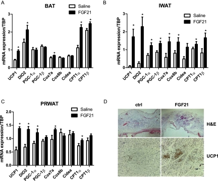
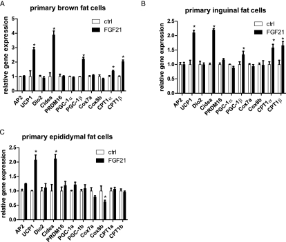
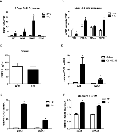
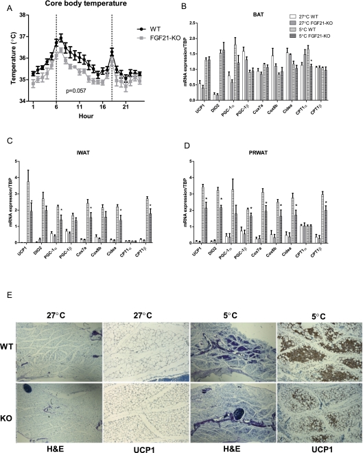
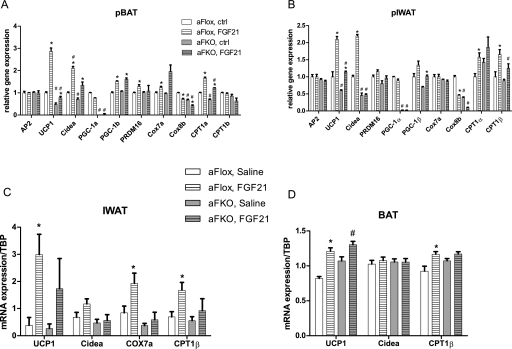
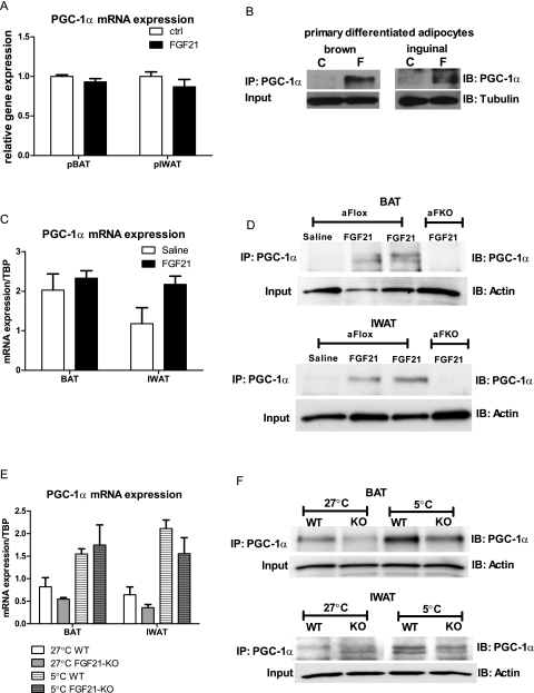
Certain white adipose tissue (WAT) depots are readily able to convert to a "brown-like" state with prolonged cold exposure or exposure to β-adrenergic compounds. This process is characterized by the appearance of pockets of uncoupling protein 1 (UCP1)-positive, multilocular adipocytes and serves to increase the thermogenic capacity of the organism. We show here that fibroblast growth factor 21 (FGF21) plays a physiologic role in this thermogenic recruitment of WATs. In fact, mice deficient in FGF21 display an impaired ability to adapt to chronic cold exposure, with diminished browning of WAT. Adipose-derived FGF21 acts in an autocrine/paracrine manner to increase expression of UCP1 and other thermogenic genes in fat tissues. FGF21 regulates this process, at least in part, by enhancing adipose tissue PGC-1α protein levels independently of mRNA expression. We conclude that FGF21 acts to activate and expand the thermogenic machinery in vivo to provide a robust defense against hypothermia.


Figures

Figure 1.
Figure 1.
FGF21 induces adipose thermogenic gene expression and browning of WAT. Mice were chronically treated with saline (n = 7) or FGF21 (n = 9) for 72 h. Real-time PCR analysis of thermogenic gene expression is shown from BAT (A), IWAT (B), and PRWAT (C). (D) H&E and UCP1 immunostaining in IWAT. Data are shown ±SEM; saline versus FGF21, (*) P < 0.05.
Figure 2.
Figure 2.
FGF21 regulates thermogenic gene expression in primary WAT and BAT adipocytes. Differentiated primary adipocytes were treated with 50 nM FGF21 for 48 h. Gene expression profiles are shown from adipocytes sourced from BAT (A), IWAT (B), and EWAT (C). Data are shown ±SEM; control versus FGF21, (*) P < 0.05.
Figure 3.
Figure 3.
Adipose FGF21 expression and secretion are regulated by cold exposure and β3-adrenergic signaling. Wild-type mice were kept for a 72-h period at 5°C, and tissues were subsequently dissected for further analysis. (A) FGF21 mRNA expression in multiple adipose tissue depots following cold exposure; 5°C (n = 5) versus 27°C (n = 5), (*) P < 0.05. Hepatic FGF21 and gluconeogenic gene expression (B) and FGF21 serum (C) levels. The effect of β-adrenergic stimulation on adipose FGF21 expression in mice (D) (saline vs. CL316243, [*] P < 0.05) and primary adipocytes (E). (F) Medium FGF21 levels from primary adipocytes treated with isoproterenol; saline versus isoproterenol, (*) P < 0.05. Data are shown ±SEM.
Figure 4.
Figure 4.
FGF21-KO display an impaired response to a cold challenge. Wild-type (n = 5) and FGF21-KO (n = 5) mice were implanted with activity- and temperature-measuring telemetry probes and were kept for a 72-h period at 5°C. (A) Diurnal core body temperature in wild-type and FGF21-KO mice. Real-time quantitative PCR analysis of thermogenic gene expression is shown from BAT (B), IWAT (C), and PRWAT (D). (E) H&E and UCP1 immunostaining in IWAT from wild-type and FGF21-KO mice. Data are shown ±SEM; 5°C wild type versus 5°C FGF21-KO, (*) P < 0.05.
Figure 5.
Figure 5.
PGC-1α is required for the FGF21-mediated induction of thermogenic genes. To assess whether PGC-1α mediates the thermogenic effects of FGF21 in vitro, primary adipocytes sourced from αFKO mice were treated with FGF21 for 48 h. Real-time quantitative PCR analysis is shown from primary brown (A) and inguinal white (B) adipocytes. To test the effects in vivo, αFlox and αFKO mice were treated for 3 d with either saline or FGF21. mRNA expression of PGC-1α target genes is shown from IWAT (C) and BAT (D). Data are shown ±SEM. Saline (n = 8–9) versus FGF21 (n = 11–12), (*) P < 0.05; αFlox versus αFKO, (#) P < 0.05.
Figure 6.
Figure 6.
FGF21 regulates PGC-1α protein content of adipocytes in vitro and in vivo. In vitro PGC-1α mRNA (A) and protein (B) levels are shown from primary BAT and IWAT adipocytes treated with FGF21. In vivo PGC-1α mRNA (C) and protein (D) levels (n = 2 mice per lane) are shown from BAT and IWAT of FGF21-treated mice. In vivo PGC-1α mRNA (E) and protein (F) levels (n = 3 mice per lane) from BAT and IWAT of wild-type and FGF21-KO mice cold-exposed for 72 h. Data are shown ±SEM where applicable. (C) Control; (F) FGF21.

References

    1. Arner P, Pettersson A, Mitchell PJ, Dunbar JD, Kharitonenkov A, Ryden M 2008. FGF21 attenuates lipolysis in human adipocytes—a possible link to improved insulin sensitivity. FEBS Lett 582: 1725–1730 - PubMed
    1. Badman MK, Pissios P, Kennedy AR, Koukos G, Flier JS, Maratos-Flier E 2007. Hepatic fibroblast growth factor 21 is regulated by PPARα and is a key mediator of hepatic lipid metabolism in ketotic states. Cell Metab 5: 426–437 - PubMed
    1. Badman MK, Koester A, Flier JS, Kharitonenkov A, Maratos-Flier E 2009. Fibroblast growth factor 21-deficient mice demonstrate impaired adaptation to ketosis. Endocrinology 150: 4931–4940 - PMC - PubMed
    1. Barbatelli G, Murano I, Madsen L, Hao Q, Jimenez M, Kristiansen K, Giacobino JP, De Matteis R, Cinti S 2010. The emergence of cold-induced brown adipocytes in mouse white fat depots is determined predominantly by white to brown adipocyte transdifferentiation. Am J Physiol Endocrinol Metab 298: E1244–E1253 doi: 10.1152/ajpendo.00600.2009 - PubMed
    1. Barbera MJ, Schluter A, Pedraza N, Iglesias R, Villarroya F, Giralt M 2001. Peroxisome proliferator-activated receptor α activates transcription of the brown fat uncoupling protein-1 gene. A link between regulation of the thermogenic and lipid oxidation pathways in the brown fat cell. J Biol Chem 276: 1486–1493 - PubMed

Publication types

MeSH terms

Substances