. 2012 Apr 6;287(15):12293-308.
doi: 10.1074/jbc.M111.294702. Epub 2012 Feb 2.

Protein interacting with C kinase 1 (PICK1) reduces reinsertion rates of interaction partners sorted to Rab11-dependent slow recycling pathway

Affiliations

Protein interacting with C kinase 1 (PICK1) reduces reinsertion rates of interaction partners sorted to Rab11-dependent slow recycling pathway

Kenneth L Madsen et al. J Biol Chem. .

Abstract

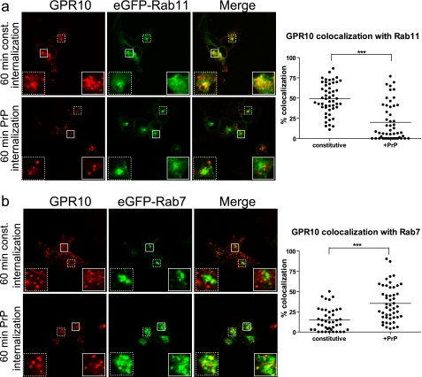
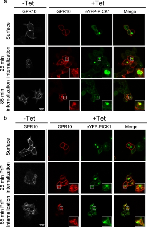
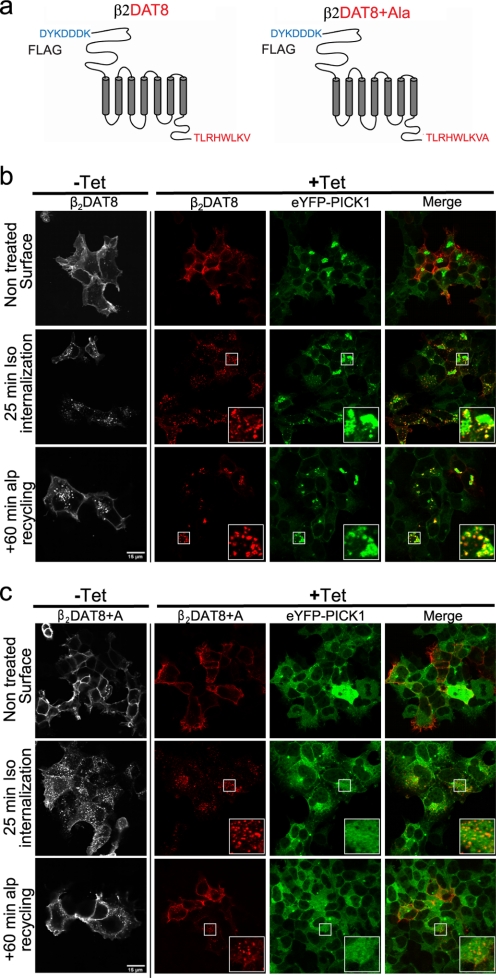
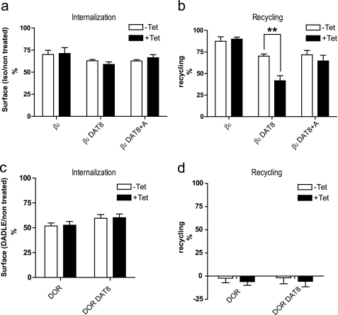
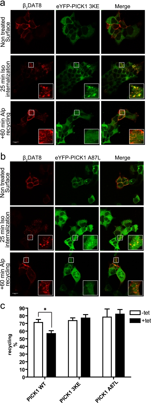
The scaffolding protein PICK1 (protein interacting with C kinase 1) contains an N-terminal PSD-95/Discs large/ZO-1 (PDZ) domain and a central lipid-binding Bin/amphiphysin/Rvs (BAR) domain. PICK1 is thought to regulate trafficking of its PDZ binding partners but different and even opposing functions have been suggested. Here, we apply ELISA-based assays and confocal microscopy in HEK293 cells with inducible PICK1 expression to assess in an isolated system the ability of PICK1 to regulate trafficking of natural and engineered PDZ binding partners. The dopamine transporter (DAT), which primarily sorts to degradation upon internalization, did not form perinuclear clusters with PICK1, and PICK1 did not affect DAT internalization/recycling. However, transfer of the PICK1-binding DAT C terminus to the β(2)-adrenergic receptor, which sorts to recycling upon internalization, led to formation of PICK1 co-clusters in Rab11-positive compartments. Furthermore, PICK1 inhibited Rab11-mediated recycling of the receptor in a BAR and PDZ domain-dependent manner. In contrast, transfer of the DAT C terminus to the δ-opioid receptor, which sorts to degradation, did not result in PICK1 co-clusters or any change in internalization/recycling. Further support for a role of PICK1 determined by its PDZ cargo was obtained for the PICK1 interaction partner prolactin-releasing peptide receptor (GPR10). GPR10 co-localized with Rab11 and clustered with PICK1 upon constitutive internalization but co-localized with the late endosomal marker Rab7 and did not cluster with PICK1 upon agonist-induced internalization. Our data suggest a selective role of PICK1 in clustering and reducing the recycling rates of PDZ domain binding partners sorted to the Rab11-dependent recycling pathway.


Figures

FIGURE 1.
FIGURE 1.
PICK1 co-clusters with Tac DAT C24 but not with TacDAT. a, schematic representation of the fusion proteins Tac DAT C24 and TacDAT. TacDAT C24 is a fusion protein between Tac and the 24 C-terminal residues of DAT with an N-terminal M1 antibody FLAG epitope. TacDAT is a head-to-tail fusion of Tac (α-subunit of the IL2 receptor) (green) with an N-terminal M1 antibody FLAG epitope (DYKDDDDK) and full-length DAT (blue) (43). b, confocal laser scanning micrographs of Flp-In T-REx 293 eYFP-PICK1 cells induced with tetracycline and transiently expressing TacDAT C24 (top panel) or TacDAT (bottom panel). Cells were surface-labeled with Alexa Fluor 568-conjugated anti-FLAG antibody to visualize surface TacDAT C24 (top middle panel) and TacDAT (middle bottom panel) in transfected cells. The eYFP-PICK1 signal (green) is shown in the left panels and illustrates the lack of clustering and partial plasma membrane recruitment in TacDAT-transfected cells. In contrast, profound eYFP-PICK1 clustering was seen in TacDAT C24-transfected cells. Nuclei were highlighted in the top panel to demonstrate the juxtanuclear localization of the eYFP-PICK1 clusters. c, confocal laser scanning micrographs of Flp-In T-REx 293 eYFP-PICK1 cells induced with tetracycline and transiently expressing TacDAT. Cells were surface-labeled with Alexa Fluor 568-conjugated anti-FLAG antibody and internalized with PMA for 25 min (top middle panel). Subsequently, the cells were treated with staurosporine (Stau; 1 μm) for 60 min to allow potential recycling (bottom middle panel). The eYFP-PICK1 signal (green) is shown in the left panel and illustrates the lack of marked clustering in the TacDAT-internalized cells. Representative images are shown from ∼15 cells visualized per condition in each experiment and over three separate experiments. The small squares mark areas that are shown enlarged inside large squares. Scale bar, 15 μm.
FIGURE 2.
FIGURE 2.
Agonist-induced internalization of GPR10 by PrP redirects endocytic sorting away from Rab11-positive pathway toward Rab7-positive pathway. a, left, confocal laser scanning micrographs of HEK293 cells transiently expressing GPR10 and GFP-Rab11. Cells were surface-labeled with Alexa Fluor 568-conjugated anti-FLAG M1 antibody at 4 °C to label GPR10 before 60 min of constitutive (const.) internalization at 37 °C (top panel) or 60 min of agonist-induced internalization (1 nm PrP) (lower panel) at 37 °C. The GPR10 signal is shown in red, and the eGFP-Rab11 signal is in green. Small white squares mark areas that are shown enlarged inside large white squares. Right, quantification of colocalization between internalized GPR10 and eGFP-Rab11 after 60 min of constitutive (n = 52) and agonist-induced internalization (n = 52). Each dot represents a single cell, and horizontal bars indicate means. ***, p < 0.001, unpaired t test. Data were collected on three separate experimental days. b, left, confocal laser scanning micrographs of HEK293 cells expressing eGFP-Rab7 and GPR10. Cells were surface-labeled with Alexa Fluor 568-conjugated anti-FLAG M1 antibody at 4 °C to label GPR10 before 60 min of constitutive internalization at 37 °C (top panel) or 60 min of agonist-induced internalization (1 nm PrP) (lower panel) at 37 °C. The GPR10 signal is shown in red, and the eGFP-Rab7 signal is in green. Small white squares mark areas that are shown enlarged inside large white squares. Right, quantification of colocalization between internalized GPR10 and eGFP-Rab7 after 60 min of constitutive (n = 42) and agonist-induced internalization (n = 50). Each dot represents a single cell, and horizontal bars indicate means. ***, p < 0.001, unpaired t test. Data were collected on three separate experimental days.
FIGURE 3.
FIGURE 3.
Constitutively internalized GPR10 co-clusters with eYFP-PICK1 in contrast to GPR10 internalized by agonist stimulation. a, confocal laser scanning micrographs of Flp-In T-REx 293 eYFP-PICK1 cells transiently expressing GPR10. The pictures show constitutive internalization of GPR10 that was surface-labeled with Alexa Fluor 568-conjugated anti-FLAG antibody at 4 °C without tetracycline (−Tet) and with (+Tet) induced expression of eYFP-PICK1 (green) prior to constitutive internalization for 25 or 85 min at 37 °C. Panels starting from the left show the GPR10 signal in non-induced cells, GPR10 signal in tetracycline-induced cells (red), eYFP-PICK1 signal (green), and merge of the red and green signals. b, confocal laser scanning micrographs of Flp-In T-REx 293 eYFP-PICK1 cells transiently expressing GPR10. The pictures show agonist-induced internalization of GPR10 that was surface-labeled with Alexa Fluor 568-conjugated anti-FLAG M1 antibody at 4 °C without (−Tet) and with (+Tet) induced expression of eYFP-PICK1 (green). Internalization was stimulated by agonist treatment (1 nm PrP) for 25 or 85 min at 37 °C. Panels starting from the left show the GPR10 signal in non-induced cells, GPR10 signal in tetracycline-induced cells (red), eYFP-PICK1 signal (green), and merge of red and green signals. Representative images are shown from ∼15 cells visualized per condition in each experiment and over three separate experiments. The small squares mark areas that are shown enlarged inside the large squares. Scale bars, 15 μm.
FIGURE 4.
FIGURE 4.
PICK1-binding chimeric receptor β2DAT8 clusters eYFP-PICK1 and traffics to PICK1-positive clusters upon agonist-induced internalization. a, schematic representation of β2DAT8 and β2DAT8 +Ala showing the N-terminal FLAG tag sequence followed by the full-length β2 adrenergic receptor and the last 8 PICK1-binding C-terminal residues of DAT. In β2DAT8 +Ala, PICK1 binding is disrupted due to the addition of an extra C-terminal alanine. b, confocal laser scanning micrographs of Flp-In T-REx 293 eYFP-PICK1 cells transiently expressing β2DAT8. The pictures show agonist (isoproterenol)-induced internalization of β2DAT8 that was surface-labeled with Alexa Fluor 568-conjugated anti-FLAG M1 antibody at 4 °C without (−Tet) and with (+Tet) induced expression of eYFP-PICK1 (green). Internalization was stimulated by 10 μm Iso for 25 min at 37 °C or by 10 μm Iso for 25 min followed by a 60-min incubation at 37 °C with the antagonist Alp to allow recycling of receptors. Panels starting from the left show the β2DAT8 signal in non-induced cells, β2DAT8 signal in tetracycline-induced cells (red), eYFP-PICK1 signal (green), and merge of red and green signals. c, confocal laser scanning micrographs of Flp-In T-REx 293 eYFP-PICK1 cells transiently expressing β2DAT8 +Ala. The pictures show conditions similar to those described for β2DAT8 above. Notably, β2DAT8 +Ala does not promote formation of perinuclear eYFP-PICK1 clusters. Pictures are representative of ∼25 cells visualized per condition in each experiment and over six separate experiments. The small squares mark areas that are shown enlarged inside the large squares. Scale bars, 15 μm.
FIGURE 5.
FIGURE 5.
Chimeric receptor DOR DAT8 does not cluster eYFP-PICK1. a, schematic representation of DOR DAT8 showing the N-terminal FLAG tag sequence followed by the full-length DOR and the last 8 PICK1 binding C-terminal residues of DAT. b, confocal laser scanning micrographs of Flp-In T-REx 293 eYFP-PICK1 cells transiently expressing DOR DAT8. The pictures show agonist-induced internalization of DOR DAT8 (compared with untreated control) that was surface-labeled with Alexa Fluor 568-conjugated anti-FLAG M1 antibody at 4 °C without (−Tet) and with (+Tet) induced expression of eYFP-PICK1 (green). Internalization was stimulated by 10 μm δ-opioid receptor agonist peptide DADLE for 25 min at 37 °C followed by a 60-min incubation at 37 °C with 10 μm antagonist Naxo to allow potential recycling of receptors. Panels starting from the left show the DOR DAT8 signal in non-induced cells, DOR DAT8 signal in tetracycline-induced cells (red), eYFP-PICK1 signal (green), and merge of red and green signals. Small white squares mark areas that are shown enlarged inside large white squares. Pictures are representative of ≈15 cells visualized per condition in each experiment and over five separate experiments. Scale bars, 15 μm.
FIGURE 6.
FIGURE 6.
PICK1 impairs recycling of agonist-internalized β2DAT8 but not DOR DAT8, which sorts to degradation. a and b, flp-In T-REx 293 eYFP-PICK1 cells transiently expressing β2, β2DAT8, or β2DAT8 +Ala with (black bars; +Tet) and without (white bars; −Tet) tetracycline-induced expression of eYFP-PICK1 were surface-labeled with anti-FLAG M1 antibody prior to stimulation of internalization with agonist (10 μm Iso for 25 min). Subsequently, cells were treated with the antagonist Alp (60 min at 10 μm) to allow recycling. Surface receptor immunoreactivity was determined by surface ELISA as described under “Experimental Procedures.” Internalization (a) refers to the fractional reduction of surface receptor in response to 25 min of agonist exposure compared with non-treated cells. Recycling (b) refers to the fractional recovery of surface receptor following antagonist incubation for 1 h. Data represent means ± S.E. from four independent experiments. c and d, Flp-In T-REx 293 eYFP-PICK1 cells transiently expressing DOR or DOR DAT8 with (black bars; +Tet) and without (white bars; −Tet) tetracycline-induced expression of eYFP-PICK1 were surface-labeled with anti-FLAG M1 antibody prior to stimulation of internalization with 10 μm δ-opioid receptor agonist peptide DADLE for 25 min followed by a 60-min incubation with 10 μm antagonist Naxo to allow potential recycling of receptors. Surface receptor immunoreactivity was determined by surface ELISA as described under “Experimental Procedures.” Internalization (c) refers to the fractional reduction of surface receptor in response to 25 min of agonist exposure compared with non-treated cells. Recycling (d) refers to the fractional recovery of surface receptor following antagonist incubation for 1 h. Data represent means ± S.E. from four independent experiments. **, p < 0.01, unpaired t test.
FIGURE 7.
FIGURE 7.
Impairment of β2DAT8 recycling by PICK1 is dependent on both intact PDZ binding crevice and BAR domain. a, confocal laser scanning micrographs of Flp-In T-REx 293 eYFP-PICK1 3KE cells induced with tetracycline and transiently expressing β2DAT8. The pictures show untreated cells (top) and agonist (isoproterenol at 10 μm)-induced internalization of β2DAT8 that was surface-labeled with Alexa Fluor 568-conjugated anti-FLAG M1 antibody at 4 °C prior to stimulation of internalization at 37 °C by 10 μm Iso for 25 min (middle) or with 10 μm Iso for 25 min followed by a 60-min incubation with the antagonist Alp (bottom) to allow recycling of receptors. Left panels, β2DAT8 signal (red); middle panels, eYFP-PICK1 3KE signal (green); right panels, merge of red and green signals. b, confocal laser scanning micrographs of Flp-In T-REx 293 eYFP-PICK1 A87L cells induced with tetracycline and transiently expressing β2DAT8. The pictures show conditions similar to those described for eYFP-PICK1 3KE above. Pictures are representative of ≈20 cells visualized per condition in each experiment and over four separate experiments. Scale bars, 15 μm. Small white squares mark areas that are shown enlarged inside large white squares. c, Flp-In T-REx 293 eYFP-PICK1 cells, Flp-In T-REx 293 eYFP-PICK1 3KE cells, or Flp-In T-REx HEK293 eYFP-PICK1 A87L cells transiently expressing β2DAT8 with (black bars; +Tet) and without (white bars; −Tet) tetracycline induction were surface-labeled with anti-FLAG M1 antibody prior to stimulation of internalization with agonist (10 μm Iso for 25 min). Subsequently, cells were treated with the antagonist Alp (60 min at 10 μm) to allow recycling. Surface receptor immunoreactivity was determined by surface ELISA as described under “Experimental Procedures.” Data are shown as recycling expressed as the fractional recovery of surface receptor following antagonist incubation for 1 h. Neither PICK1 3KE nor PICK1 A87L retained the ability to impede recycling of β2DAT8. Data represent means ± S.E. from four independent experiments. *, p < 0.05, unpaired t test.
FIGURE 8.
FIGURE 8.
Putative model illustrating PDZ cargo-determined function of PICK1 in regulating trafficking of its binding partners. a, PICK1 PDZ binding partners (“PDZ cargo”), such as the DAT and agonist-internalized GPR10 as well as the DOR engineered to bind PICK1 (DOR DAT8), are sorted upon internalization primarily to late endosomes and subsequently to lysosomal degradation. In contrast, PICK1 PDZ binding partners, such as constitutively internalized GPR10 and AMPAR as well as β2AR engineered to bind PICK1 (β2DAT8), are sorted to the Rab11-dependent long loop recycling pathway. b, PICK1 will be recruited to the plasma membrane by its different PDZ cargos independently of their postendocytic sorting pattern. At the plasma membrane, PICK1 might serve a variety of different functions in relation to these cargos including e.g. bringing PKCα in close proximity to regulate their phosphorylation or bind other PDZ cargos. Our data provide no evidence that PICK1 affects internalization of its PDZ cargo, and our data do not provide any evidence that PICK1 acts as a sorting module and thus affects their postendocytic sorting pattern. Also, our data suggest that PICK1 does not reside by itself in any recycling pathway but is brought there by its cargo. Thus, the role of PICK1 in regulating recycling might be explained simply by a stabilization of PICK1 in complex with its PDZ cargo in a Rab11-positive recycling compartment in a BAR domain-dependent fashion. According to the previously proposed autoinhibition hypothesis for the PICK1 BAR domain, this recruitment to a membrane compartment by its cargo would unmask the membrane binding capacity of the BAR domain, leading to clustering of PICK1 with its cargo. In this way, PICK1 might function as a compartment-specific anchor only activated by interaction partners sorted to Rab11-dependent recycling. It is possible that this function also involves other proteins, such as the small GTPases ARF1/3 or neuronal calcium sensor 1 (NCS1), and/or direct interaction with actin filaments, but further studies are required to clarify this issue. Finally, it should be considered that via the lipid deforming capacity of the BAR domain, PICK1 once brought to the recycling endosomes and activated might also be capable of affecting the shape of the membranes, thereby affecting fusion or fission events in this compartment in general.

Similar articles

Cited by

References

    1. Dev K. K. (2007) PDZ domain protein-protein interactions: a case study with PICK1. Curr. Top. Med. Chem. 7, 3–20 - PubMed
    1. Xu J., Xia J. (2006) Structure and function of PICK1. Neurosignals 15, 190–201 - PubMed
    1. Staudinger J., Lu J., Olson E. N. (1997) Specific interaction of the PDZ domain protein PICK1 with the COOH terminus of protein kinase C-α. J. Biol. Chem. 272, 32019–32024 - PubMed
    1. Staudinger J., Zhou J., Burgess R., Elledge S. J., Olson E. N. (1995) PICK1: a perinuclear binding protein and substrate for protein kinase C isolated by the yeast two-hybrid system. J. Cell Biol. 128, 263–271 - PMC - PubMed
    1. Madsen K. L., Beuming T., Niv M. Y., Chang C. W., Dev K. K., Weinstein H., Gether U. (2005) Molecular determinants for the complex binding specificity of the PDZ domain in PICK1. J. Biol. Chem. 280, 20539–20548 - PubMed

Publication types

MeSH terms

LinkOut - more resources