. 2012 Mar 23;287(13):10200-10209.
doi: 10.1074/jbc.M111.318733. Epub 2012 Feb 2.

Curcumin suppresses T cell activation by blocking Ca2+ mobilization and nuclear factor of activated T cells (NFAT) activation

Affiliations

Curcumin suppresses T cell activation by blocking Ca2+ mobilization and nuclear factor of activated T cells (NFAT) activation

Christian Kliem et al. J Biol Chem. .

Abstract

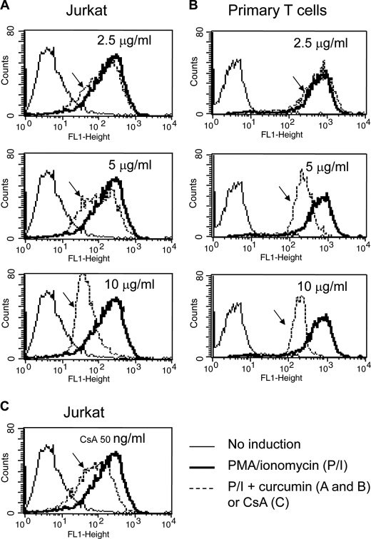
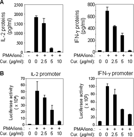
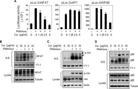
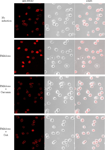
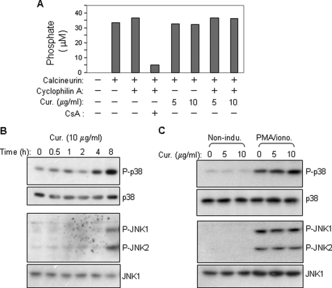
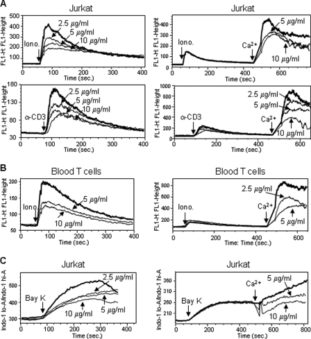
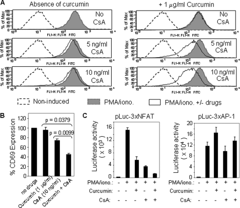
Curcumin is the active ingredient of the spice turmeric and has been shown to have a number of pharmacologic and therapeutic activities including antioxidant, anti-microbial, anti-inflammatory, and anti-carcinogenic properties. The anti-inflammatory effects of curcumin have primarily been attributed to its inhibitory effect on NF-κB activity due to redox regulation. In this study, we show that curcumin is an immunosuppressive phytochemical that blocks T cell-activation-induced Ca(2+) mobilization with IC(50) = ∼12.5 μM and thereby prevents NFAT activation and NFAT-regulated cytokine expression. This finding provides a new mechanism for curcumin-mediated anti-inflammatory and immunosuppressive function. We also show that curcumin can synergize with CsA to enhance immunosuppressive activity because of different inhibitory mechanisms. Furthermore, because Ca(2+) is also the secondary messenger crucial for the TCR-induced NF-κB signaling pathway, our finding also provides another mechanism by which curcumin suppresses NF-κB activation.


Figures

FIGURE 1.
FIGURE 1.
Curcumin suppresses CD69 expression in activated T cells. A and B, curcumin suppresses CD69 expression in T cells. Jurkat (A) and freshly isolated human peripheral blood T cells (B) were stimulated with PMA (5 ng/ml) and ionomycin (1 μm) in the absence or the presence of 2.5 to 10 μg/ml curcumin as indicated. After 24 h stimulation, the cells were analyzed for CD69 surface expression levels by FACS. C, CsA suppresses CD69 expression. Jurkat T cells were stimulated with PMA and ionomycin in the absence or presence of CsA (50 ng/ml) as in A and B. The reduced CD69 expression is indicated by arrows. Results are representative of two independent experiments.
FIGURE 2.
FIGURE 2.
Curcumin suppresses cytokine expression in T cells by down-regulation of promoter activity. A, curcumin inhibits IL-2 and IFN-γ protein expression in primary peripheral T cells. Freshly isolated human peripheral blood T cells were stimulated with PMA (5 ng/ml) and ionomycin (0.5 μm) in the absence or presence of different concentrations of curcumin as indicated. After 24 h stimulation, the supernatants were analyzed for IL-2 and IFN-γ protein production by ELISA. Results are representative of two independent experiments. B, curcumin down-regulates promoter activities of IL-2 and IFN-γ. Luciferase constructs containing the human IL-2 and IFN-γ promoter were transfected into Jurkat T cells. After overnight recovery, transfected cells were split and pre-incubated with different concentrations of curcumin as indicated for 1 h and then stimulated with PMA and ionomycin or left unstimulated for 8 h. The promoter activity was given as luciferase activity. Results are representative of two independent experiments each in triplicate transfections.
FIGURE 3.
FIGURE 3.
Curcumin inhibits NFAT transcriptional activity by blocking its nuclear expression. A, curcumin inhibits NFAT-mediated transcription. Jurkat T cells were transfected with luciferase constructs containing multiple copies of the NFAT binding element derived from the IL-2 promoter or the consensus DNA binding element of either AP1 or NF-κB. After overnight recovery, the cells were split and pre-incubated with the indicated amounts of curcumin for 0.5 h and then stimulated with PMA (5 ng/ml) and ionomycin (0.5 μm) or left unstimulated. Luciferase activity was determined after 8 h stimulation. Data are representative of three independent experiments performed in triplicate. B, curcumin prevents stimulation-induced nuclear expression of NFAT determined by Western blot. Jurkat T cells were stimulated with PMA and ionomycin for 2 h in the absence or presence of different concentrations of curcumin as indicated. Nuclear proteins were isolated and the expression levels of NFAT were analyzed by Western blot with the specific mAb against NFATc1. YY1 was used for an equal loading control. Total cell lysates were used as an additional control for total amounts of NFAT expression in the experiment. C and D, as controls, Western blot was also carried out in parallel with antibodies against Jun and NF-κB (p50/p65), respectively. Total cell lysates were used as additional controls for total amounts of Jun and NF-κB expression in the experiment.
FIGURE 4.
FIGURE 4.
Curcumin prevents stimulation-induced nuclear expression of NFAT determined by confocal LSM. Jurkat T cells were treated as described in Fig. 3B. The cellular and nuclear expression levels of NFATc1 were visualized by confocal LSM.
FIGURE 5.
FIGURE 5.
Curcumin does not directly inhibit calcineurin phosphatase activity and does also not enhance activities of the NFAT kinases p38 and JNK in activated T cells. A, curcumin has no direct inhibitory effect on calcineurin phosphatase. Calcineurin phosphatase activity was measured using an in vitro calcineurin assay kit in the absence or presence of curcumin. B, kinetic analysis of the effect of curcumin on p38 and JNK activity. Jurkat T cells were treated with curcumin (10 μg/ml) for different time periods. Total cell lysates were subjected to Western blot with antibodies against phosphorylated p38 (p-p38) and JNK (p-JNK1/2). C, curcumin does not enhance PMA/ionomycin-induced phosphorylation of p38 and JNK. Jurkat T cells were stimulated with PMA (5 ng/ml) plus ionomycin (1 μm) for 2 h in the absence or presence of different concentrations of curcumin. Total cell lysates were analyzed by Western blot. All data are representative of two independent experiments.
FIGURE 6.
FIGURE 6.
Curcumin blocks Ca2+ mobilization in activated T cells. A, effect of curcumin on Ca2+ mobilization in Jurkat T cells. Jurkat T cells were loaded with 1 μm fluorescent Ca2+-indicator Fluo-4 in PBS in the absence of extracellular Ca2+ followed by pretreated with different doses of curcumin for 10 min. Cells were washed with PBS. The release of Ca2+ from the intracellular stores was measured by flow cytometry after adding ionomycin (1 μm) or αCD3 (OKT3 10 μg/ml) (left panels). For determination of influx of extracellular Ca2+, the intracellular Ca2+ stores were first depleted in the absence of extracellular Ca2+ by ionomycin or by αCD3 for 7 min. The cells were then treated with or without curcumin for 30 s. The Ca2+ influx was measured after adding 1 mm Ca2+ (right panels). B, effect of curcumin on Ca2+ mobilization in peripheral blood T cells. Blood T cells were treated as in A. C, effect of curcumin on Ca2+ mobilization in Jurkat T cells stimulated with (±)-Bay K 8644. Jurkat T cells were loaded with 1 μm indol-1 and pretreated without or with curcumin as in A. The Ca2+ levels were monitored by flow cytometry after adding 100 μm (±)-Bay K 8644. All data are representative of at least two independent experiments.
FIGURE 7.
FIGURE 7.
Curcumin enhances the immunosuppressive activities of CsA. A, curcumin and CsA synergize to inhibit CD69 expression in activated T cells. Jurkat T cells were stimulated with PMA/ionomycin in the absence or the presence of either CsA alone or CsA plus 1 μg/ml curcumin. After 17 h stimulation, the cells were analyzed for CD69 surface expression levels by FACS. B, data from A are presented as bar charts. C, curcumin and CsA cooperatively inhibit NFAT transcriptional activity. The NFAT and AP1 luciferase reporter plasmids were transfected into Jurkat T cells. After overnight culture, the cells were split and pre-incubated with solvents, curcumin, CsA, or curcumin plus CsA for 30 min and then stimulated with PMA/ionomycin or left unstimulated. Luciferase activity was determined 8 h after stimulation. Data are representative of three independent experiments performed in triplicate transfections.

Similar articles

Cited by

References

    1. Ammon H. P., Wahl M. A. (1991) Pharmacology of Curcuma longa. Planta Med. 57, 1–7 - PubMed
    1. Araújo C. C., Leon L. L. (2001) Biological activities of Curcuma longa L. Mem. Inst. Oswaldo. Cruz. 96, 723–728 - PubMed
    1. Jurenka J. S. (2009) Anti-inflammatory properties of curcumin, a major constituent of Curcuma longa: a review of preclinical and clinical research. Altern. Med. Rev. 14, 141–153 - PubMed
    1. Goel A., Kunnumakkara A. B., Aggarwal B. B. (2008) Curcumin as “Curecumin”: from kitchen to clinic. Biochem. Pharmacol. 75, 787–809 - PubMed
    1. Palkowitsch L., Marienfeld U., Brunner C., Eitelhuber A., Krappmann D., Marienfeld R. B. (2011) The Ca2+-dependent phosphatase calcineurin controls the formation of the Carma1-Bcl10-Malt1 complex during T cell receptor-induced NF-κB activation. J. Biol. Chem. 286, 7522–7534 - PMC - PubMed

MeSH terms

LinkOut - more resources