. 2002:1:e0039.
doi: 10.1199/tab.0039. Epub 2002 Mar 27.

The Arabidopsis thaliana-pseudomonas syringae interaction

The Arabidopsis thaliana-pseudomonas syringae interaction

Fumiaki Katagiri et al. Arabidopsis Book. 2002.
No abstract available


Figures

Figure 1.
Figure 1.
A transmission electron microscope image of Pseudomonas syringae pv. tomato DC3000. Note that DC3000 produces polar flagella (15 nm in diameter) and a few Hrp pili (8 nm in diameter). The flagella and Hrp pili are indicated with arrows. Flagella enable bacteria to swim toward or away from specific chemical stimuli. Hrp pili are involved in type III secretion of avirulence and virulence proteins.
Figure 2.
Figure 2.
Disease symptoms in Arabidopsis leaves caused by DC3000 infection. Leaves (indicated with arrows) were syringe-infiltrated with 5 × 10⁵ cfu/mL of Pst DC3000 and pictures were taken four days after inoculation. The whole plant is shown in (A). A close-up of a diseased leaf is shown in (B).
Figure 3.
Figure 3.
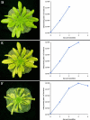
Disease symptom development in a susceptible Arabidopsis plant 1, 2, 3 and 4 days after inoculation. Leaves were vacuum infiltrated with 1 × 10⁶ bacteria/ml of DC3000. A picture was taken before inoculation (A) immediately after vacuum infiltration (B) and every day for 4 days (C to F). To the right of each picture is a plot of the level of bacteria present within the leaves at that particular time. Note, water-soaking symptoms, appeared at 48 to 60 hours. Significant chlorosis and necrosis occurred at 72 to 96 hours after inoculation. Note that bacteria multiplied to a near maximum level before chlorosis or massive cell death appeared.
Figure 3.
Figure 3.
Continued.
Figure 4.
Figure 4.
A scanning electron microscopic image of a cross section of an Arabidopsis (ecotype Columbia; susceptible) leaf infected with DC3000. HC: host cells. Ba: Bacteria. Arrows indicate the direction of type III secretion from bacteria in the apoplast into the host cell interior. Note that the host cell wall remains intact, physically separating bacteria and host cells until the very late stages of the interaction, when host cells collapse.
Figure 5.
Figure 5.
The RPM1 resistance gene-dependent HR induced by the expression of the P. syringae avrB gene directly in Arabidopsis. Left panel: An Arabidopsis rps3-1 (an rpm1mutant; Columbia background) seedling expressing avrB under the 35S promoter. No HR is present. Right panel: An F1 seedling from a cross between the rps3-1/avrB plant and a wild-type Columbia plant (RPM1+). Arrow indicates dark HR necroses on the cotyledon leaf. This seedling died before true leaves emerged because of systemic development of the HR.
Figure 6.
Figure 6.
The ligand-receptor model of R gene and avr gene interaction. A specific signal molecule is directly or indirectly generated by the avr gene in P. syringae. The signal molecule is recognized by the receptor encoded by the corresponding R gene in Arabidopsis. This moleuclar recognition leads to rapid induction of defense response.
Figure 7.
Figure 7.
The guard model of R gene and avr gene interaction. When a plant does not have an appropriate R gene (r background; left), a virulence factor derived from P. syringae interacts with the plant virulence target molecule. The virulence target molecule has a role in defense response induction in the plant cell, and this function is inhibited by the interacting virulence factor. When a plant has the appropriate R gene (R+ background; right), the virulence target is guarded by the R protein. When the target is attacked by the virulence factor, the R protein senses the attack and rapidly induces defense response.
Figure 8.
Figure 8.
A hypothetical model of the potential targets of type III effector proteins in the host cell. P. syringae is an extracellular pathogen, living and multiplying in the leaf apoplast. Some effector proteins must therefore be involved in releasing water, carbohydrates and other nutrients from the host cell. Other effector proteins are likely involved in suppressing or evading host defense responses. In the top right corner is a scanning electron microscopic image of a cross section of an Arabidopsis leaf infected with DC3000 (see Figure 4).
Figure 9.
Figure 9.
The structure of coronatine.
Figure 10.
Figure 10.
Arabidopsis grown in pots with mesh. (A) A pot of four-week-old Arabidopsis plants. (B) A pot of six-week-old Arabidopsis plants.
Figure 11.
Figure 11.
Syringe infiltration of Arabidopsis leaves. (A) The abaxial (under) side of the Arabidopsis leaf to be syringe-infiltrated. (B) Placement of the syringe on the right side of the leaf, avoiding the midvein. (C) Gentle infiltration of a portion of the leaf's intercellular space. (D) The syringe-infiltrated leaf. Note that the infiltrated area appears water-soaked.
Figure 12.
Figure 12.
Spray inoculation of Arabidopsis plants. (A) Arabidopsis plants before inoculation. (B) The same pot of plants after spray-inoculation. (C) The spray bottle. (D) Spraying inoculum onto the plants.
Figure 13.
Figure 13.
The vacuum-infiltration apparatus. The vacuum pump, refrigerated condensation trap, vacuum pressure gauge, bell jar, and valve with stopcock are indicated by arrows.
Figure 14.
Figure 14.
Vacuum infiltration procedure steps 3 through 6. (A) The plants and bacterial suspension before infiltration. (B) Inverting the pot of Arabidopsis plants in the bacterial suspension. (C) Vacuum infiltration of the plants while in the sealed bell jar. (D) Release of the vacuum pressure by removal of the valve stopcock. Note that the surface of the bacterial suspension and the leaf surface are covered with bubbles before the vacuum pressure is released. (E) Removal of the pot of plants from the bacterial suspension. (F) Comparison of uninoculated (left) and vacuum-infiltrated plants (right). The vacuum-infiltrated leaves have inoculum within their intercellular space and appear water-soaked.
Figure 15.
Figure 15.
Disease symptoms following vacuum infiltration. Plants 4 days after inoculation with different densities of Pst DC3000 are shown. Plants vacuum infiltrated with 1 × 10⁴ cfu/mL (A) 1 × 10⁵ cfu/mL (B) 1 × 10⁶ cfu/mL (C) and 1 × 10⁷ cfu/mL (D).
Figure 16.
Figure 16.
Multiplication of P. syringae pv. tomato DC3000 strains in Arabidopsis leaves. Leaves were inoculated with 1 × 10⁵ cfu/mL of bacteria and in planta bacterial populations were determined daily. Multiplication of P. syringae pv. tomato DC3000 (virulent), DC3000/avrRpm1 (avirulent), and the DC3000 hrpH mutant (nonpathogenic), in Arabidopsis Columbia leaves is plotted on a log scale. The error bars indicate the standard deviation within the 3 replicate samples for each treatment.
Figure 17.
Figure 17.
Determination of the bacterial population in inoculated leaf tissue. (A) A square plate containing agar medium with the appropriate antibiotics was spotted six times with 10 µL of six 10-fold dilutions of a homogenate of Pst DC3000-inoculated Arabidopsis leaves. The plate was incubated at 28°C for 2 days. (B) A close-up of a portion of the plate from (A) is shown. The dilution factor of each sample is indicated. Countable colonies are visible in spots from sample dilutions of 10−4 and/or 10−5.

Similar articles

Cited by

References

    1. Aarts N., Metz M., Holub E., Staskawicz B. J., Daniels M. J. Different requirements for EDS1 and NDR1 by disease resistance genes define at least two R gene-mediated signaling pathways in Arabidopsis. Proc. Natl. Acad. Sci. USA. 1998;951(1):10306–10311. - PMC - PubMed
    1. Agrios G. N. Plant Pathology. 1997. (San Diego: Academic Press)
    1. Alfano J. R., Collmer A. Bacterial pathogens in plants: Life up against the wall. Plant Cell. 1996;81(1):1683–1698. - PMC - PubMed
    1. Alfano J. R., Collmer A. The type III (Hrp) secretion pathway of plant pathogenic bacteria: Trafficking harpins, Avr proteins, and death. J. Bacteriol. 1997;1791(1):5655–5662. - PMC - PubMed
    1. Alfano J. R., Kim H-S., Delaney T. P., Collmer A. Evidence that the Pseudomonas syringae pv. syringae hrp-linked hrmA gene encodes an Avr-like protein that acts in an hrp-dependent manner within tobacco cells. Mol. Plant-Microbe Interact. 1997;101(1):580–588. - PubMed

LinkOut - more resources