. 2011:9:e0148.
doi: 10.1199/tab.0148. Epub 2011 Aug 29.

Phytochrome signaling mechanisms

Phytochrome signaling mechanisms

Jigang Li et al. Arabidopsis Book. 2011.

Abstract

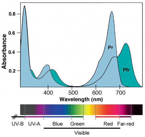
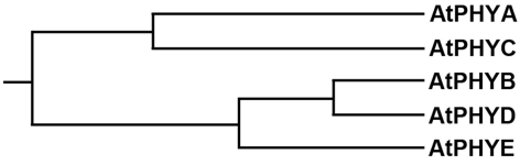
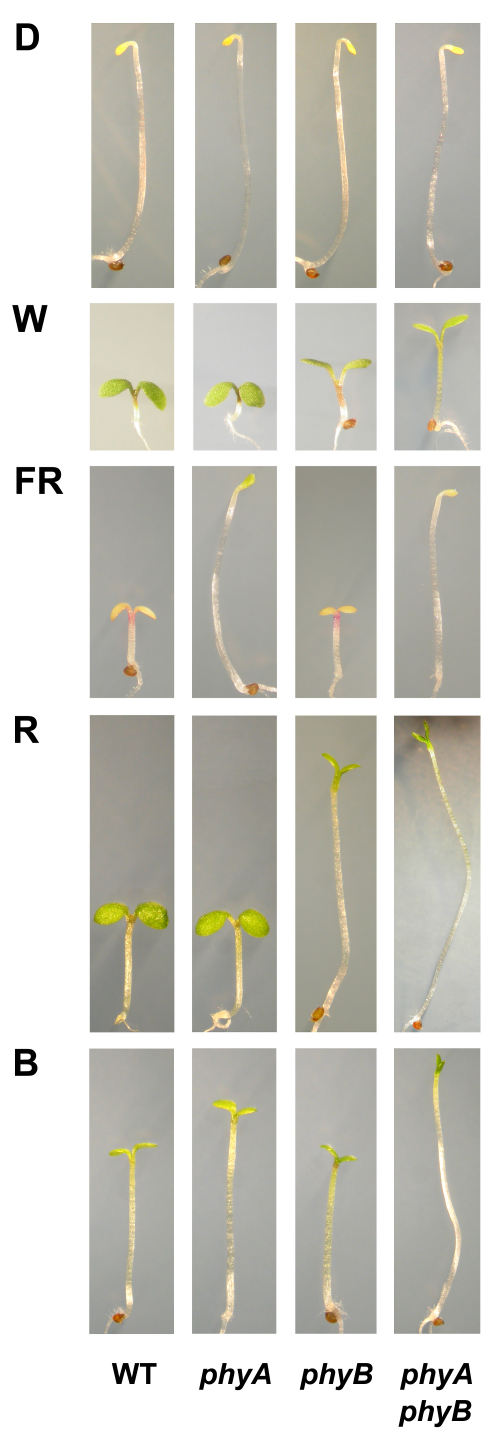
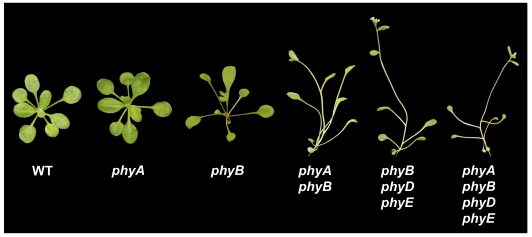
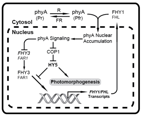
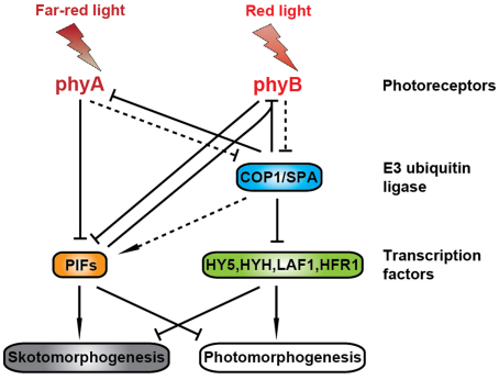
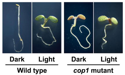
Phytochromes are red (R)/far-red (FR) light photoreceptors that play fundamental roles in photoperception of the light environment and the subsequent adaptation of plant growth and development. There are five distinct phytochromes in Arabidopsis thaliana, designated phytochrome A (phyA) to phyE. phyA is light-labile and is the primary photoreceptor responsible for mediating photomorphogenic responses in FR light, whereas phyB-phyE are light stable, and phyB is the predominant phytochrome regulating de-etiolation responses in R light. Phytochromes are synthesized in the cytosol in their inactive Pr form. Upon light irradiation, phytochromes are converted to the biologically active Pfr form, and translocate into the nucleus. phyB can enter the nucleus by itself in response to R light, whereas phyA nuclear import depends on two small plant-specific proteins FAR-RED ELONGATED HYPOCOTYL 1 (FHY1) and FHY1-LIKE (FHL). Phytochromes may function as light-regulated serine/threonine kinases, and can phosphorylate several substrates, including themselves in vitro. Phytochromes are phosphoproteins, and can be dephosphorylated by a few protein phosphatases. Photoactivated phytochromes rapidly change the expression of light-responsive genes by repressing the activity of CONSTITUTIVE PHOTOMORPHOGENIC 1 (COP1), an E3 ubiquitin ligase targeting several photomorphogenesis-promoting transcription factors for degradation, and by inducing rapid phosphorylation and degradation of Phytochrome-Interacting Factors (PIFs), a group of bHLH transcription factors repressing photomorphogenesis. Phytochromes are targeted by COP1 for degradation via the ubiquitin/26S proteasome pathway.


Figures

Figure 1.
Figure 1.
Absorption spectra of phytochromes. Absorption spectra of the two forms (Pr and Pfr) of phytochromes. The Pr form absorbs maximally at 660 nm, while the Pfr form absorbs maximallyat 730 nm. The visible light range of the human eye is approximately 380–700 nm. The light spectrum was adapted from Kami et al. (2010). Reprinted with permission from Elsevier.
Figure 2.
Figure 2.
Arabidopsis phytochrome chromophore. (A) The biosynthesis pathway of Arabidopsis phytochrome chromophore. Image adapted from Kohchi et al. (2001). (B) Red (R) light triggers a “Z” to “E” isomerization in the C15–C16 double bond between the C and D rings of the linear tetrapyrrole (upper panel), which is accompanied by rearrangement of the apoprotein backbone (lower panel; adapted from Bae and Choi, 2008). This results in the photoconversion of phytochromes from the Pr form to the Pfr form. Please note that the chromophore ring A rather than D is rotated during photoconversion according to a recent NMR analysis (Ulijasz et al., 2010). The discrepancy needs to be resolved in future studies. Far-red (FR) light converts the Pfr form back to the Pr form. Upper panel image reprinted from Bae and Choi (2008) with permission, from the Annual Review of Plant Biology, Volume 59 © 2008 by Annual Reviews (www.annualreviews.org).
Figure 3.
Figure 3.
The phylogenetic tree of the five phytochrome species from Arabidopsis thaliana. PHYB and PHYD share ∼80% amino acid sequence identity, and constitute a branch of the gene family. PHYE itself, PHYA and PHYC form two other branches of the evolutionary family tree. Image adapted from Clack et al. (1994). Reprinted with permission from Springer.
Figure 4.
Figure 4.
The domain structure of Arabidopsis phyA and phyB molecules. H, hinge; NTE, N-terminal extension; PAS, Per (period circadian protein), Arnt (Ah receptor nuclear translocator protein), and Sim (single-minded protein); GAF, cGMP-stimulated phosphodiesterase, Anabaena adenylate cyclases and Escherichia coli FhlA; PHY, phytochrome; PRD, PAS-related domain; HKRD, histidine kinase—related domain. The chromophore is attached to a conserved cysteine residue in the GAF domain. The numbers indicate the positions of each domain. Image adapted from Bae and Choi (2008). Reprinted with permission, from the Annual Review of Plant Biology, Volume 59 © 2008 by Annual Reviews (www.annualreviews.org).
Figure 5.
Figure 5.
Phenotypes of 4-d-old wild-type (WT), phyA, phyB and phyA phyB mutant plants grown in darkness (D) or under continuous white (W), far-red (FR), red (R) and blue (B) light conditions.
Figure 6.
Figure 6.
Phenotypes of 3-week-old wild-type (WT), phyA, phyB, phyA phyB, phyB phyD phyE, phyA phyB phyD phyE plants grown under white light conditions (16-h light/8-h dark).
Figure 7.
Figure 7.
Control of FHY1/FHL expression and phyA nuclear accumulation. FHY1 and FHL are required for phyA nuclear accumulation (Hiltbrunner et al., 2005, 2006; Genoud et al., 2008). FHY3 and FAR1 are two transposase-derived transcription factors that directly activate FHY1/FHL transcription, and thus indirectly regulate phyA nuclear accumulation and subsequent responses (Lin et al., 2007). phyA is localized exclusively in the cytosol in darkness in its inactive Pr form. Upon light exposure, the Pfr form of phyA is imported into the nucleus by FHY1/FHL, and thus triggers phyA signaling leading to multiple light responses, including the reduction of COP1 in the nucleus and accumulation of HY5 (Osterlund and Deng, 1998; Osterlund et al., 2000), and feedback regulation of FHY3 and FAR1 transcript levels (Lin et al., 2007). HY5 plays dual roles in phyA signaling: promoting photomorphogenesis, and down-regulating FHY1/FHL transcript levels by modulating the activities of the transcriptional activators FHY3 and FAR1 (Li et al., 2010). FHY3 and FHY1 (indicated by larger letters) are the more predominant players in the phyA signaling process compared to their respective homologs FAR1 and FHL. Pr: R-absorbing form of phyA (inactive); Pfr: FR-absorbing form of phyA (active). Arrow, positive regulation; bar, negative regulation. Image adapted from Li et al. (2010).
Figure 8.
Figure 8.
Structural comparison of Arabidopsis phytochromes and the bacterial phytochrome Cph1 (adapted from Yeh and Lagarias, 1998). HKD, histidine kinase domain. The percent amino acid identities between the HKD domain of Cph1 and both PRD and HKRD domains of Arabidopsis phytochromes are indicated. Image adapted from Yeh and Lagarias (1998). Reprinted with permission from the National Academy of Sciences.
Figure 9.
Figure 9.
A simplified model of the phytochrome signaling pathway. phyA is the primary photoreceptor responsible for perceiving and mediating various responses to FR light, whereas phyB is the predominant phytochrome regulating responses to R light. Under light conditions, these photoreceptors act to suppress two main branches of light signaling: COP1-TFs and PIFs.COP1, whose activity is repressed by phytochromes in light conditions, is an E3 ubiquitin ligase targeting several photomorphogenesis-promoting transcription factors (such as HY5, HYH, LAF1 and HFR1) for degradation. PIFs are a subset of bHLH transcription factors required for skotomorphogenesis. Photo-activated phytochromes directly interact with PIFs, resulting in PIFs' phosphorylation and degradation, while COP1 positively regulates PIFs' protein levels. Phytochromes are targeted for degradation by COP1, and PIFs contribute to the degradation of phyB by promoting COP1/phyB interaction. Arrow, positive regulation; bar, negative regulation; solid line, direct regulation; dotted line, indirect regulation. Image adapted from Lau and Deng (2010). Reprinted with permission from Elsevier.
Figure 10.
Figure 10.
Dark-grown cop1 mutant seedlings phenotypically mimic light-grown wild-type seedlings.

References

    1. Ahmad M., Cashmore A.R. The blue-light receptor cryptochrome 1 shows functional dependence on phytochrome A or phytochrome B in Arabidopsis thaliana. Plant J. 1997;11:421–427. - PubMed
    1. Ahmad M., Jarillo J.A., Smirnova O., Cashmore A.R. The CRY1 blue light photoreceptor of Arabidopsis interacts with phytochrome A in vitro. Mol. Cell. 1998;1:939–948. - PubMed
    1. Allen T., Koustenis A., Theodorou G., Somers D.E., Kay S.A., Whitelam G.C., Devlin P.F. Arabidopsis FHY3 specifically gates phytochrome signaling to the circadian clock. Plant Cell. 2006;18:2506–2516. - PMC - PubMed
    1. Al-Sady B., Kikis E.A., Monte E., Quail P.H. Mechanistic duality of transcription factor function in phytochrome signaling. Proc. Natl. Acad. Sci. USA. 2008;105:2232–2237. - PMC - PubMed
    1. Al-Sady B., Ni W., Kircher S., Schafer E., Quail P.H. Photoactivated phytochrome induces rapid PIF3 phosphorylation prior to proteasome-mediated degradation. Mol. Cell. 2006;23:439–446. - PubMed