. 2012;7(1):e30841.
doi: 10.1371/journal.pone.0030841. Epub 2012 Jan 27.

Coxiella burnetii induces apoptosis during early stage infection via a caspase-independent pathway in human monocytic THP-1 cells

Affiliations

Coxiella burnetii induces apoptosis during early stage infection via a caspase-independent pathway in human monocytic THP-1 cells

Yan Zhang et al. PLoS One. 2012.

Abstract

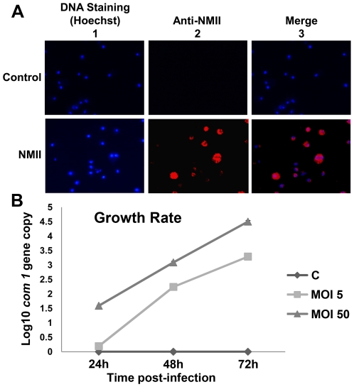
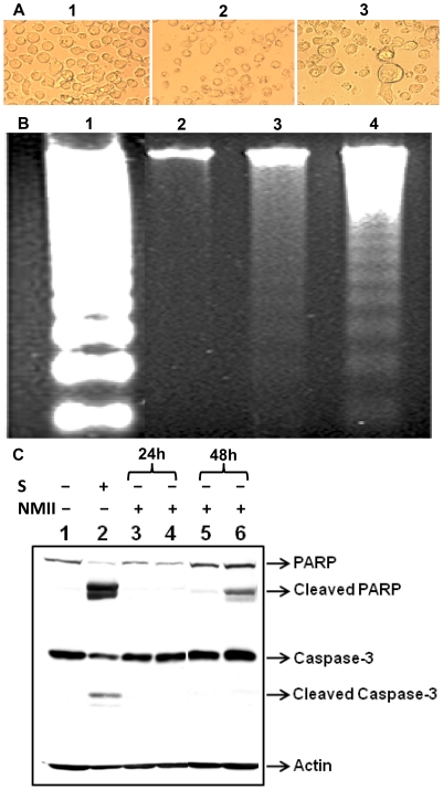
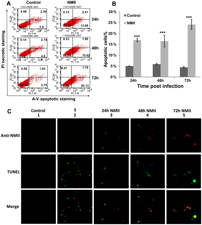
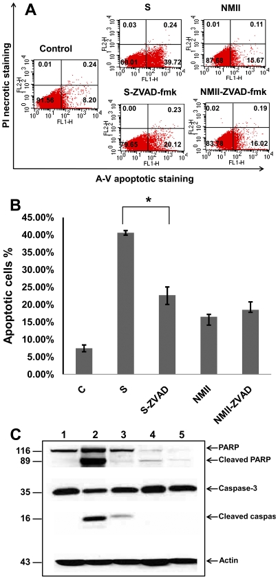
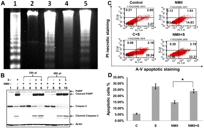
The ability of Coxiella burnetii to modulate host cell death may be a critical factor in disease development. In this study, human monocytic THP-1 cells were used to examine the ability of C. burnetii Nine Mile phase II (NMII) to modulate apoptotic signaling. Typical apoptotic cell morphological changes and DNA fragmentation were detected in NMII infected cells at an early stage of infection. FACS analysis using Annexin-V-PI double staining showed the induction of a significant number of apoptotic cells at an early stage of NMII infection. Double staining of apoptotic cell DNA and intracellular C. burnetii indicates that NMII infected cells undergoing apoptosis. Interestingly, caspase-3 was not cleaved in NMII infected cells and the caspase-inhibitor Z-VAD-fmk did not prevent NMII induced apoptosis. Surprisingly, the caspase-3 downstream substrate PARP was cleaved in NMII infected cells. These results suggest that NMII induces apoptosis during an early stage of infection through a caspase-independent pathway in THP-1 cells. In addition, NMII-infected monocytes were unable to prevent exogenous staurosporine-induced apoptotic death. Western blot analysis indicated that NMII infection induced the translocation of AIF from mitochondria into the nucleus. Cytochrome c release and cytosol-to-mitochondrial translocation of the pore-forming protein Bax in NMII infected cells occurred at 24 h post infection. These data suggest that NMII infection induced caspase-independent apoptosis through a mechanism involving cytochrome c release, cytosol-to-mitochondrial translocation of Bax and nuclear translocation of AIF in THP-1 monocytes. Furthermore, NMII infection increased TNF-α production and neutralization of TNF-α in NMII infected cells partially blocked PARP cleavage, suggesting TNF-α may play a role in the upstream signaling involved in NMII induced apoptosis. Antibiotic inhibition of C. burnetii RNA synthesis blocked NMII infection-induced PARP activation. These results suggest that both intracellular C. burnetii replication and secreted TNF-α contribute to NMII infection-triggered apoptosis during an early stage of infection.


Conflict of interest statement

Competing Interests: The authors have declared that no competing interests exist.

Figures

Figure 1
Figure 1. Growth rate and indirect immunofluorescence assay (IFA) staining of NMII in THP-1 cells.
Cells were infected with NMII at MOI of 50 for 24 h. Infected cells were then washed and incubation was continued for 48 h. Infected and uninfected THP-1 cells were fixed with paraformaldehyde and permeabilized. Intracellular C. burnetii were stained by IFA with rabbit anti-Coxiella polyclonal antibodies and viewed using a fluorescence microscope. Panel 1A, IFA staining of NMII infected THP-1 cells. Upper panel: uninfected control cells; lower panel: NMII infected cells. 1 Hoechst staining for host cell DNA; 2 Cells stained with anti-Coxiella antibodies; and 3 Merge. Panel 1B, growth rate of NMII in THP-1 cells. NMII infected and uninfected monocytic THP-1 cells were directly lysed at 24, 48 or 72 h post infection. DNA was extracted and used as a template to quantify the number of C. burnetii com1 gene copies by real-time PCR.
Figure 2
Figure 2. C. burnetii NMII infection induced cell death in THP-1 cells.
NMII (MOI 50) infected and control cells were cultured in 24 well plates. Uninfected control cells were treated with staurosporine (1 µm) for 4 h. Panel 2A, morphological changes in NMII infected THP-1 cell at 48 h post infection. 1, control cells; 2, normal cells with staurosporine-treatment; and 3, NMII infected cells. Typical apoptotic morphological changes were displayed in staurosporine-treated and NMII infected cells. Arrow indicates large vacuoles that contain intracellular organisms in NMII infected cells. Panel 2B, pattern of DNA fragmentation during NMII infection. Genomic DNA from NMII infected and control cells were extracted at 48 h post infection. DNA was separated on 1.8% agarose gels and stained with ethidium bromide. Lane 1, U937 apoptotic cells as positive control; Lane 2, Uninfected THP-1 cell control; Lane 3, NMII infected THP-1 cells; Lane 4, Staurosporine treated THP-1 cells. Panel 2C, Western blot of PARP and caspase-3 activities in NMII induced apoptosis. Lane 1, normal cells; Lane 2, staurosporine treated positive control cells; Lane 3, NMII (MOI 5) infected cells at 24 h post infection; Lane 4, NMII (MOI 50) infected cells at 24 h post infection; Lane 5, NMII (MOI 5) infected cells at 48 h post infection; Lane 6, NMII (MOI 50) infected cells at 48 h post infection.
Figure 3
Figure 3. FACS Analysis of Annexin-V staining of NMII infected THP-1 cells.
Approximately 1×10⁶ NMII infected or uninfected cells were double stained with Annexin-V-FITC and PI. Panel 3A, representative FACS scatter plots of THP-1 cells. Fluorescence was detected using a fluorescence-activated cell sorter to analyze necrotic (PI+), non-apoptotic (negative for both dyes), early apoptotic (Annexin+/PI−), and late apoptotic cells (Annexin+/PI+). Panel 3B, percentages of apoptotic cells in NMII infected THP-1 cells. Data shown represents the Mean±SE from at least three independent experiments. *denotes significant differences (***p<0.001) between infected and uninfected cells at each time point post infection. Panel 3C, double staining of intracellular C. burnetii and apoptotic cell DNA. Intracellular C. burnetii was stained by IFA with rabbit anti-Coxiella polyclonal antibodies and apoptotic host nuclei were stained with TUNEL staining. Upper panel: Cells stained with anti-Coxiella antibodies; Middle panel: TUNEL stating of apoptotic host nuclei; Lower panel: Merge. From left to right 1) Normal cell control; 2) Staurosporine (1 µm) treated apoptotic control cell; 3) NMII infected cells at 24 h post infection; 4) NMII infected cells at 48 h post infection; 5) NMII infected cells at 72 h post infection.
Figure 4
Figure 4. The caspase inhibitor ZVAD-fmk failed to inhibit NMII induced cell death.
Panel 4A, FACS analysis of Annexin-V staining in ZVAD-fmk treated NMII infected THP-1 cells. Control cells were treated with staurosporine (1 µm) or staurosporine with ZVAD-fmk (50 µm) for 4 h. NMII infected THP-1 cells were treated with ZVAD-fmk (50 µm) and refreshed daily up to 48 h post infection. Fluorescence was detected using a fluorescence-activated cell sorter to analyze necrotic (PI+), non-apoptotic (negative for both dyes), early apoptotic (Annexin+/PI−), and late apoptotic cells (Annexin+/PI+). Panel 4B, percentage of apoptotic cells in Z-VAD-fmk treated NMII infected THP-1 cells. Data shown are the Mean±SE from at least three independent experiments. *denotes significant differences (*p<0.05). Panel 4C, Western blot of Caspase-3 and PARP activities in ZVAD-fmk treated NMII infected cells. Lane 1, normal THP-1 cells; lane 2, staurosporine treated THP-1 cells; lane 3, staurosporine with ZVAD-fmk treated THP-1 cells; lane 4, NMII infected THP-1 cells; lane 5, NMII infected THP-1 cells treated with ZVAD-fmk.
Figure 5
Figure 5. Inability of C. burnetii infection to inhibit exogenously induced apoptosis.
THP-1 cells were infected with NMII at MOI 5 or 50 and left untreated or treated with staurosporine (1 µm) for 4 h. Panel 5A, genomic DNA from 48 h post infected cells and control cells were run on 1.8% agarose gels and stained with ethidium bromide. Lane 1, U937 apoptotic cells as positive control; Lane 2, Uninfected THP-1 cell control; Lane 3, THP-1 cells treated with 1 µm staurosporine for 4 h. Lane 4, NMII infected THP-1 cells; Lane 5, NMII infected cells treated with 1 µm staurosporine for 4 h. Panel 5B, FACS Analysis of staurosporine-treated NMII infected THP-1 cells. Fluorescence was detected using a fluorescence-activated cell sorter to analyze necrotic (PI+), non-apoptotic (negative for both dyes), early apoptotic (Annexin+/PI−), and late apoptotic cells (Annexin+/PI+). Panel 5C, percentage of apoptotic cells in staurosporine treated NMII infected THP-1 cells. Data shown are the Mean±SE from at least three independent experiments. *denotes significant differences (*p<0.05). Panel 5D, Western blot of caspase-3 and PARP activities in staurosporine treated NMII infected THP-1 cells. Lane 1, normal cells; Lane 2, staurosporine treated cells; Lane 3, NMII (MOI 5) infected cells at 24 h post infection; Lane 4, NMII (MOI 50) infected cells at 24 h post infection; Lane 5, staurosporine treated NMII (MOI 5) infected cells at 24 h post infection; Lane 6, staurosporine treated NMII (MOI 50) infected cells at 24 h post infection; Lane 7, NMII (MOI 5) infected cells at 48 h post infection; Lane 8, NMII (MOI 50) infected cells at 48 h post infection; Lane 9, staurosporine treated NMII (MOI 5) infected cells at 48 h post infection; Lane 10, staurosporine treated NMII (MOI 50) infected cells at 48 h post infection.
Figure 6
Figure 6. NMII induced apoptosis involved in AIF nuclear translocation and changes in mitochondrial membrane permeability.
Panel 6A, Western blot of AIF translocation in NMII infected THP-1 cells. Infected cells were harvested at different time points post infection. Mitochondrial or nuclear fraction was prepared for analysis of AIF translocation. Lane 1, control cells; lane 2, NMII (MOI 5) infected cells at 24 h post infection; lane 3, NMII (MOI 50) infected cells at 24 h post infection; lane 4, NMII (MOI 5) infected cells at 48 h post infection; lane 5, NMII (MOI 50) infected cells at 48 h post infection. Panel 6B, Western blot analysis of cytochrome c release and translocation of Bax. Lane 1, control cells; lane 2, NMII (MOI 5) infected cells at 24 h post infection; lane 3, NMII (MOI 50) infected cells at 24 h post infection; lane 4, NMII (MOI 5) infected cells at 48 h post infection; lane 5, NMII (MOI 50) infected cells at 48 h post infection.
Figure 7
Figure 7. Neutralization of secreted TNF-α partially blocked PARP cleavage.
Panel 7A, concentration of TNF-α in NMII infected THP-1 cell supernatants as measured by ELISA. LPS treated THP-1 cells were used as positive controls. THP-1 cells were infected with different MOIs (5 or 50) of NMII. LPS (100 ng/ml) treated or NMII infected THP-1 cells were treated with anti-human TNF-α/TNFSF1A (0.05 µg/ml). Supernatants were collected at 24, 48 and 72 h post-treatment or infection, respectively. Panel 7B, Western blot of PARP activity in TNF-α neutralized NMII infected THP-1 cells. Lane 1, control cells; lane 2, LPS treated cells at 48 h; lane 3, LPS treated cells with anti-human TNF-α at 48 h; lane 4, NMII (MOI 5) infected cells at 48 h post infection; lane 5, NMII (MOI 5) infected cells with anti-human TNF-α at 48 h post infection; lane 6, NMII (MOI 50) infected cells at 48 h post infection; lane 7, NMII (MOI 50) infected cells with anti-human TNF-α at 48 h post infection.
Figure 8
Figure 8. NMII induced apoptosis depends on C. burnetii replication.
NMII infected THP-1 cells were treated with 10 µg/ml rifampin up to 72 h post infection. Panel 8A, quantification of C. burnetii com1 gene copies by real-time PCR. Panel 8B, Western blot of rifampin inhibition in NMII infected THP-1 cells. Lane 1, normal cells; Lane 2, staurosporine treated positive control cells; Lane 3, NMII (MOI 5) infected cells at 24 h post infection; Lane 4, NMII (MOI 50) infected cells at 24 h post infection; Lane 5, NMII (MOI 5) infected cells with rifampin treatment at 24 h post infection; Lane 6, NMII (MOI 50) infected cells with rifampin treatment at 24 h post infection; Lane 7, NMII (MOI 5) infected cells at 48 h post infection; lane 8, NMII (MOI 50) infected cells at 48 h post infection; Lane 9, NMII (MOI 5) infected cells with rifampin at 48 h post infection; Lane 10, NMII (MOI 50) infected cells with rifampin treatment at 48 h post infection.
Figure 9
Figure 9. Hypothetic model of C. burnetii induced caspase-independent apoptosis in monocytic THP-1 cells.
C. burnetii infection increases TNF-α release. The ligation of TNFR-1 results in recruitment of proteins containing death domains (DD), including FADD, TRADD and RIP. FADD can stimulate caspase-independent cell death through mitochondrial ROS. Bacterial factors may release during intracellular C. burnetii replication and may direct target on PARP that subsequently cause translocation of pore-forming protein Bax/Bak to the mitochondrial, leading to the release of AIF and cytochrome c. PARP activation is a sensitive indicator of DNA damage that may also be induced by oxidative stress via TNF signaling. Overactivation of PARP transduces signaling (PAR polymer and PARP-bound proteins) to cause AIF to translocate from mitochondria to the nucleus, where response for the nuclear condensation and fragmentation. PARP-mediated apoptosis result in spread of intracellular bacterial to neighboring cells.

Similar articles

Cited by

References

    1. Maurin M, Raoult D. Q fever. Clin Microbiol Rev. 1999;12:518–553. - PMC - PubMed
    1. Gao LY, Kwaik YA. The modulation of host cell apoptosis by intracellular bacterial pathogens. Trends Microbiol. 2000;8:306–313. - PubMed
    1. Baca OG, Paretsky D. Q fever and Coxiella burnetii: a model for host-parasite interactions. Microbiol. Rev. 1983;47:127–149. - PMC - PubMed
    1. Beare PA, Samuel JE, Howe D, Virtaneva K, Porcella SF, et al. Genetic diversity of the Q fever agent, Coxiella burnetii, assessed by microarray-based whole-genome comparisons. J Bacteriol. 2006;188:2309–2324. - PMC - PubMed
    1. Hackstadt T. The role of lipopolysaccharides in the virulence of Coxiella burnetii. Ann NY Acad Sci. 1990;590:27–32. - PubMed

Publication types

MeSH terms