. 2012;7(1):e31049.
doi: 10.1371/journal.pone.0031049. Epub 2012 Jan 27.

PKCθ synergizes with TLR-dependent TRAF6 signaling pathway to upregulate MUC5AC mucin via CARMA1

Affiliations

PKCθ synergizes with TLR-dependent TRAF6 signaling pathway to upregulate MUC5AC mucin via CARMA1

Hirofumi Jono et al. PLoS One. 2012.

Abstract

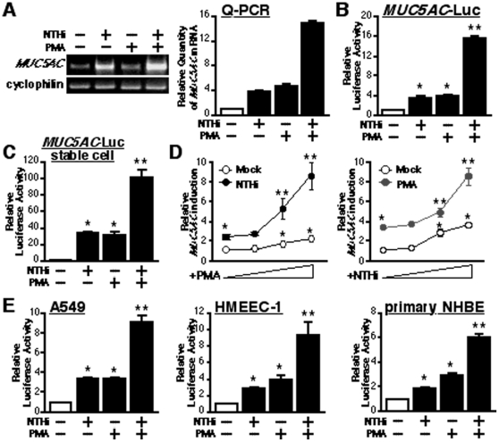
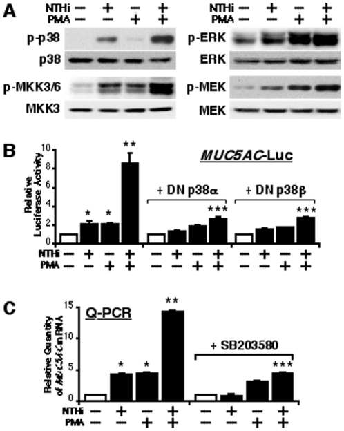
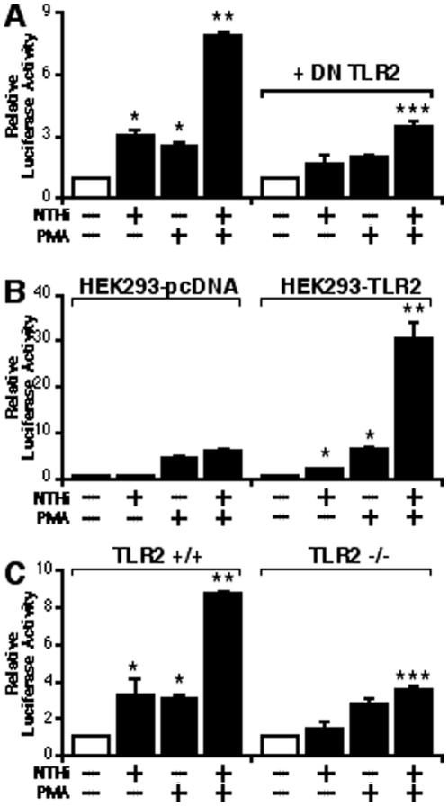
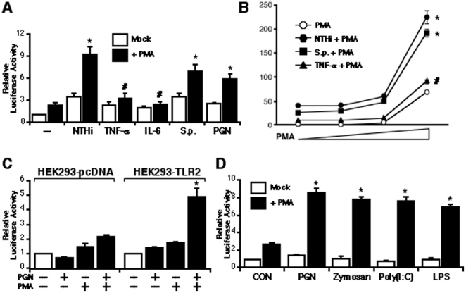
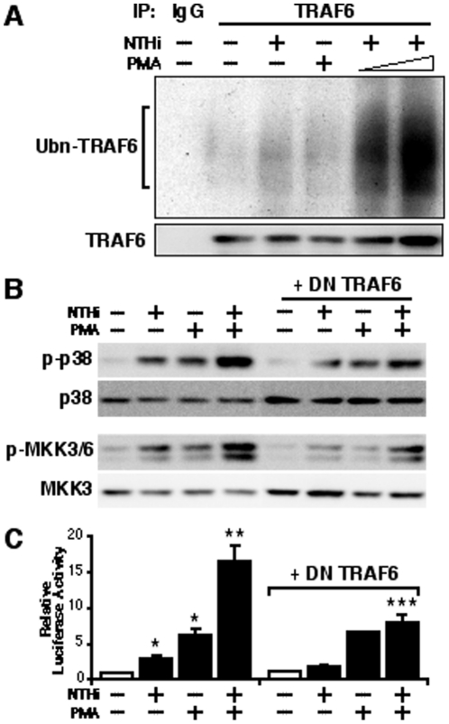
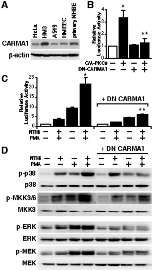
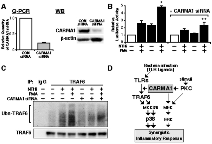
CARD-containing MAGUK protein 1 (CARMA1) plays a crucial role in regulating adaptive immune responses upon T-cell receptor (TCR) activation in T cells. Its role in regulating host mucosal innate immune response such as upregulation of mucin remains unknown. Here we show that CARMA1 acts as a key signaling mediator for synergistic upregulation of MUC5AC mucin by bacterium nontypeable Haemophilus influenzae (NTHi) and phorbol ester PMA in respiratory epithelial cells. NTHi-induced TLR-dependent TRAF6-MKK3-p38 MAPK signaling pathway synergizes with PKCθ-MEK-ERK signaling pathway. CARMA1 plays a crucial role in mediating this synergistic effect via TRAF6, thereby resulting in synergistic upregulation of MUC5AC mucin. Thus our study unveils a novel role for CARMA1 in mediating host mucosal innate immune response.


Conflict of interest statement

Competing Interests: The authors have declared that no competing interests exist.

Figures

Figure 1
Figure 1. PMA synergizes with NTHi to induce MUC5AC expression in human epithelial cell.
(A) PMA synergistically enhanced NTHi-induced MUC5AC expression at the mRNA level in human epithelial HM3 cell, as assessed by performing RT-PCR (left panel) and real-time quantitative PCR (Q-PCR) analysis (right panel). Cyclophilin was used as a control for amount of RNA used in each reaction. (B) PMA synergized with NTHi to induce MUC5AC expression at the transcriptional level in human epithelial cells, as assessed by MUC5AC-dependent promoter Luciferase assay. (C) Synergistic induction of MUC5AC expression by NTHi and PMA was also observed in HM3 cells stably transfected with pMUC5AC 3.7 kb-luc. (D) PMA synergizes with NTHi to induce MUC5AC transcription in a dose-dependent manner and vice versa. (E) PMA synergistically enhanced NTHi-induced MUC5AC expression at the transcriptional level in human airway A549, middle ear HMEEC-1 and primary bronchial epithelial NHBE cells, as assessed by MUC5AC-dependent promoter assays. Values are the means ± S.D. (n = 3). *p<0.05 vs. control; **p<0.05 vs. NTHi alone. The data shown are representative of three independent experiments. −, absence of; +, presence of; NTHi, nontypeable Haemophilus influenzae.
Figure 2
Figure 2. PMA synergistically enhances NTHi-induced MUC5AC expression via MKK3/6-p38 MAPK pathway.
(A) PMA synergistically enhanced NTHi-induced phosphorylation of p38 MAPK and MKK3/6, but not ERK and MEK1. (B) The synergistic induction of MUC5AC transcription by NTHi and PMA was inhibited by overexpressing DN mutant forms of p38α and p38β in human epithelial cells, as assessed by MUC5AC-dependent promoter Luciferase assay. Cells were transfected with 0.8 µg of DN p38α, DN p38β, or control vector, and treated with NTHi with or without PMA. Relative luciferase activity of MUC5AC was measured from the cell lysate. (C) SB203580, a specific inhibitor for p38 MAPK signaling, attenuated the synergistic induction of MUC5AC expression by NTHi and PMA at the mRNA level as assessed by Q-PCR. Cells were pre-treated with 10 µM of SB203580 or vehicle control, and treated with NTHi with or without PMA. mRNA expression level of MUC5AC was measured by Q-PCR. Values are the means ± S.D. (n = 3). *p<0.05 vs. control; **p<0.05 vs. NTHi alone; ***p<0.05 vs NTHi with PMA in control vector transfected (B) or vehicle treated (C) cells. The data shown are representative of three independent experiments. −, absence of; +, presence of; DN, dominant negative; NTHi, nontypeable Haemophilus influenzae.
Figure 3
Figure 3. TLR2 is involved in the synergistic enhancement of NTHi-induced MUC5AC expression by PMA.
(A) Overexpression of a DN mutant form of TLR2 inhibited the synergistic induction of MUC5AC transcription by NTHi and PMA in human epithelial cells, as assessed by MUC5AC-dependent promoter Luciferase assay. Cells were transfected with 0.8 µg of DN TLR2 or control vector, and treated with NTHi with or without PMA. Relative luciferase activity of MUC5AC was measured from the cell lysate. (B) The synergistic enhancement of NTHi-induced MUC5AC transcription by PMA was observed in HEK293-TLR2 cells, but not in HEK293-pcDNA cells. (C) PMA did not synergize with NTHi to induce MUC5AC expression in TLR2−/− MEF cells. Values are the means ± S.D. (n = 3). *p<0.05 vs. control; **p<0.05 vs. NTHi alone; ***p<0.05 vs NTHi with PMA in control vector transfected cells (A) or TLR2+/+ cells (C). The data shown are representative of three independent experiments. −, absence of; +, presence of; NTHi, nontypeable Haemophilus influenzae; DN, dominant negative; +/+, wild-type; −/−, knock-out.
Figure 4
Figure 4. PMA synergistically enhances up-regulation of MUC5AC induced by activation of TLR signaling.
(A) PMA synergistically enhanced MUC5AC expression induced only by NTHi, S.p. and PGN, but not by TNF-α and IL-6, as assessed by MUC5AC-dependent promoter Luciferase assay. Values are the means ± S.D. (n = 3). *p<0.05 vs. NTHi, S.p., or PGN alone; #p>0.05 vs. TNF-α or IL-6 alone. (B) PMA synergized with either NTHi or S.p., but not with TNF-α to enhance MUC5AC expression in a dose-dependent manner. Values are the means ± S.D. (n = 3). *p<0.05 vs. PMA alone; #p>0.05 vs. PMA alone. (C) PGN-induced MUC5AC transcription was synergistically enhanced by PMA in HEK293-TLR2 cells, but not in HEK293-pcDNA cells. Values are the means ± S.D. (n = 3). *p<0.05 vs. PGN alone. (D) Synergistic enhancement of MUC5AC expression by PMA was also observed in the cells treated with TLR ligands, such as PGN, Zymosan, Poly(I:C) and LPS, respectively. Values are the means ± S.D. (n = 3). *p<0.05 vs. PGN, Zymosan, Poly(I:C), or LPS alone. The data shown are representative of three independent experiments. −, absence of; +, presence of; S.p., Streptococcus pneumoniae; NTHi, nontypeable Haemophilus influenzae.
Figure 5
Figure 5. PMA synergistically enhances TLR-dependent MUC5AC induction by NTHi via TRAF6.
(A) PMA synergistically enhanced NTHi-induced polyubiquitination of TRAF6 in human epithelial cells. Cells were transfected with TRAF6, and were treated with NTHi and PMA as indicated. Whole cell extracts were subjected to co-immunoprecipitation (IP) with either control IgG or anti-TRAF6 antibodies and immunoblotting with anti-ubiquitin antibody. The same blots were re-probed with anti-TRAF6. (B) Overexpression of a DN mutant form of TRAF6 blocked the synergistic phosphorylation of p38 MAPK and MKK3/6 induced by NTHi and PMA. Cells were transfected with 0.8 µg of DN-TRAF6 or control vector, and treated with NTHi with or without PMA. Cell lysate was blotted with antibodies as indicated in the figure. (C) The synergistic enhancement of NTHi-induced MUC5AC transcription by PMA was inhibited by overexpression of DN-TRAF6 in human epithelial cells, as assessed by MUC5AC-dependent promoter Luciferase assay. Cells were transfected with 0.8 µg of DN-TRAF6 or control vector, and treated with NTHi with or without PMA. Relative luciferase activity of MUC5AC was measured from the cell lysate. Values are the means ± S.D. (n = 3). *p<0.05 vs. control; **p<0.05 vs. NTHi alone; ***p<0.05 vs. NTHi with PMA in control vector transfected cells. The data shown are representative of three independent experiments. −, absence of; +, presence of; NTHi, nontypeable Haemophilus influenzae; DN, dominant negative; Ubn, ubiquitin.
Figure 6
Figure 6. TLR-TRAF6-dependent synergistic MUC5AC induction by NTHi and PMA is mediated by PKCθ.
(A) Rottlerin, a specific PKC™/θ inhibitor, blocked the synergistic induction of MUC5AC transcription by NTHi and PMA in human epithelial cells, as assessed by MUC5AC-dependent promoter Luciferase assay. Cells were pre-treated with 20 µM of Rottlerin or vehicle control, and treated with NTHi with or without PMA. Relative luciferase activity of MUC5AC was measured from the cell lysate. Values are the means ± S.D. (n = 3). *p<0.05 vs. control; **p<0.05 vs. NTHi alone; ***p<0.05 vs. NTHi with PMA in vehicle treated cells. (B) The synergistic induction of MUC5AC expression was also attenuated by Rottlerin at the mRNA level, as assessed by performing Q-PCR. Cells were pre-treated with 20 µM of Rottlerin or vehicle control, and treated with NTHi with or without PMA. mRNA expression level of MUC5AC was measured by Q-PCR. (C) Co-expressing WT-PKCθ enhanced, whereas DN-PKCθ inhibited, the synergistic induction of MUC5AC transcription by NTHi and PMA. Cells were transfected with 0.3 µg of WT- PKCθ, 0.6 µg of DN PKCθ, or control vector, and treated with NTHi with or without PMA. Relative luciferase activity of MUC5AC was measured from the cell lysate. Values are the means ± S.D. (n = 3). *p<0.05 vs. control; **p<0.05 vs. NTHi alone; ***p<0.05 vs. NTHi with PMA in control vector transfected cells. (D) C/A-PKC-induced MUC5AC expression was synergistically enhanced by NTHi in human epithelial cells. Cells were transfeced with 0.3 µg of C/A PKCθ or control vecttor, and treated with NTHi. Relative luciferase activity of MUC5AC was measured from the cell lysate. Values are the means ± S.D. (n = 3). *p<0.05 vs. NTHi alone. (E) C/A-PKCθ synergized with WT-TRAF6 to induce MUC5AC expression in a dose-dependent manner. Cells were transfected with 0.1, 0.3, or 0.6 µg of C/A PKCθ with or without 0.3 µg of WT-TRAF6. mRNA expression level of MUC5AC was measured by Q-PCR. Values are the means ± S.D. (n = 3). *p<0.05 vs. control; **p<0.05 vs. C/A PKCθ transfected cells. The data shown are representative of three independent experiments. −, absence of; +, presence of; NTHi, nontypeable Haemophilus influenzae; WT, wild-type; DN, dominant negative; C/A, constitutively active form.
Figure 7
Figure 7. CARMA1 acts downstream of PKCθ in mediating PMA-induced synergistic enhancement of TLR-TRAF6-dependent MUC5AC expression.
(A) CARMA1 was expressed in a variety of epithelial cells, such as in human cervix HeLa, colon HM3, airway A549, middle ear HMEEC-1 and primary bronchial epithelial NHBE cells, as assessed by WB using antibody against CARMA1. (B) Overexpression of a DN mutant form of CARMA1 attenuated C/A-PKCθ-induced MUC5AC expression. Cells were transfeced with 0.3 µg of C/A PKCθ with or without 0.8 µg of DN-CARMA1. Relative luciferase activity of MUC5AC was measured from the cell lysate. Values are the means ± S.D. (n = 3). *p<0.05 vs. control; **p<0.05 vs. C/A PKCθ transfected cells. (C) The synergistic induction of MUC5AC transcription by NTHi and PMA was potently inhibited by overexpression of DN-CARMA1. Cells were transfeced with 0.8 µg of DN-CARMA1 or control vector, and treated with NTHi with or without PMA. Relative luciferase activity of MUC5AC was measured from the cell lysate. Values are the means ± S.D. (n = 3). *p<0.05 vs. NTHi alone; **p<0.05 vs. control vector transfected cells. (D) Overexpression of a DN mutant form of CARMA1 greatly inhibited not only synergistic phosphorylation of p38 MAPK and MKK3/6 induced by NTHi and PMA, but also ERK and MEK phosphorylation. Cells were transfeced with 0.8 µg of DN-CARMA1 or control vector, and treated with NTHi with or without PMA. Cell lysate was blotted with antibodies indicated in the figure. The data shown are representative of three independent experiments. −, absence of; +, presence of; NTHi, nontypeable Haemophilus influenzae; DN, dominant negative; C/A, constitutively active form.
Figure 8
Figure 8. CARMA1 mediates TLR-dependent synergistic MUC5AC induction by NTHi and PMA via cross-talk with TRAF6.
(A) CARMA1 knockdown by CARMA1-siRNA efficiently reduced the endogenous CARMA1 expression at both the mRNA and protein level, as assessed by Q-PCR and WB, respectively. (B) CARMA1-siRNA inhibited the synergistic induction of MUC5AC transcription by NTHi and PMA. Values are the means ± S.D. (n = 3). *p<0.05 vs. NTHi alone; **p<0.05 vs. control siRNA transfeced cells. (C) Synergistic enhancement of NTHi-induced TRAF6 polyubiquitination by PMA was attenuated by CARMA1 knockdown. (D) Schematic representation depicting how CARMA1 mediates the synergistic enhancement of MUC5AC expression in human epithelial cells. The data shown are representative of three independent experiments. CON, control; −, absence of; +, presence of; NTHi, nontypeable Haemophilus influenzae; Ubn, ubiquitin; TLRs, toll-like receptors; TRAF6, TNF receptor associated factor 6; PKC, protein kinase C.

Similar articles

Cited by

References

    1. Liew FY, Xu D, Brint EK, O'Neill LA. Negative regulation of toll-like receptor-mediated immune responses. Nat Rev Immunol. 2005;5:446–458. - PubMed
    1. Dunne A, O'Neill LA. The interleukin-1 receptor/Toll-like receptor superfamily: signal transduction during inflammation and host defense. Sci STKE. 2003;2003:re3. - PubMed
    1. Akira S, Takeda K. Functions of toll-like receptors: lessons from KO mice. C R Biol. 2004;327:581–589. - PubMed
    1. Beutler B. Inferences, questions and possibilities in Toll-like receptor signalling. Nature. 2004;430:257–263. - PubMed
    1. Jono H, Shuto T, Xu H, Kai H, Lim DJ, et al. Transforming growth factor-beta -Smad signaling pathway cooperates with NF-kappa B to mediate nontypeable Haemophilus influenzae-induced MUC2 mucin transcription. J Biol Chem. 2002;277:45547–45557. - PubMed

Publication types

MeSH terms