

Binding of filamentous actin to anthrax toxin receptor 1 decreases its association with protective antigen
- PMID: 22303962
- PMCID: PMC3286128
- DOI: 10.1021/bi2016469
Binding of filamentous actin to anthrax toxin receptor 1 decreases its association with protective antigen
Abstract
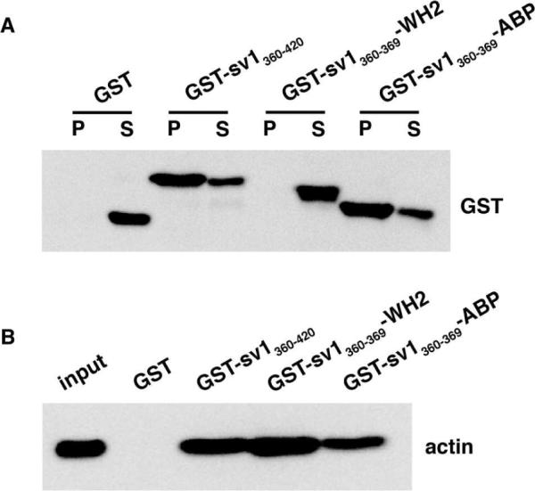
ANTXR1 is a type I membrane protein that binds the protective antigen (PA) component of anthrax toxin. The cytosolic domain of ANTXR1 has a novel actin-binding region that influences the interaction of the ectodomain with PA. Here, we have investigated features of the cytosolic domain of ANTXR1 that reduce the association of the receptor with PA. We mutated a stretch of conserved acidic amino acids adjacent to the actin-binding region and found that the mutation increased the affinity for monomeric actin in vitro. ANTXR1 bearing this mutation exhibited increased association with the cytoskeleton and bound less PA compared to the wild-type receptor, confirming the inverse correlation between the two interactions. To determine whether binding of actin is sufficient to regulate the ectodomain, we replaced the actin-binding region of ANTXR1 with that from the yeast protein abp140 and with the WH2 domain of WAVE2. Although both of these domains bound monomeric actin in vitro, only the sequence from abp140 reduced binding of PA to a hybrid receptor. The actin binding regions of ANTXR1 and abp140, but not the WH2 domain, colocalized with actin stress fibers, which suggested that filamentous actin regulates ANTXR1. Consistent with this notion, disruption of actin filaments using latrunculin A increased the amount of PA bound to cells. This work provides evidence that cytoskeletal dynamics regulate ANTXR1 function.
Figures


Similar articles
-
Changing the receptor specificity of anthrax toxin.mBio. 2012 May 1;3(3):e00088-12. doi: 10.1128/mBio.00088-12. Print 2012. mBio. 2012. PMID: 22550037 Free PMC article.
-
Direct interaction between anthrax toxin receptor 1 and the actin cytoskeleton.Biochemistry. 2009 Nov 10;48(44):10577-81. doi: 10.1021/bi9015296. Biochemistry. 2009. PMID: 19817382 Free PMC article.
-
Anthrax toxin receptor 1/tumor endothelial marker 8: mutation of conserved inserted domain residues overrides cytosolic control of protective antigen binding.Biochemistry. 2010 Aug 31;49(34):7403-10. doi: 10.1021/bi100887w. Biochemistry. 2010. PMID: 20690680 Free PMC article.
-
Anthrax toxin: receptor binding, internalization, pore formation, and translocation.Annu Rev Biochem. 2007;76:243-65. doi: 10.1146/annurev.biochem.75.103004.142728. Annu Rev Biochem. 2007. PMID: 17335404 Review.
-
Receptors of anthrax toxin and cell entry.Mol Aspects Med. 2009 Dec;30(6):406-12. doi: 10.1016/j.mam.2009.08.007. Epub 2009 Sep 2. Mol Aspects Med. 2009. PMID: 19732789 Free PMC article. Review.
Cited by
-
Bacillus cereus Certhrax ADP-ribosylates vinculin to disrupt focal adhesion complexes and cell adhesion.J Biol Chem. 2014 Apr 11;289(15):10650-10659. doi: 10.1074/jbc.M113.500710. Epub 2014 Feb 26. J Biol Chem. 2014. PMID: 24573681 Free PMC article.
-
Cell autonomous ANTXR1-mediated regulation of extracellular matrix components in primary fibroblasts.Matrix Biol. 2017 Oct;62:105-114. doi: 10.1016/j.matbio.2016.12.002. Epub 2016 Dec 20. Matrix Biol. 2017. PMID: 28011198 Free PMC article.
-
Converging physiological roles of the anthrax toxin receptors.F1000Res. 2019 Aug 12;8:F1000 Faculty Rev-1415. doi: 10.12688/f1000research.19423.1. eCollection 2019. F1000Res. 2019. PMID: 31448094 Free PMC article. Review.
-
The role of anthrax toxin protein receptor 1 as a new mechanosensor molecule and its mechanotransduction in BMSCs under hydrostatic pressure.Sci Rep. 2019 Sep 2;9(1):12642. doi: 10.1038/s41598-019-49100-5. Sci Rep. 2019. PMID: 31477767 Free PMC article.
References
-
- Tournier JN, Rossi Paccani S, Quesnel-Hellmann A, Baldari CT. Anthrax toxins: A weapon to systematically dismantle the host immune defenses. Mol Aspects Med. 2009 - PubMed
-
- Duesbery NS, Webb CP, Leppla SH, Gordon VM, Klimpel KR, Copeland TD, Ahn NG, Oskarsson MK, Fukasawa K, Paull KD, Vande Woude GF. Proteolytic inactivation of MAP-kinase-kinase by anthrax lethal factor. Science. 1998;280:734–737. - PubMed
Publication types
MeSH terms
Substances
Grants and funding
LinkOut - more resources
Full Text Sources
