. 2012 Feb 28;51(8):1730-9.
doi: 10.1021/bi201159b. Epub 2012 Feb 10.

Amsacrine as a topoisomerase II poison: importance of drug-DNA interactions

Affiliations

Amsacrine as a topoisomerase II poison: importance of drug-DNA interactions

Adam C Ketron et al. Biochemistry. .

Abstract

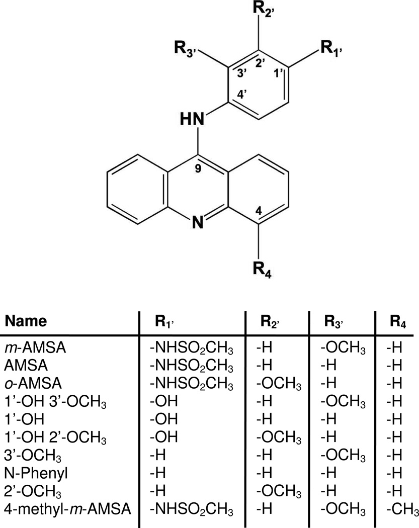
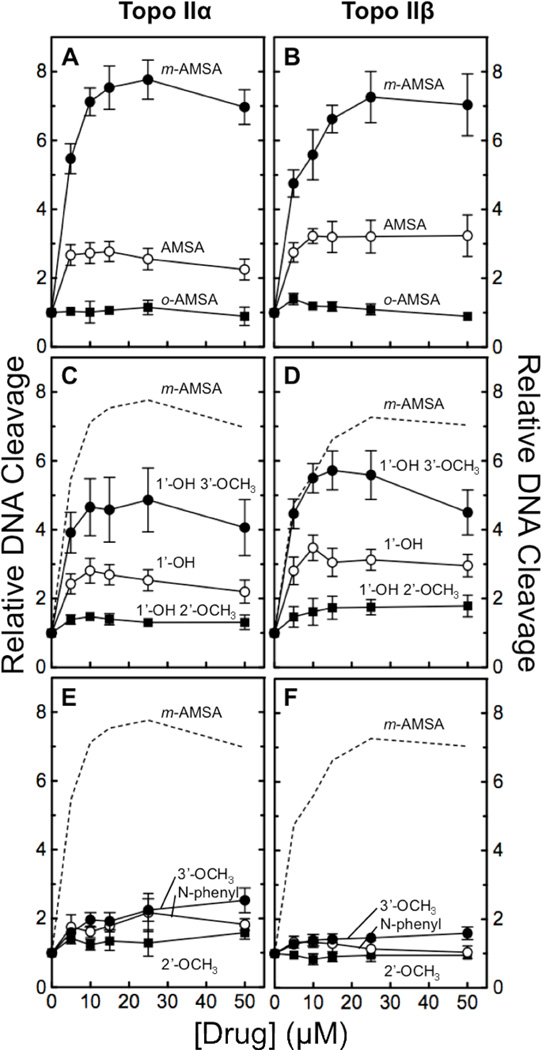
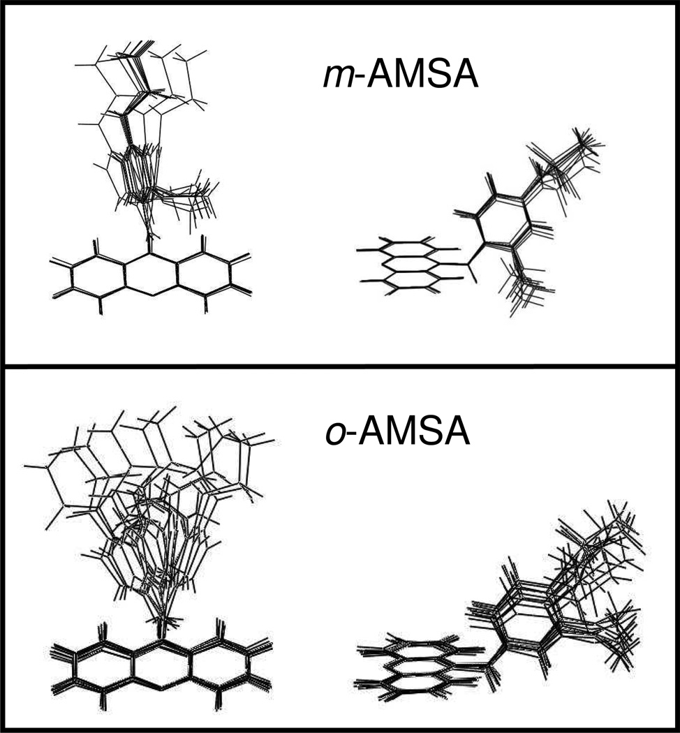
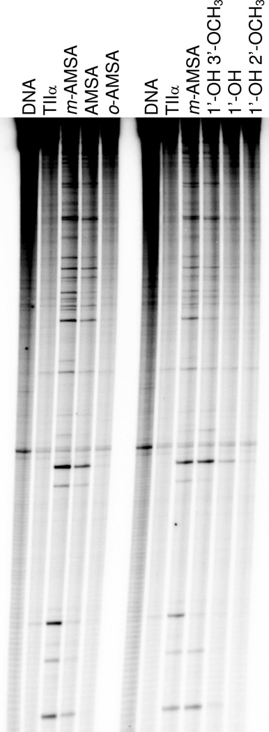
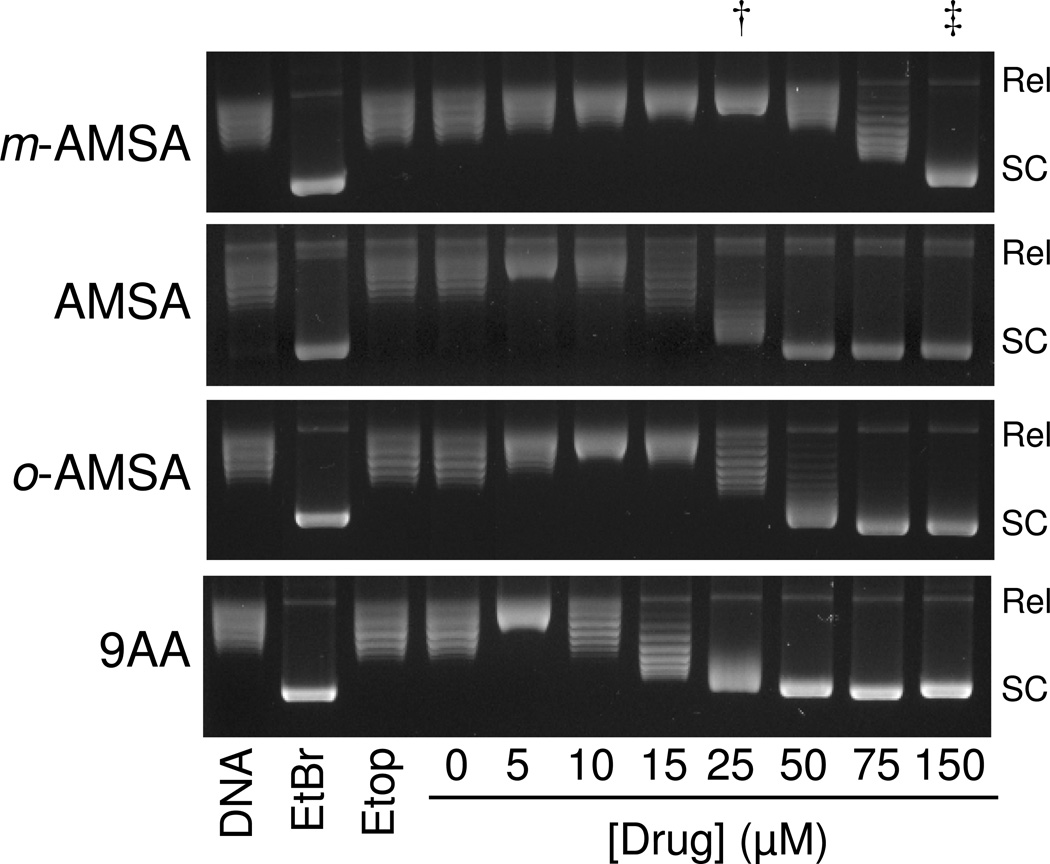
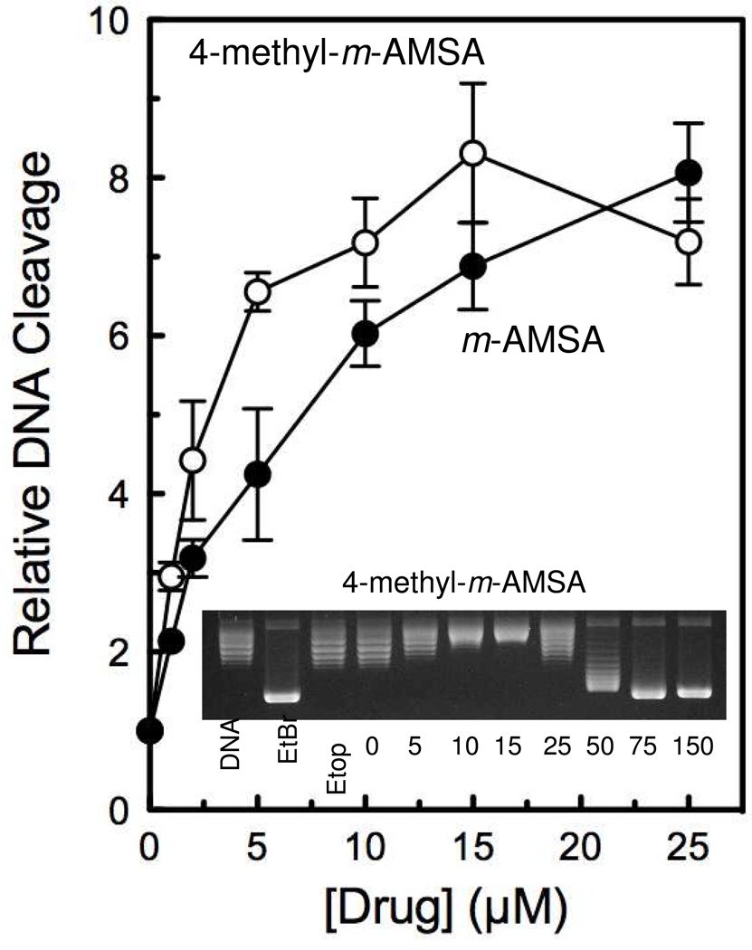
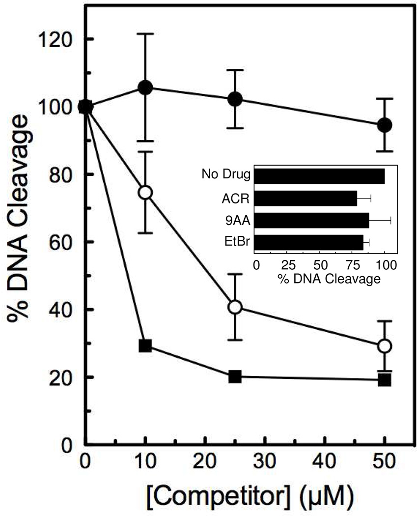
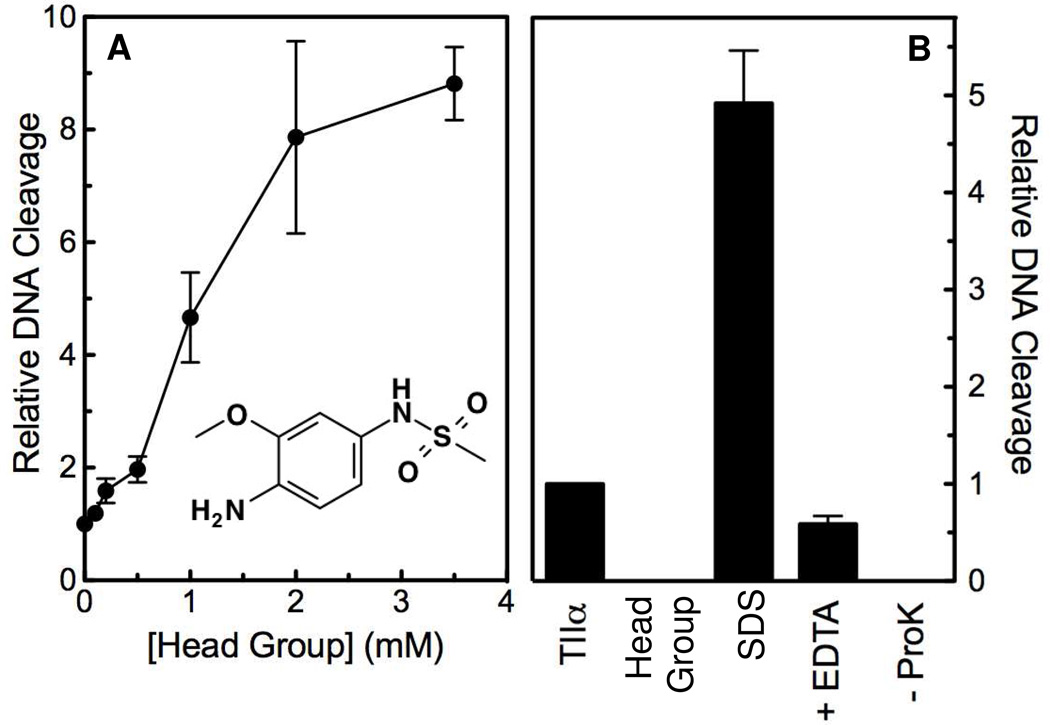
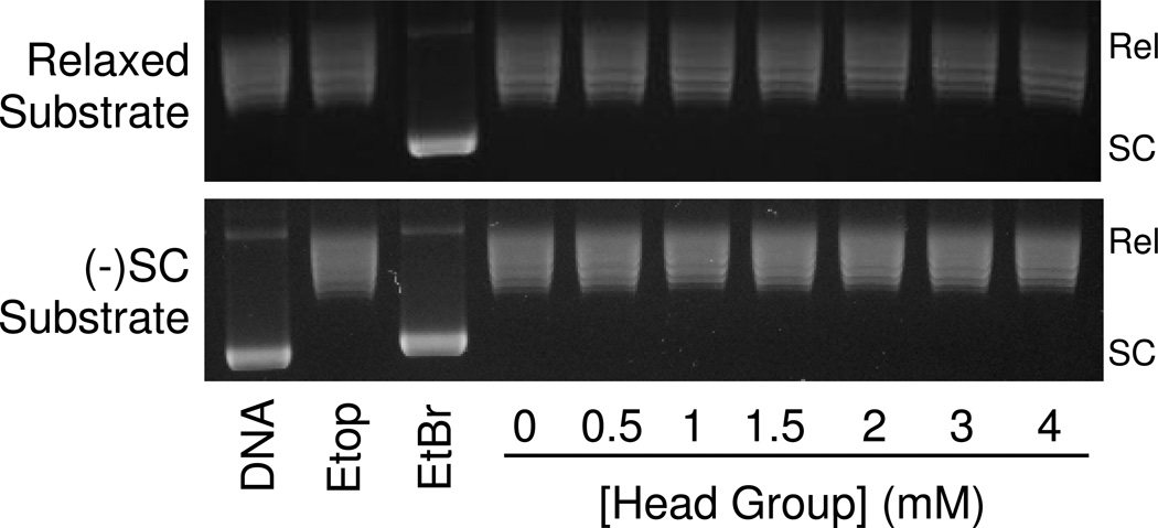
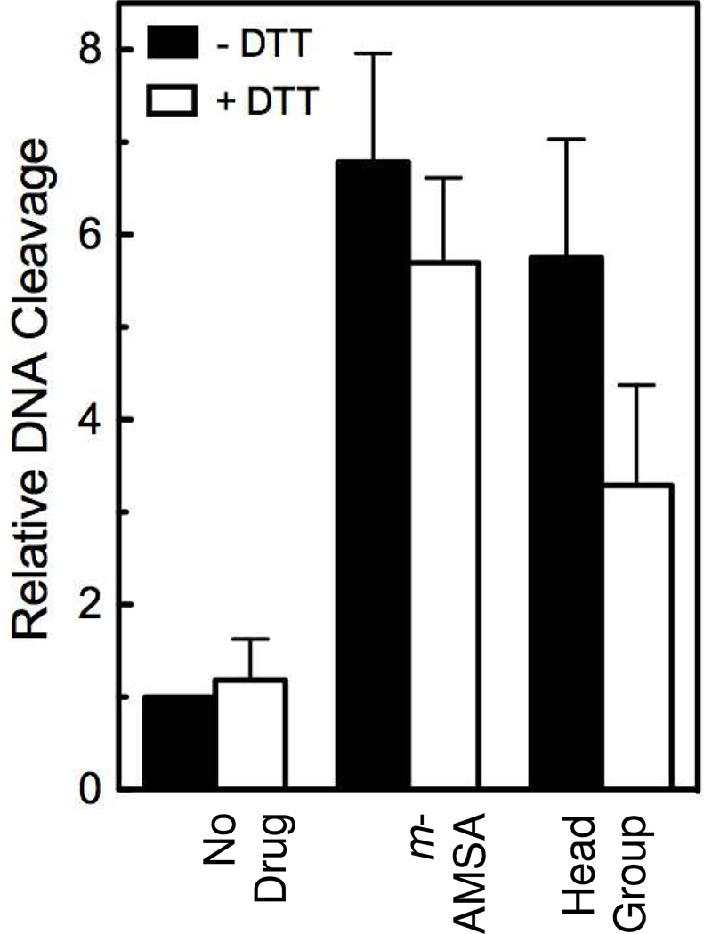
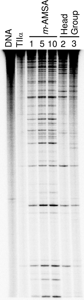
Amsacrine (m-AMSA) is an anticancer agent that displays activity against refractory acute leukemias as well as Hodgkin's and non-Hodgkin's lymphomas. The drug is comprised of an intercalative acridine moiety coupled to a 4'-amino-methanesulfon-m-anisidide headgroup. m-AMSA is historically significant in that it was the first drug demonstrated to function as a topoisomerase II poison. Although m-AMSA was designed as a DNA binding agent, the ability to intercalate does not appear to be the sole determinant of drug activity. Therefore, to more fully analyze structure-function relationships and the role of DNA binding in the action of m-AMSA, we analyzed a series of derivatives for the ability to enhance DNA cleavage mediated by human topoisomerase IIα and topoisomerase IIβ and to intercalate DNA. Results indicate that the 3'-methoxy (m-AMSA) positively affects drug function, potentially by restricting the rotation of the headgroup in a favorable orientation. Shifting the methoxy to the 2'-position (o-AMSA), which abrogates drug function, appears to increase the degree of rotational freedom of the headgroup and may impair interactions of the 1'-substituent or other portions of the headgroup within the ternary complex. Finally, the nonintercalative m-AMSA headgroup enhanced enzyme-mediated DNA cleavage when it was detached from the acridine moiety, albeit with 100-fold lower affinity. Taken together, our results suggest that much of the activity and specificity of m-AMSA as a topoisomerase II poison is embodied in the headgroup, while DNA intercalation is used primarily to increase the affinity of m-AMSA for the topoisomerase II-DNA cleavage complex.


Figures

Figure 1
Figure 1
Structure of m-AMSA and derivatives.
Figure 2
Figure 2
Enhancement of topoisomerase II-mediated DNA cleavage by m-AMSA and derivatives. The effects of m-AMSA (closed circles), AMSA (open circles), and o-AMSA (squares) (panels A and B), 1’-OH 3’-OCH3 (closed circles), 1’-OH (open circles) and 1’-OH 2’-OCH3 (squares) (panels C and D), and 3’-OCH3 (closed circles), N-phenyl (open circles), and 2’-OCH3 (squares) (panels E and F) on the cleavage of negatively supercoiled plasmid DNA by human topoisomerase IIα (panels A, C, and E) and topoisomerase IIβ (panels B, D, and F) were determined. Error bars represent the standard deviation of three independent experiments. Data for m-AMSA are included as dashed lines in panels C–F for comparison.
Figure 3
Figure 3
Energy minimization models of m-AMSA and o-AMSA. Low-energy structures (within ± 5 kcal/mol of the optimized structures for m-AMSA (top) and o-AMSA (bottom) are shown. Front and side views are shown at left and right, respectively.
Figure 4
Figure 4
DNA cleavage site specificity and utilization by human topoisomerase IIα in the presence of m-AMSA and derivatives. A singly end-labeled linear 4332 bp fragment of pBR322 was used as the cleavage substrate. An autoradiogram of a polyacrylamide gel is shown. DNA cleavage reactions were carried out in the absence of drug (TIIα), or in the presence of 10 µM m-AMSA; 25 µM AMSA, 1'-OH 3’-OCH3, or 1’-OH; or 100 µM o-AMSA or 1'-OH 2’-OCH3. DNA standards (DNA) also are shown. Results are representative of three independent experiments.
Figure 5
Figure 5
DNA intercalation by m-AMSA and derivatives. The abilities of 0–150 µM m-AMSA, AMSA, o-AMSA, and 9-aminoacridine (9AA) to intercalate into DNA were determined using a topoisomerase I-based supercoiling assay. Representative ethidium bromide (EtBr)-stained agarose gels are shown. The effects of 10 µM EtBr and 100 µM etoposide (Etop) are included as positive and negative controls, respectively. Relaxed DNA standards (DNA) also are shown. Results are representative of three independent experiments. As described in Table 1, concentrations of intercalators required to yield “fully relaxed” (Rel) and “fully supercoiled” (SC) plasmid are used for comparative purposes. Lanes that include these concentrations for m-AMSA are indicated by a dagger (†) and double dagger (‡), respectively.
Figure 6
Figure 6
Enhancement of topoisomerase IIα-mediated DNA cleavage by 4-methyl-m-AMSA. The effects of 4-methyl-m-AMSA (open circles) on the cleavage of negatively supercoiled plasmid DNA by human topoisomerase IIα are compared to those of m-AMSA (closed circles). Error bars represent the standard deviation of three independent experiments. The inset shows a representative topoisomerase I-based intercalation assay for 4-methyl-m-AMSA.
Figure 7
Figure 7
Inhibition of m-AMSA-induced topoisomerase IIα-mediated DNA cleavage by intercalators. The abilities of 0–50 µM acridine (ACR, closed circles), 9-aminoacridine (9AA, open circles), and ethidium bromide (EtBr, squares) to inhibit DNA cleavage induced by 25 µM m-AMSA were determined. A singly end-labeled 50-mer oligonucleotide substrate was used as the cleavage substrate. The level of DNA cleavage in the presence of 25 µM m-AMSA and the absence of competitor was set to 100%. The inset shows the effects of 50 µM ACR, 9AA, and EtBr on DNA cleavage mediated by human topoisomerase IIα in the absence of m-AMSA. The baseline level of DNA cleavage in the absence of intercalators was set to 100%. Error bars represent the standard deviation of three independent experiments for both the figure and the inset.
Figure 8
Figure 8
Enhancement of topoisomerase IIα-mediated DNA cleavage by the detached m-AMSA head group. Panel A shows the effects of the isolated m-AMSA head group (structure shown as inset) on the cleavage of negatively supercoiled plasmid DNA by human topoisomerase IIα. determined. Panel B shows a series of control experiments that confirm that DNA cleavage induced by the isolated head group is mediated by human topoisomerase IIα. Reactions contained DNA and enzyme in the absence of m-AMSA head group (TIIα), DNA and 3 mM head group in the absence of enzyme, or complete reaction mixtures treated with SDS prior to adding EDTA (SDS). The reversibility of DNA cleavage induced by 3 mM head group was determined by incubating reactions with EDTA prior to trapping cleavage complexes with SDS (EDTA). To determine whether DNA cleavage induced by 3 mM head group was protein-linked, proteinase K treatment was omitted (−ProK). Error bars for both panels represent standard deviations for three independent experiments.
Figure 9
Figure 9
The isolated m-AMSA head group does not intercalate in DNA. Representative topoisomerase I-based DNA intercalation assay gels are shown for the isolated m-AMSA head group. Assays starting with either relaxed or negatively supercoiled DNA plasmid substrates are included. Etoposide (Etop, 100 µM) and ethidium bromide (EtBr, 10 µM) are included as positive and negative controls, respectively. DNA standards (DNA) also are shown. Results are representative of three independent experiments.
Figure 10
Figure 10
Covalent linkage of the head group to the acridine moiety is necessary for the high potency of m-AMSA as a topoisomerase II poison. The ability of the detached head group (HG, closed circles) or a 1:1 mixture of head group + acridine (HG + ACR, open circles) to stimulate topoisomerase IIα-mediated DNA cleavage is shown. A control experiment assessing the effects of acridine alone on the DNA cleavage activity of topoisomerase IIα (ACR, squares) also is shown. Error bars represent the standard deviation of three independent experiments.
Figure 11
Figure 11
Partial redox-dependence of the isolated m-AMSA head group as a topoisomerase II poison. The effects of 25 µM m-AMSA or 3 mM m-AMSA head group on the cleavage of negatively supercoiled plasmid DNA by human topoisomerase IIα in the absence (closed bars) or presence (open bars) of 3 mM dithiothreitol (DTT) are shown. A control experiment carried out in the absence of drugs also is shown (No Drug). DNA cleavage levels are relative to those induced by the enzyme in the absence of drug or DTT. Error bars represent the standard deviation of three independent experiments.
Figure 12
Figure 12
DNA cleavage site specificity and utilization by human topoisomerase IIα in the presence of the m-AMSA head group. A singly end-labeled linear 4332 bp fragment of pBR322 was used as the cleavage substrate. An autoradiogram of a polyacrylamide gel is shown. DNA cleavage reactions were carried out in the absence of drug (TIIα), or in the presence of 1, 5, or 10 µM m-AMSA or 2 or 3 mM head group. A DNA standard (DNA) also is shown. Results are representative of three independent experiments.

Similar articles

Cited by

References

    1. Deweese JE, Osheroff N. The DNA cleavage reaction of topoisomerase II: wolf in sheep's clothing. Nucleic Acids Res. 2009;37:738–748. - PMC - PubMed
    1. Pommier Y, Leo E, Zhang H, Marchand C. DNA topoisomerases and their poisoning by anticancer and antibacterial drugs. Chem. Biol. 2010;17:421–433. - PMC - PubMed
    1. National Cancer Institute. Clinical Trials. 2011 http://www.cancer.gov/clinicaltrials/search/results?protocolsearchid=923....
    1. Jehn U, Heinemann V. New drugs in the treatment of acute and chronic leukemia with some emphasis on m-AMSA. Anticancer Res. 1991;11:705–711. - PubMed
    1. Kell J. Treatment of relapsed acute myeloid leukaemia. Rev. Recent Clin. Trials. 2006;1:103–111. - PubMed

Publication types

MeSH terms