. 2012 Jul 17;45(7):1122-31.
doi: 10.1021/ar200253u. Epub 2012 Feb 3.

Molecular mechanisms of RNA-triggered gene silencing machineries

Affiliations
Free PMC article

Molecular mechanisms of RNA-triggered gene silencing machineries

Zhonghan Li et al. Acc Chem Res. .
Free PMC article

Abstract

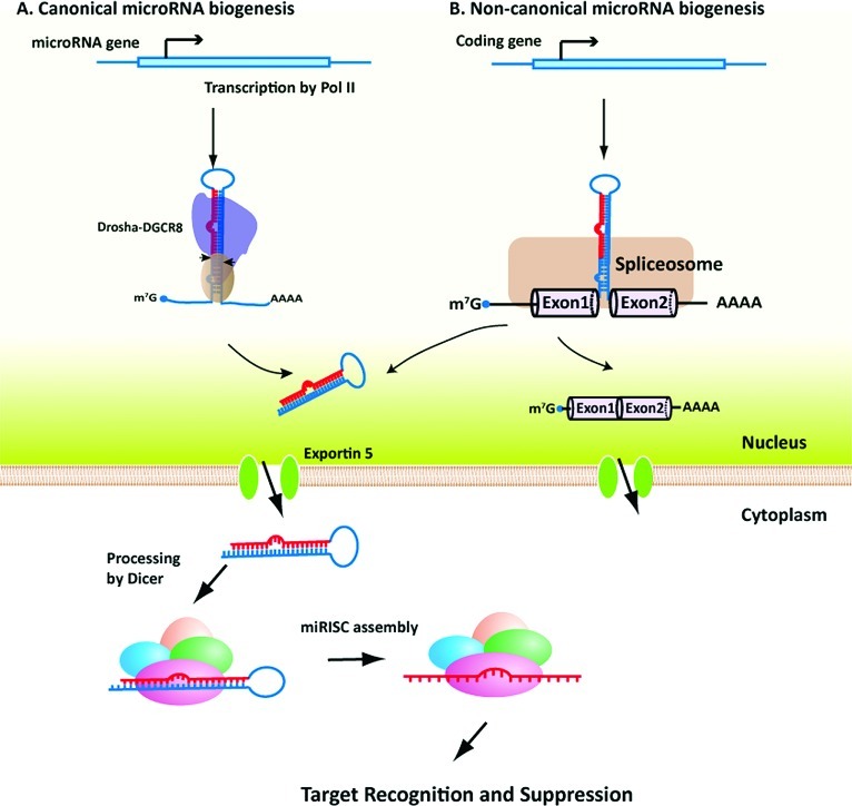
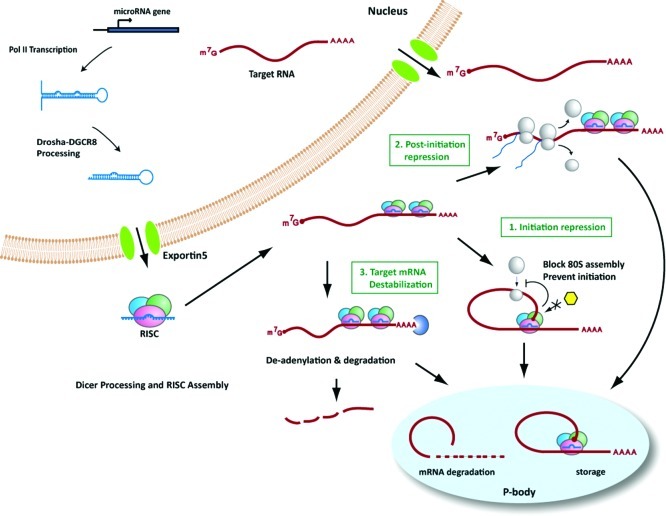
Gene silencing by RNA triggers is an ancient, evolutionarily conserved, and widespread phenomenon. This process, known as RNA interference (RNAi), occurs when double-stranded RNA helices induce cleavage of their complementary mRNAs. Because these RNA molecules can be introduced exogenously as small interfering RNAs (siRNAs), RNAi has become an everyday experimental tool in laboratory research. In addition, the number of RNA-based therapeutics that are currently in clinical trials for a variety of human diseases demonstrate the therapeutic potential of RNAi. In this Account, we focus on our current understanding of the structure and function of various classes of RNAi triggers and how this knowledge has contributed to our understanding of the biogenesis and catalytic functions of siRNA and microRNA in mammalian cells. Mechanistic studies to understand the structure and function of small RNAs that induce RNAi have illuminated broad functions of the ancient RNAi machinery in animals and plants. In addition, such studies have provided insight to identify endogenous physiological gene silencing RNA triggers that engage functional machineries similar to siRNAs. Several endogenous small RNA species have been identified: small noncoding RNAs (microRNAs), piwi-interacting RNAs (piRNAs), and endogenous siRNAs (endo-siRNAs). microRNAs are the most widespread class of small RNAs in mammalian cells. Despite their importance in biology and medicine, the molecular and cellular mechanisms of microRNA biogenesis and function are not fully understood. We provide an overview of the current understanding of how these molecules are synthesized within cells and how they act on gene targets. Interesting questions remain both for understanding the effects of modifications and editing on microRNAs and the interactions between microRNAs and other cellular RNAs such as long noncoding RNAs.


Figures

Scheme 1
Scheme 1
Figure 1
Figure 1
Steps in RISC function. Double-stranded (ds) or short hairpin (sh) RNAs are first bound and cleaved by Dicer into small interfering RNAs (siRNAs; ∼21 nt) with 2 nt overhangs and 5′ phosphates. These siRNAs are then loaded into protein complexes termed RNA-induced silencing complex (RISCs). Ago2, a component of RISCs, binds the double-stranded siRNAs and cleaves the passenger strand, which induces its dissociation from the RISC complex and degradation. The remaining guide strand then leads the activated RISCs to find target mRNAs that contain perfectly matched complementary sequences to the guide strand. Binding of RISCs to the target mRNAs induces conformational changes and results in cleavage of the mRNA by Ago2. Cleaved mRNAs are then subject to mRNA decay or degradation, thus silencing the target gene expression.
Figure 2
Figure 2
Crystal Structure of T. thermophilus Ago (Asn478) bound with 21 nucleotide guide DNA and 19 nucleotide target RNA. Two views of the 2.8 A crystal structure of the ternary complex. The structure was generated using mutant Ago of T. thermophilus, which is unable to cleave the target RNA, thus facilitating detailed examination of the cleavage site at position 10–11. The guide strand DNA (red) is traced for nucleotides 1–16, which are perfectly matched with its target mRNA (blue). Target RNA is traced for nucleotides 2′–16′. Only the 5′ end of the guide strand is anchored in this ternary complex. The two strands retain the conformation to one turn of A-form helix (12 nt) upon binding,, and the cleavage site of nt 10–11 stack on each other in a catalytically competent conformation. The N-domain of Ago seems to block the interaction between the guide strand and target mRNA beyond position 16, thus the 3′ end could be released from the PAZ domain. Adapted from ref (17).
Figure 3
Figure 3
Kinetics of RISC assembly and function. The assembly and function of RISCs can be divided into at least two catalytic steps; for simplicity, only two checkpoints are considered here. The first checkpoint is RISC loading. siRNA binding by RISCs is denoted as K1, and assembly of functional activated RISCs is denoted as K2. K2 can be affected by the thermodynamics of siRNAs. The second checkpoint involves target recognition and cleavage. After guide strands of siRNAs are loaded into RISCs, the protein complex is activated and led by the guide strand to target mRNAs. Target mRNAs are bound by functional RISCs, change their conformation to A-form helices and are finally cleaved by Ago2 at nt position 10–11 from the 5′ end of the guide strand. The target mRNA recognition by RISCs is denoted as K3, and mRNA cleavage is denoted as K4. K3 could be affected by several factors such as the secondary structure of target mRNAs.
Figure 4
Figure 4
Canonical and noncanonical microRNA biogenesis pathways. Depending on the origin of miRNAs, two pathways have been proposed for miRNA biogenesis in vivo. (a) Canonical miRNA biogenesis. In this pathway, miRNA-encoding genes are first transcribed, usually through the Pol II promoter, into primary-miRNA-containing mRNAs. Hairpin structures within these mRNAs are then detected and bound by the Drosha–DGCR8 protein complex. Drosha cleaves the hairpin and generates ∼70 nt long miRNA precursors, called pre-microRNAs. Pre-miRNAs are then transported from the nucleus into the cytoplasm through exportin 5, and are further processed by the Dicer complex. Processing by Dicer generates ∼21 nt mature miRNAs which are then loaded by Ago2 to form functional RISCs and carry out downstream functions. (b) Noncanonical microRNA biogenesis. In this pathway, miRNAs are usually encoded in the intron regions of protein-coding genes, called mirtrons. After transcription, primary mRNAs are bound and processed by spliceosome protein complexes, which give rise to mature protein coding mRNAs and ∼70 nt pre-miRNAs after debranching. Pre-miRNAs generated in this way then join the ones from the canonical pathway for transportation and Dicer processing.
Figure 5
Figure 5
microRNA function. After loading with Ago proteins to form functional RISCs, miRNA-guided RISCs bind to the target mRNAs and inhibit target gene expression. Currently, there are at least three mechanisms that have been linked to miRNA-mediated gene silencing. (1) Repression of translation initiation. In this case, miRISCs inhibit initiation of translation by affecting the eIF4F-cap recognition, 40S small ribosomal subunit recruitment, and/or by inhibiting incorporation of the 60S subunit and formation of the 80S ribosomal complex. Some of the target mRNAs bound by miRISC is transported into P-bodies for storage and may re-enter the translation phase when induced. (2) Postinitiation translational repression. miRISCs could interfere after translation has been initiated by inhibiting elongation of ribosomes, causing ribosome drop-off from mRNAs, and/or by facilitating degradation of newly synthesized nascent peptides. (3) Destabilization of target mRNAs. miRISCs could cause destabilization of target mRNAs by directly interacting with CCR4-containing deadenylation complexes and facilitating the deadenylation of poly A tails of target mRNAs. Following deadenylation, the 5′ end-capping structures of target mRNAs are also removed by the DCP1–DCP2 complex.

Similar articles

Cited by

References

    1. Fire A.; Xu S.; Montgomery M. K.; Kostas S. A.; Driver S. E.; Mello C. C. Potent and specific genetic interference by double-stranded RNA in Caenorhabditis elegans. Nature 1998, 391, 806–811. - PubMed
    1. Kim V. N.; Han J.; Siomi M. C. Biogenesis of small RNAs in animals. Nat. Rev. Mol. Cell Biol. 2009, 10, 126–139. - PubMed
    1. Krol J.; Loedige I.; Filipowicz W. The widespread regulation of microRNA biogenesis, function and decay. Nat. Rev. Genet. 2010, 11, 597–610. - PubMed
    1. Ambros V. MicroRNAs and developmental timing. Curr. Opin. Genet. Dev. 2011, 21, 511–7. - PMC - PubMed
    1. Bagga S.; Bracht J.; Hunter S.; Massirer K.; Holtz J.; Eachus R.; Pasquinelli A. E. Regulation by let-7 and lin-4 miRNAs results in target mRNA degradation. Cell 2005, 122, 553–563. - PubMed

Publication types