. 2012 Feb 3;148(3):568-82.
doi: 10.1016/j.cell.2012.01.024.

Ret is a multifunctional coreceptor that integrates diffusible- and contact-axon guidance signals

Affiliations

Ret is a multifunctional coreceptor that integrates diffusible- and contact-axon guidance signals

Dario Bonanomi et al. Cell. .

Abstract

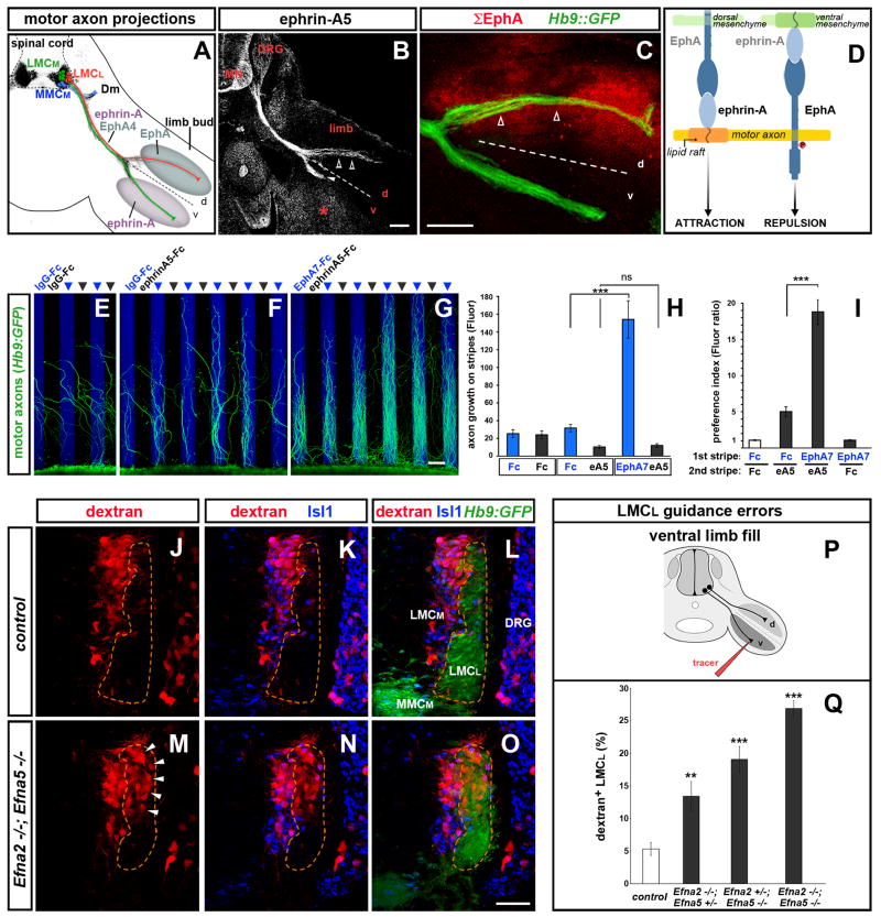
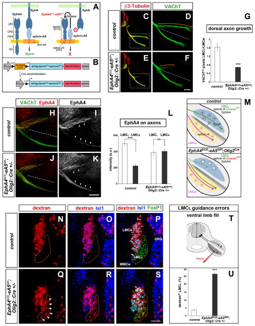
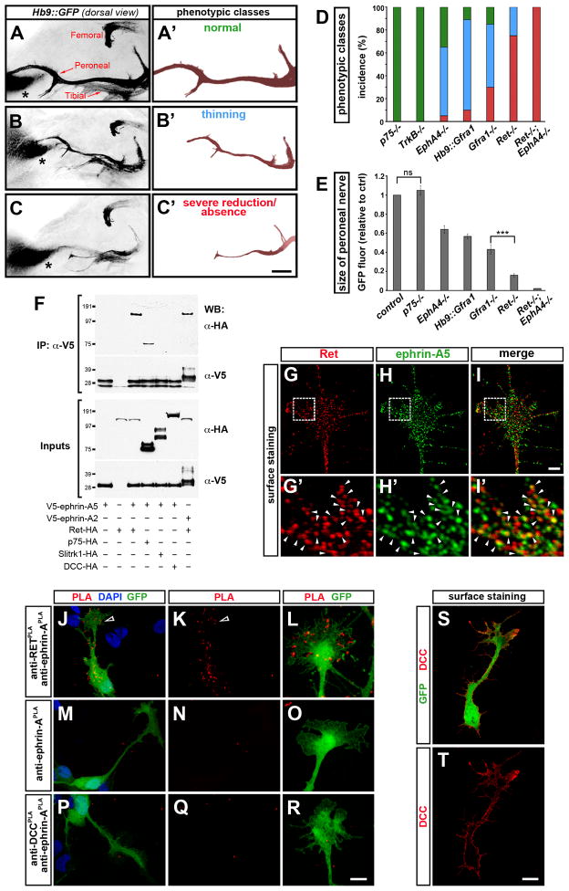
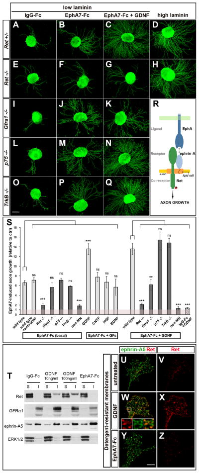
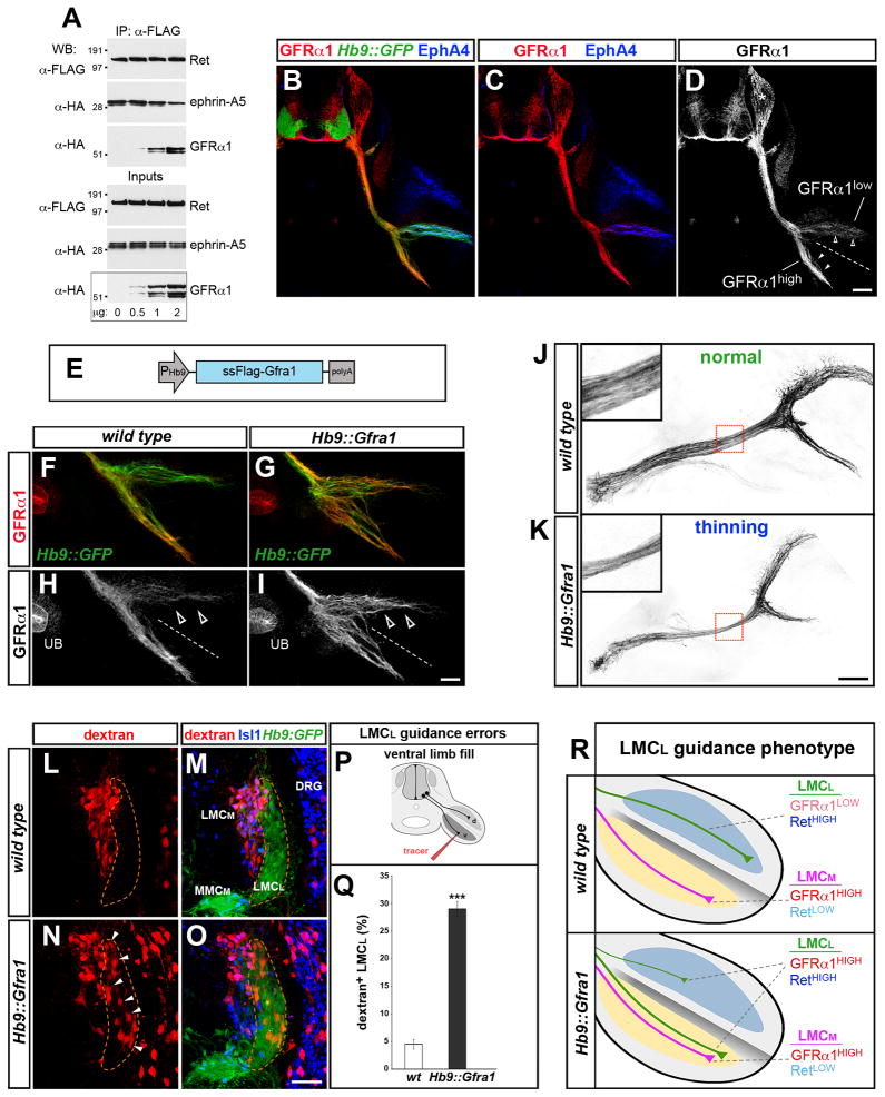
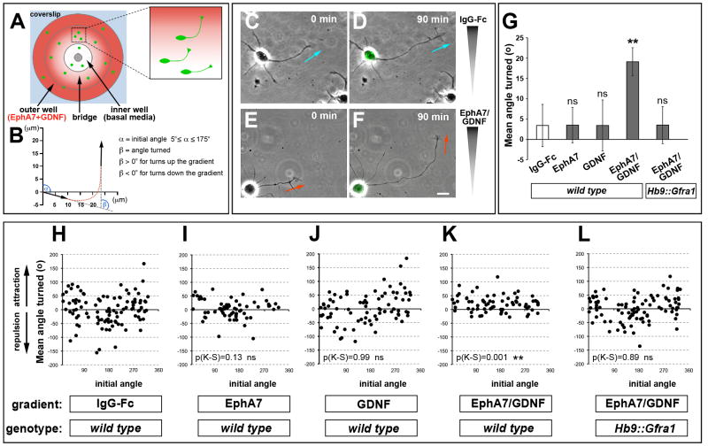
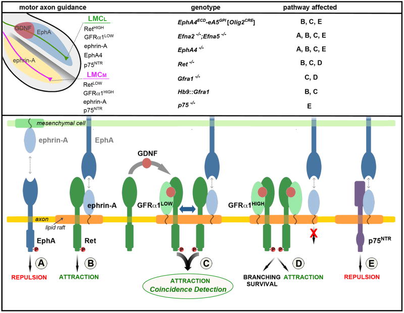
Growing axons encounter multiple guidance cues, but it is unclear how separate signals are resolved and integrated into coherent instructions for growth cone navigation. We report that glycosylphosphatidylinositol (GPI)-anchored ephrin-As function as "reverse" signaling receptors for motor axons when contacted by transmembrane EphAs present in the dorsal limb. Ephrin-A receptors are thought to depend on transmembrane coreceptors for transmitting signals intracellularly. We show that the receptor tyrosine kinase Ret is required for motor axon attraction mediated by ephrin-A reverse signaling. Ret also mediates GPI-anchored GFRα1 signaling in response to GDNF, a diffusible chemoattractant in the limb, indicating that Ret is a multifunctional coreceptor for guidance molecules. Axons respond synergistically to coactivation by GDNF and EphA ligands, and these cooperative interactions are gated by GFRα1 levels. Our studies uncover a hierarchical GPI-receptor signaling network that is constructed from combinatorial components and integrated through Ret using ligand coincidence detection.


Figures

Figure 1
Figure 1. Coordination of forward and reverse ephrin-A signaling enhances the fidelity of axon pathway selection
(A) Schematic of motor neuron axon projections superimposed on transverse section of an e11.5 Hb9::GFP+ (black) embryo at hindlimb level. Lateral LMC (LMCL, red) and medial LMC (LMCM, green) axons bifurcate at the base of the limb. Dashed line here and in following figures divides the dorsal (d) and ventral (v) halves of the limb. LMCL axons co-express EphA4 and ephrin-As, avoid ephrin-As in the ventral limb and project into the EphA-rich dorsal mesenchyme. Medial motor neurons (MMCM, blue) extend to the axial musculature (demomyotome, Dm). (B) Ephrin-A5 staining on e11.5 LMCL axons (open arrowheads), LMCM axons (arrowhead), ventral limb mesenchyme (asterisk) and other embryonic tissues including motor neurons (MN) and dorsal root ganglia (DRG). (C) Cumulative distribution of EphA proteins (Σ EphA, red) in limb and motor axons detected with ephrin-A5-AP on e11.5 Hb9::GFP embryos. LMCL axons (open arrowheads) extend through the EphA-rich mesenchyme. (D) Ephrin-A reverse and forward signaling emanate from distinct membrane domains and exert opposite effects on motor axons. (E–I) Stripe assay with Hb9::GFP+ lumbar mouse motor neuron explants. (E) Motor axons on control IgG-Fc stripes. (F) Axons avoid ephrin-A5 stripes. (G) Axon growth is enhanced on EphA7-Fc stripes. (H) Quantification of GFP signal on each set of stripes. (I) Ratio between motor axons (GFP signal in H) on the first and second set of stripes. Mean ± SEM, N explants: IgG-Fc/IgG-Fc, 12; IgG-Fc/ephrin-A5-Fc, 11; EphA7-Fc/ephrin-A5-Fc, 12; EphA7-Fc/IgG-Fc, 10 (***) p<0.001; (ns) p=0.52 unpaired t test. (J–Q) Rhodamine-dextran (red) fills of ventral-projecting motor neurons reveals LMCL guidance errors in Efna2/Efna5 e13.5 mutant embryos. (J–L) Ventral shank injection selectively labels LMCM neurons (Hb9::GFPlow; Isl1high) in WT and Hets. (M–O) Ventral fills in Efna2/Efna5 mutants label LMCM and misguided LMCL neurons (Hb9::GFPhigh; Isl1low, arrowheads). (P) Schematic of the ventral limb fill. (Q) Proportion (%) of LMCL neurons labeled by the ventral tracer. Mean ± SEM, N cells (from N embryos): Control, 985 (9); Efna2−/−;Efna5+/−, 775 (6); Efna2+/−;Efna5−/−, 395 (4); Efna2−/−; Efna5−/−, 1027 (8); (***) p<0.001; (**) p<0.01 Dunnett’s test vs control. Scale bars: A–C: 100μm; E–G: 100μm; J–O: 50μm. See also Figure S1.
Figure 2
Figure 2. Ephrin-A reverse signaling is required for LMCL dorsal limb innervation
(A) The EphA4ECD-ephrinA5GPI chimeric masking construct is targeted to lipid rafts where it binds endogenous ephrin-As via cis-interactions. The cis-binding abolishes ephrin-Eph trans-interactions thereby blocking ephrin-A reverse signaling. (B) Schematic of the transgenic mouse strategy. Cre removes a 3× polyA-STOP cassette allowing expression of FLAG-tagged EphA4ECD-ephrinA5GPI. An internal ribosome entry site (ires) enables simultaneous translation of the monomeric, membrane-associated red fluorescent protein (RFP) MmCherry. (C–G) Reduced motor axon growth into the dorsal limb at e11.5 following motor neuron-specific expression of EphA4ECD-ephrinA5GPI activated by Olig2::Cre (orange trace adjacent to dorsal branch). The ventral motor axon appears normal. Motor neurons are marked by VAChT (green). Boxed regions in C and E are enlarged in D and F. (G) Ratio of VAChT+ axons (LMCL/LMCM) in dorsal and ventral limb of transgenics and controls (WT or Olig2::Cre+/− littermates). Mean ± SEM, N limbs: Control, 22; Transgenic, 26; (***) p<0.001 unpaired t test. (H–M) EphA4+ LMCL axons (open arrowheads) project exclusively to the dorsal limb in e11.5 controls, but extend in both dorsal and ventral nerve branches in EphA4ECD-ephrinA5GPI;Olig2::Cre+/− embryos (arrowheads). (L) Quantification of EphA4 signal in LMCL and LMCM axons of transgenic and control embryos. mean ± SEM, N limbs: Control, 12; Transgenic, 14 ; (***) p<0.001; (ns) p=0.76 unpaired t test. (M) Top, control: EphA in the dorsal limb mesenchyme attracts (+) while ephrin-A in the ventral limb repels (−) LMCL axons. Bottom, EphA4ECD-ephrinA5GPI;Olig2::Cre+/−: silencing of ephrin-A reverse signaling in motor neurons leads to the aberrant projection of LMCL axons into the ventral limb. (N–P) Rhodamine-dextran (red) injection in the ventral shank exclusively labels LMCM neurons (FoxP1+; Isl1+) in control e12.5 embryos. (Q–S) Ventral fills in EphA4ECD-ephrinA5GPI;Olig2::Cre+/− transgenic embryos labels LMCM and misguided LMCL neurons (FoxP1+; Isl1, arrowheads). (T) Schematic of the ventral fill experiment. (U) Proportion (%) of LMCL neurons labeled by the ventral tracer. Mean ± SEM, N cells (from N embryos): Control, 1179 (15); Transgenic, 1538 (11); (***) p<0.001 unpaired t test. Scale bars: C, E: 200μm; D, F: 100μm; H–K: 100μm; N–S: 50μm. See also Figure S2.
Figure 3
Figure 3. Genetic studies of dorsal limb innervation and Ret association with ephrin-A
(A–C) Dorsal view of motor axon projections into the hindlimb of whole-mount e12.5 mouse embryos (Hb9::GFP+, black). The dorsal peroneal nerve and ventral tibial nerve arise from the sacral (sciatic) plexus, while the dorsal femoral nerve extends from the more rostral lumbar (femoral) plexus. The non-neuronal site of GFP expression in the dorsal limb (asterisk) serves as a reference for comparing nerve growth between embryos. (A′–C′) Peroneal nerve shown in isolation with examples of phenotype classes. (D) Incidence of the phenotypic classes in mutants (green, normal; blue, thinning; red: severe reduction/absence). (E) Quantification of GFP signal from the entire peroneal nerve in mutant embryos normalized to controls (mix of WT and Het littermates). Mean ± SEM, N limbs: Control, 110; p75−/−, 20; TrkB−/−, 25; EphA4−/−, 50; Hb9::Gfra1, 70; Gfra1−/−, 30; Ret−/−, 60; Ret−/−;EphA4−/−, 10; (ns) p=0.41 p75−/− vs control; other mutants were significantly different from control: p<0.001 Dunnett s test; (***) p<0.001 Gfra1−/− vs Ret−/−, unpaired t test. The femoral nerve that innervates proximal dorsal muscles was unaffected in all the mutant genotypes analyzed. (F) Immunoprecipitation (IP) of V5-tagged ephrin-A2 and ephrin-A5 proteins in AD293 cells followed by Western blot (WB). Ephrin-A5 and ephrin-A2 interact with Ret. Ret was recovered with higher efficiency from ephrin-A immunocomplexes than positive control p75. Ephrin-A5 does not associate with Slitrk-1 or DCC. (G–I′) Colocalization of Ret and ephrin-A5 (arrowheads) on the growth cone of non-permeabilized chick motor neurons transfected with tagged proteins [Pearson’s colocalization coefficient: 0.57±0.02, N=20 growth cones]. Boxed area in G–I is enlarged in G′–I′. (J–R) Detection of Ret (transfected) and ephrin-A (endogenous) in close-association by PLA (red) on the surface of non-permeabilized GFP+ chick motor neurons (arrowheads mark growth cone). Only sporadic background signal is visible in negative controls: (M–O) anti-Ret antibody is omitted; (P–R) PLA between DCC and ephrin-A in DCC-transfected neurons. (S, T) The anti-DCC antibody used in P–R detects the receptor on the surface of DCC-transfected, non-permeabilized GFP+ chick motor neurons. Scale bars, A–C′: 200μm; G–I: 2μm; G′–I′: 0.44μm; J,K,M,N,P,Q,S,T: 10μm; L,O,R: 4.3μm. See also Figure S3.
Figure 4
Figure 4. Ret mediates attractive ephrin-A reverse signaling
(A–Q) Hb9::GFP+ lumbar LMC mouse motor neurons cultured on control IgG-Fc or EphA7-Fc (low laminin), in the presence or absence of GDNF. Ephrin-A reverse signaling is activated by the EphA7-Fc substrate. (A, B, E, F) Axonal growth stimulated by ephrin-A reverse signaling is impaired in Ret−/− explants; but is intact in (I, J) Gfra1−/−, (L, M) p75−/−, (O–P) and TrkB−/− explants. (C, N, Q) GDNF potentiates the growth-promoting effects of EphA-Fc in control, p75−/− and TrkB−/− explants. (G, K) GDNF fails to enhance axonal growth on EphA-Fc with Ret−/− and Gfra1−/− explants. (D, H) Ret−/− neurite outgrowth on a permissive substrate (high laminin) is unaffected. (R) Schematic: Ret mediates ephrin-A reverse signaling that promotes motor axon growth. (S) Quantification of the outgrowth of GFP+ motor axons on EphA7-Fc relative to control IgG-Fc in basal media or with growth factors (GFs) [GDNF, CNTF, HGF, BDNF] or anti-GDNF antibody. Light bars are control explants (WT and Het mutants), and dark bars mutants. ‘non-MN’ corresponds to GFP axons (see Figure S4D, H). The wild type/EphA7+GDNF bar is duplicated from the EphA7-Fc+GFs condition to facilitate comparison. The striped bar shows motor axon growth on IgG-Fc in the presence of GDNF. Number of explants on (IgG-Fc) and [EphA7-Fc] substrates. Basal MN media: Control genotypes, (63)/[69]; Ret−/− (58)/[59]; Gfra1−/− (16)/[17]; p75−/− (18)/[35]; TrkB−/− (9)/[14]; ‘non-MN’ (15)/[15]. GDNF media: Control, (51)/[82]; Ret−/− (33)/[34]; Gfra1−/− (13)/[12]; p75−/− (61)/[74]; TrkB−/− (10)/[12]; ‘non-MN’ (32)/[33]. Other treatments: anti-GDNF (10)/[12]; CNTF (6)/[10]; HGF (12)/[13]; BDNF (7)/[8]. (ns) p>0.05; (**) p<0.01; (***) p<0.001 Dunnett’s test vs WT in either EphA7-Fc (basal) or EphA7-Fc+GDNF conditions. (T) Triton X-100 soluble (S) and insoluble (I) fractions prepared from MCF-7 cells expressing endogenous Ret and GFRα1 and transfected V5-ephrin-A5. Ephrin-A5 and GFRα1 are enriched in the detergent-resistant I fraction. Ret is recruited to the I fraction after GDNF stimulation (10ng/ml or 100ng/ml for 20min) but not after EphA7-Fc stimulation (5μg/ml for 20min). Enrichment of the cytoplasmic protein ERK1/2 in the S fraction serves as a control. (U–V) In resting conditions, Ret is associated with detergent-soluble membranes. (W–X) GDNF stimulation (50ng/ml for 15min) induces the translocation of Ret into detergent-resistant rafts where it colocalizes with ephrin-A5 (insets in W). (Y–Z) Ret translocation is not observed upon EphA7-Fc stimulation (5μg/ml for 15min). Scale bars, A–Q: 200μm; U–Z: 5μm; 1.6 μm for insets in W. See also Figure S4.
Figure 5
Figure 5. LMCL axon guidance is influenced by GFRα1 levels
(A) AD293 cells transfected with increasing GFRα1 plasmid and constant amounts of Ret and ephrin-A5 were subjected to Ret immunoprecipitation (IP) followed by Western blot (WB) to detect Ret/ephrin-A5 and Ret/GFRα1 complexes. The relative amount of ephrin-A5 associated with Ret declines when GFRα1 is elevated. Increasing GFRα1 levels favors Ret/GFRα1 complex formation. (B–D) Immunostaining of transverse sections of e11.5 Hb9::GFP+ embryos at hindlmb level. GFRα1 is high on LMCM axons (arrowheads) and low on EphA4+ LMCL axons (open arrowheads). GFRα1 is also expressed by sensory neurons (asterisk). The specificity of the anti-GFRα1 antibody was confirmed on Gfra1−/− sections (data not shown). (E) Schematic of Hb9::Gfra1 transgenic construct. (F–I) GFRα1 levels increase in LMCL axons (open arrowheads) from e11.5 Hb9::Gfra1 embryos. GFRα1 staining in the ureteric bud (UB), which is not affected by the Hb9::Gfra1 transgene, provides an internal standard to estimate the relative level of GFRα1 overexpression in motor axons. (J, K) Dorsal views of the peroneal nerve in e12.5 whole-mount preparations (Hb9::GFP+, black). Increased expression of GFRα1 in Hb9::Gfra1 transgenics leads to thinning of the peroneal nerve. The boxed regions are enlarged in the insets. The incidence and extent of the phenotype are quantified in Figure 3D, E. (L–R) Projection errors of LMCL axons in Hb9::Gfra1 transgenics detected by injection of rhodamine-dextran tracer (red) in the ventral shank of 13.5 embryos. (L, M) Ventral fills selectively label LMCM neurons (Hb9::GFPlow; Isl1high) in WT embryos. (N, O) Ventral fills detect misprojecting LMCL neurons (Hb9::GFPhigh; Isl1low, arrowheads) in Hb9::Gfra1 embryos. (P) Schematic of the ventral fill experiment. (Q) Proportion (%) of LMCL neurons labeled by the ventral tracer. Mean ± SEM, N cells (from N embryos): WT, 975 (9); Hb9::Gfra1, 1250 (13); (***) p<0.001 unpaired t test. (R) Schematic: The levels of Ret relative to GFRα1 differ in LMCL vs. LMCM axons. Motor neuron-specific overexpression of GFRα1 in Hb9::Gfra1 transgenics leads to LMCL guidance errors. Scale bar, A–C: 100μm; F–I: 50μm; J, K: 100μm, insets: 33μm; L–O: 50μm. See also Figure S3S, S5, S6.
Figure 6
Figure 6. Coincidence detection of EphA and GDNF promotes growth cone turning and is gated by GFRα1 levels
(A) Top view schematic of the Dunn chamber. The inner well contains control media and the outer chamber is supplemented with guidance factors. (B) Method to calculate growth cone turning during 90 min. The initial angle (α) is calculated from the distal 10μm of axon relative to the gradient at t=0. The angle turned (β) is calculated from the initial and final axon trajectories. (C–F) Brightfield images of axons that turn toward EphA7-Fc + GDNF gradient. Axons do not turn in response to control IgG-Fc. The final images at t=90 min are superimposed on Hb9::GFP signal to confirm the motor neuron identity of the cells. (G) Mean angle turned (β ± SEM) in the presence of various factors for WT and Hb9::Gfra1 motor axons [(ns) p>0.05; (**) p<0.01 unpaired t test vs control IgG-Fc]. (H–L) Scatter plots of the angle turned β versus the initial angle α for WT motor axons in the presence of IgG-Fc (N=102 axons), EphA7-Fc (N=64), GDNF (N=80), EphA7-Fc + GDNF (N=71), or Hb9::Gfra1 motor axons in the presence of EphA7-Fc + GDNF (N=94); (KS), Kolmogorov-Smirnov test vs control IgG-Fc: (ns) p>0.05; (**) p<0.01. Scale bar, 10μm. See also Figure S6.
Figure 7
Figure 7. Model for the integration of GDNF and ephrin-A reverse signaling
(Upper-left) The limb is compartmentalized into a dorsal EphA+ region (blue) and ventral ephrin-A+ region (yellow). LMCL motor axons (green) enter the dorsal limb where the diffusible ligand GDNF (red) and transmembrane ligand EphA intersect. The expression levels of guidance receptors and co-receptors EphA4, ephrin-As, GFRα1, and Ret are different on LMCL and LMCM (purple) motor neurons. (Upper-right) Summary of the mouse mutants examined and the signaling pathways (A–E, lower panel) affected by each mutation. (Lower) The assembly of receptor/co-receptor complexes with different signaling properties depends on the relative level of the components and their distribution within the membrane. Homo-dimerization and higher-order interactions are likely, but monomers are shown for simplicity. (A) Repulsive EphA forward signaling activated by trans-binding to ephrin-As in the ventral limb. (B) Ret mediates attractive ephrin-A reverse signaling that is activated by EphAs expressed in the dorsal limb. (C) Coincidence detection and amplification of GDNF and EphA signals via GFRα1-mediated recruitment of Ret into membrane rafts where ephrin-As are also located. The co-activation of GFRα1 and ephrin-A leads to a synergistic stimulation of axon attraction that occurs when cognate ligands (GDNF, EphAs) are simultaneously encountered at the base of the dorsal limb. Together GFRα1 and ephrin-As form a coincidence detector that relies on sharing Ret for signal integration (blue double arrow). (D) High levels of GFRα1 compete with ephrin-As for binding to Ret, disrupting the co-incidence detector. GFRα1/Ret function is preserved. (E) Ephrin-A interactions with p75 produce reverse signaling for axon repulsion. This condition exists in retinal cells (Lim et al., 2008; Marler et al., 2008) and may occur in LMCM motor neurons where ephrin-A could be displaced from Ret by high levels of GFRα1.

References

    1. Airaksinen MS, Saarma M. The GDNF family: signalling, biological functions and therapeutic value. Nat Rev Neurosci. 2002;3:383–394. - PubMed
    1. Bai G, Chivatakarn O, Bonanomi D, Lettieri K, Franco L, Xia C, Stein E, Ma L, Lewcock JW, Pfaff SL. Presenilin-dependent receptor processing is required for axon guidance. Cell. 2011;144:106–118. - PMC - PubMed
    1. Bonanomi D, Pfaff SL. Motor axon pathfinding. Cold Spring Harb Perspect Biol. 2010;2:a001735. - PMC - PubMed
    1. Bourne HR, Nicoll R. Molecular machines integrate coincident synaptic signals. Cell. 1993;72(Suppl):65–75. - PubMed
    1. Cacalano G, Farinas I, Wang LC, Hagler K, Forgie A, Moore M, Armanini M, Phillips H, Ryan AM, Reichardt LF, et al. GFRalpha1 is an essential receptor component for GDNF in the developing nervous system and kidney. Neuron. 1998;21:53–62. - PMC - PubMed

Publication types

MeSH terms

Substances